

Fibril structure of amyloid-β(1-42) by cryo-electron microscopy
- PMID: 28882996
- PMCID: PMC6080689
- DOI: 10.1126/science.aao2825
Fibril structure of amyloid-β(1-42) by cryo-electron microscopy
Abstract
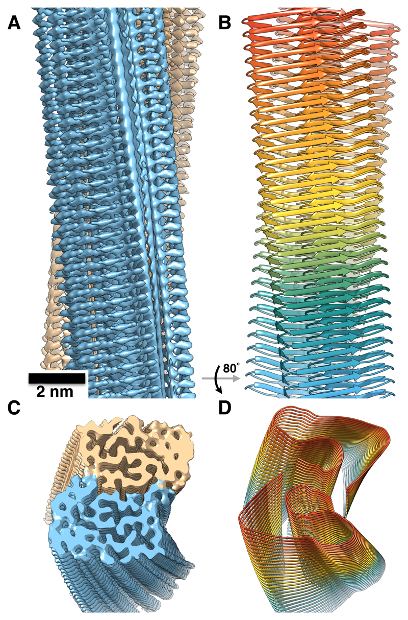
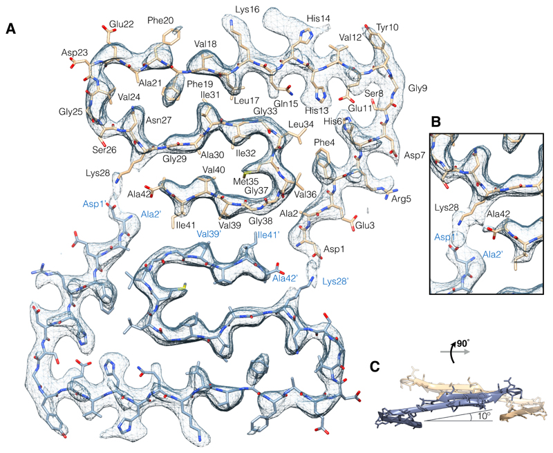
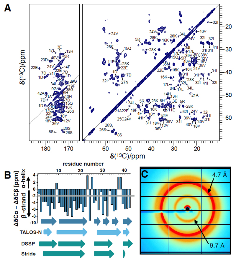
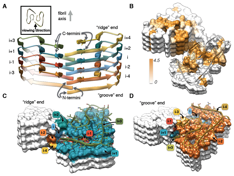
Amyloids are implicated in neurodegenerative diseases. Fibrillar aggregates of the amyloid-β protein (Aβ) are the main component of the senile plaques found in brains of Alzheimer's disease patients. We present the structure of an Aβ(1-42) fibril composed of two intertwined protofilaments determined by cryo-electron microscopy (cryo-EM) to 4.0-angstrom resolution, complemented by solid-state nuclear magnetic resonance experiments. The backbone of all 42 residues and nearly all side chains are well resolved in the EM density map, including the entire N terminus, which is part of the cross-β structure resulting in an overall "LS"-shaped topology of individual subunits. The dimer interface protects the hydrophobic C termini from the solvent. The characteristic staggering of the nonplanar subunits results in markedly different fibril ends, termed "groove" and "ridge," leading to different binding pathways on both fibril ends, which has implications for fibril growth.
Copyright © 2017 The Authors, some rights reserved; exclusive licensee American Association for the Advancement of Science. No claim to original U.S. Government Works.
Conflict of interest statement
The authors declare no competing financial interests.
Figures
Comment in
-
The molecular basis of Alzheimer's plaques.Science. 2017 Oct 6;358(6359):45-46. doi: 10.1126/science.aap8002. Science. 2017. PMID: 28983039 No abstract available.
References
Publication types
MeSH terms
Substances
Grants and funding
LinkOut - more resources
Full Text Sources
Other Literature Sources
Medical
Molecular Biology Databases
