. 2017 Sep 22;357(6357):1255-1261.
doi: 10.1126/science.aam9080. Epub 2017 Sep 7.

Dopamine oxidation mediates mitochondrial and lysosomal dysfunction in Parkinson's disease

Affiliations

Dopamine oxidation mediates mitochondrial and lysosomal dysfunction in Parkinson's disease

Lena F Burbulla et al. Science. .

Abstract

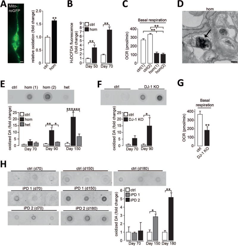
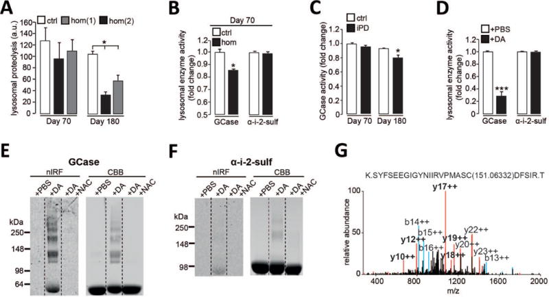
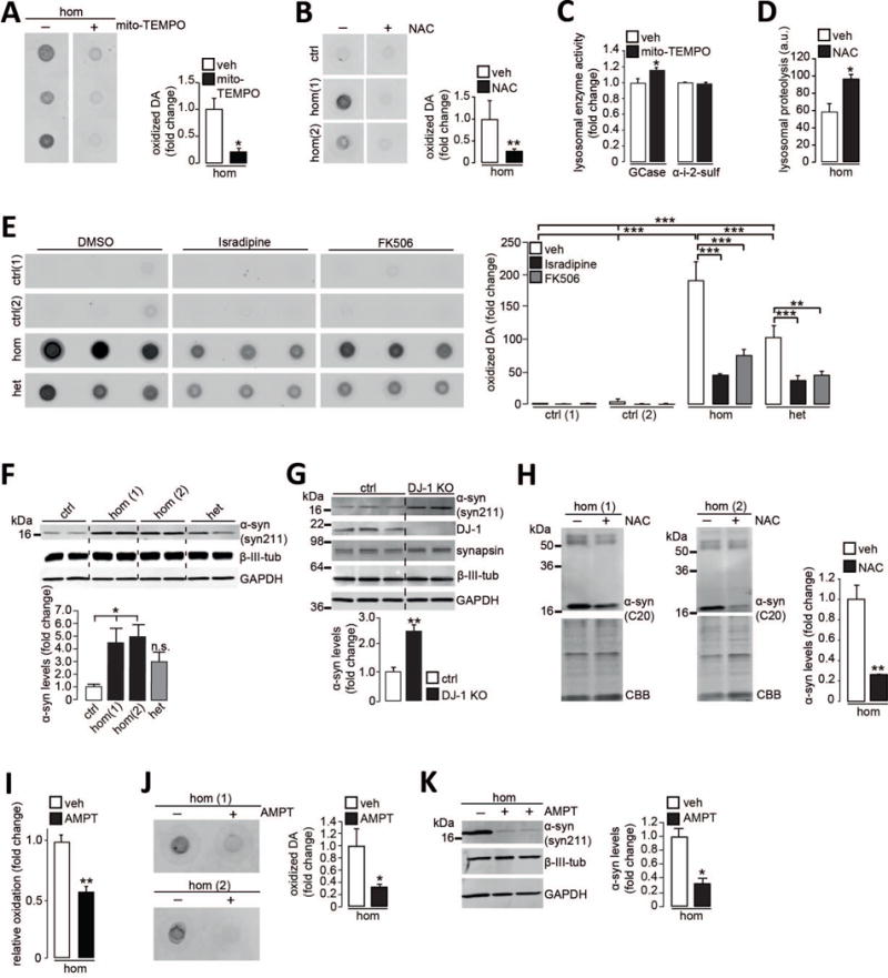
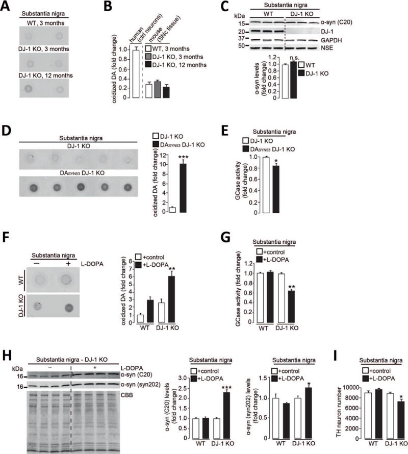
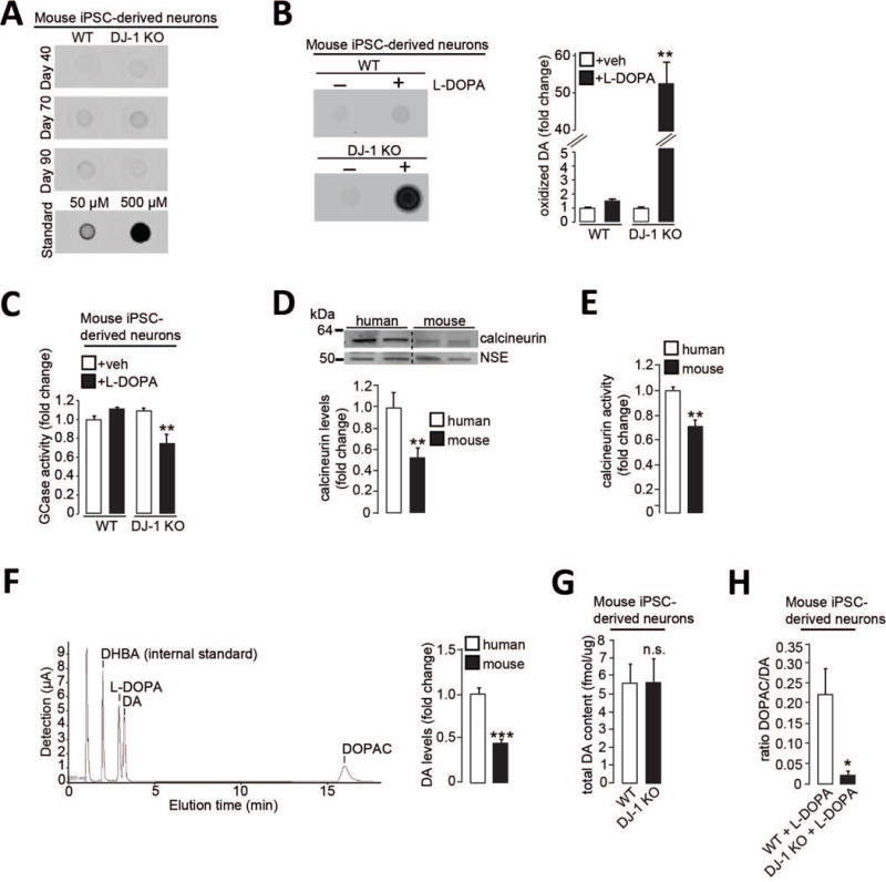
Mitochondrial and lysosomal dysfunction have been implicated in substantia nigra dopaminergic neurodegeneration in Parkinson's disease (PD), but how these pathways are linked in human neurons remains unclear. Here we studied dopaminergic neurons derived from patients with idiopathic and familial PD. We identified a time-dependent pathological cascade beginning with mitochondrial oxidant stress leading to oxidized dopamine accumulation and ultimately resulting in reduced glucocerebrosidase enzymatic activity, lysosomal dysfunction, and α-synuclein accumulation. This toxic cascade was observed in human, but not in mouse, PD neurons at least in part because of species-specific differences in dopamine metabolism. Increasing dopamine synthesis or α-synuclein amounts in mouse midbrain neurons recapitulated pathological phenotypes observed in human neurons. Thus, dopamine oxidation represents an important link between mitochondrial and lysosomal dysfunction in PD pathogenesis.


Figures

Figure 1
Figure 1. Neuromelanin and oxidized dopamine accumulate in PD patient dopaminergic neurons
(A) Neuronal expression of mito-roGFP and quantification of relative oxidation in control (ctrl) and homozygous DJ-1 mutant (hom) neurons at d50 (n=3). Scale bar, 10μm. (B) H2DCFDA fluorescence in neurons at d50 and d70 (n=3). (C) Oxygen consumption rate (OCR) in neurons under basal conditions at d50 (n=3). (D) Electron microscopy (EM) image of neuromelanin deposition (arrow) in homozygous DJ-1 mutant neuron at d90. Scale bar 200nm. (E-F) Oxidized dopamine (DA) by nIRF at d90 in (E) homozygous DJ-1 mutant (hom(1) and hom(2)) neurons and controls, quantification at d70, d90, and d150 (n=3), het for heterozygous, or (F) DJ-1 KO and isogenic control neurons and quantification at d70 and d90 (n=3-4). (G) OCR in DJ-1 KO neurons and isogenic controls under basal conditions at d50 (n=12). (H) Oxidized dopamine in control and two idiopathic PD (iPD1; iPD2) neurons at d70, d150 and d180 (n=3). Quantification for each iPD line is normalized to control for each time point. Equal protein concentrations were used for nIRF assays. Error bars, mean ± SEM. *P<0.05, **P<0.01, ***P<0.001, Student’s t test (A, B, F-H) or one-way analysis of variance (ANOVA) with Tukey post-hoc test (C, E).
Figure 2
Figure 2. Dopamine-mediated modification of GCase and lysosomal dysfunction in PD patient neurons
(A) Lysosomal proteolysis in control (ctrl) and homozygous DJ-1 mutant (hom) neurons at d70 and d180 (n=3). (B) GCase and a-i-2-sulf activity in lysosomal fractions from control and homozygous DJ-1 mutant neurons at d70 (n=3). (C) GCase activity in lysosomal fractions from control and idiopathic PD (iPD) neurons at d70 and d180 (n=3). (D) Recombinant GCase and α-i-2-sulf activity after incubation with DA or phosphate-buffered saline (PBS) (n=5). (E-F) Oxidized DA by in-gel nIRF of (E) recombinant GCase or (F) α-i-2-sulf after incubation with DA, DA+NAC or PBS. Coomassie brilliant blue (CBB) was used to visualize total protein. (G) MS/MS spectrum of GCase treated with DA. Modified cysteine is indicated with mass adduct in parentheses. Prominent b (blue) and y (red) ions and fragments containing the additional mass (bold) are indicated. Error bars, mean ± SEM. *P<0.05, ***P<0.001, one-way ANOVA with Tukey post-hoc test (A) or Student’s t test (B-D).
Figure 3
Figure 3. Mitochondrial antioxidants and calcium modulators attenuate the toxic cascade in DJ-1 mutant dopaminergic neurons
(A-B) Oxidized dopamine (DA) in homozygous DJ-1 mutant neurons (hom) treated with (A) mito-TEMPO or (B) NAC compared to vehicle (veh) at d70 (n=3). (C) GCase and α-i-2-sulf activity in lysosomal fractions from homozygous DJ-1 mutant neurons treated with mito-TEMPO or vehicle at d70 (n=3). (D) Lysosomal proteolysis in homozygous DJ-1 mutant neurons treated with NAC or vehicle at d180 (n=3). a.u., arbitrary units. (E) Oxidized DA in homozygous DJ-1 mutant, heterozygous DJ-1 carrier (het) and control neurons treated with isradipine, FK506 or vehicle (DMSO, dimethyl sulfoxide) at d90 (n=3-6). (F-G) Immunoblot analysis of α-synuclein (syn211 antibody) at d70 in Triton X-100 (T)-soluble neuronal lysates from (F) control, heterozygous DJ-1 carrier and two homozygous DJ-1 mutant lines (n=4) or (G) a gene-edited DJ-1 KO line (n=3 or 4). β-III-tubulin, synapsin, and GAPDH (glyceraldehyde-3-phosphate dehydrogenase) were used as loading controls. (H) T-insoluble α-synuclein (C20 antibody) at d70 in homozygous DJ-1 mutant neurons treated with NAC or vehicle. CBB used as loading control (n=4). (I) Homozygous DJ-1 mutant neurons expressing mito-roGFP treated with AMPT at d50 (n=3). (J-K) Homozygous DJ-1 mutant neurons treated with AMPT and analyzed at d70 for (J) oxidized DA by nIRF (n=3) or (K) T-soluble α-synuclein (syn211 antibody). GAPDH and β-III-tubulin loading controls (n=3). Treatment applied for 30 days (A-C, E, H-K) or 140 days (D). Error bars, mean ± SEM. *P<0.05, **P<0.01, ***P<0.001, Student’s t test (A-D, G-K) or one-way ANOVA with Tukey post-hoc test (E-F).
Figure 4
Figure 4. Increasing dopamine synthesis or amounts of α-synuclein in mouse midbrain neurons recapitulates pathological phenotypes observed in human neurons
(A-B) Oxidized DA in substantia nigra (SN) from DJ-1 KO and WT mice at 3 and 12 months compared to human control neurons at d70. (C) T-soluble α-synuclein (C20 antibody) in SNc of WT and DJ-1 KO mice. GAPDH and NSE *neural-specific enolase) loading controls (n=3 per group). (D-E) SN from DJ-1 KO and DASYN53 x DJ-1 KO mice analyzed at 8 months of age for (D) oxidized DA and (E) GCase activity (n=5 per group). (F-I) WT and DJ-1 KO mice were fed L-DOPA-supplemented or vehicle-treated chow for 6 months, substantia nigra analyzed at 14 months for (F) oxidized DA (n=3-4 per group), (G) GCase activity (n=3-4 per group), (H) T-insoluble α-synuclein (C20 and syn202 antibodies). CBB used as loading control (n=3-4 per group), and (I) number of DAB (3,3′-diaminobenzidine)-stained TH-positive neurons (n=3 per group). Equal protein concentrations were used for nIRF assays. Error bars, mean ± SEM. *P<0.05, **P<0.01, ***P<0.001, Student’s t test (C-E, H) or one-way ANOVA with Tukey post-hoc test (B, F-G, I,). n.s. = not significant.
Figure 5
Figure 5. Alterations in calcium homeostasis and dopamine metabolism contribute to intrinsic differences between human and mouse dopaminergic neurons
(A-C) WT and DJ-1 KO mouse iPSC-derived dopaminergic neurons were analyzed for (A) oxidized DA at d40, d70 and d90 (50μM and 500μM standards shown), or treated with L-DOPA or vehicle and analyzed at d55 for (B) oxidized DA (n=3) and (C) lysosomal GCase activity (n=3). (D-F) Control human and WT mouse iPSC-derived dopaminergic neurons analyzed for (D) T-soluble calcineurin (n=5-6), (E) calcineurin activity (n=5), and (F) DA amounts by HPLC (n=5 (mouse); n=10 (human)). HPLC chromatogram in a 4 μM standard sample is shown. Dihydroxybenzylamine (DHBA) was used as internal standard. (G) WT and DJ-1 KO mouse iPSC-derived dopaminergic neurons were analyzed for total DA content at d55 (n=5). (H) The ratio of DOPAC/DA for WT and DJ-1 KO mouse iPSC-derived dopaminergic neurons treated with L-DOPA (n=8). Equal protein concentration was used in nIRF assays. Error bars, mean ± SEM. *P<0.05, **P<0.01, ***P<0.001, Student’s t test (D-H) or one-way ANOVA with Tukey post-hoc test (B-C). n.s. = not significant.

Comment in

References

    1. Kalia LV, Lang AE. Parkinson’s disease. Lancet. 2015;386:896–912. - PubMed
    1. Surmeier DJ, Obeso JA, Halliday GM. Selective neuronal vulnerability in Parkinson disease. Nature reviews Neuroscience. 2017;18:101–113. - PMC - PubMed
    1. Dawson TM, Ko HS, Dawson VL. Genetic animal models of Parkinson’s disease. Neuron. 2010;66:646–661. - PMC - PubMed
    1. Bonifati V, et al. Mutations in the DJ-1 gene associated with autosomal recessive early-onset parkinsonism. Science. 2003;299:256–259. - PubMed
    1. Guzman JN, et al. Oxidant stress evoked by pacemaking in dopaminergic neurons is attenuated by DJ-1. Nature. 2010;468:696–700. - PMC - PubMed

Publication types

MeSH terms