Review
. 2017 Sep 1;10(9):1061-1074.
doi: 10.1242/dmm.030403.

Mouse models of metastasis: progress and prospects

Affiliations
Review

Mouse models of metastasis: progress and prospects

Laura Gómez-Cuadrado et al. Dis Model Mech. .

Abstract

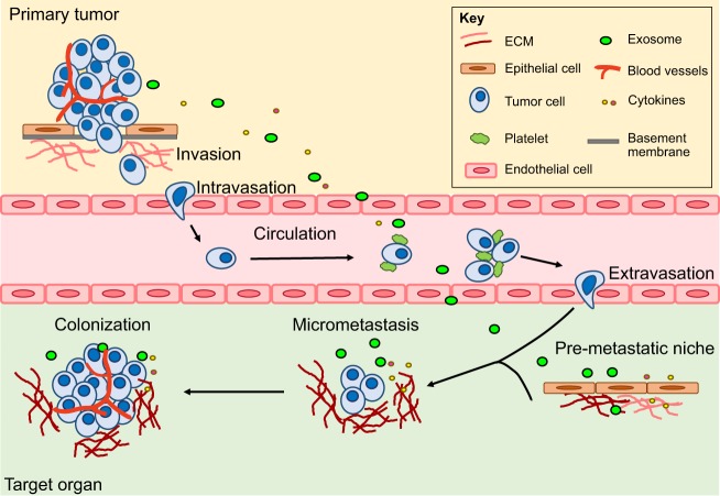
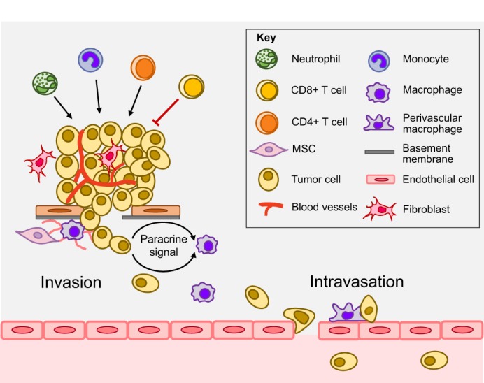
Metastasis is the spread of cancer cells from a primary tumor to distant sites within the body to establish secondary tumors. Although this is an inefficient process, the consequences are devastating as metastatic disease accounts for >90% of cancer-related deaths. The formation of metastases is the result of a series of events that allow cancer cells to escape from the primary site, survive in the lymphatic system or blood vessels, extravasate and grow at distant sites. The metastatic capacity of a tumor is determined by genetic and epigenetic changes within the cancer cells as well as contributions from cells in the tumor microenvironment. Mouse models have proven to be an important tool for unraveling the complex interactions involved in the metastatic cascade and delineating its many stages. Here, we critically appraise the strengths and weaknesses of the current mouse models and highlight the recent advances that have been made using these models in our understanding of metastasis. We also discuss the use of these models for testing potential therapies and the challenges associated with the translation of these findings into the provision of new and effective treatments for cancer patients.

Keywords: Cancer; Metastasis; Mouse models; Stroma.


Conflict of interest statement

Competing interestsThe authors declare no competing or financial interests.

Figures

Fig. 1.
Fig. 1.
Metastatic cascade. Metastasis is a multistep process. Initially, tumor cells migrate into adjacent tissues, referred to as local invasion. This involves breakdown of the basement membrane and invasion into the surrounding ECM. Intravasation then allows cells to enter the circulation. In blood vessels, CTCs exist as single cells or clusters, coated with platelets. They need to survive shear stress and evade clearance by the immune system to successfully reach distant organs. Tumor cells then attach to endothelial cells, which facilitates their extravasation. After settling in the metastatic target organ, tumor cells must survive in this foreign environment and establish micrometastases. These DTCs can remain dormant for many years before proliferating into large macrometastases in a process termed colonization. The primary site also regulates the development of metastasis via secretion of factors (such as cytokines and exosomes) that can prime a pre-metastatic niche (Box 2) and support survival of DTCs. See Glossary in Box 1 for an explanation of key terms.
Fig. 2.
Fig. 2.
Stromal influences in the primary tumor. Stromal cells such as MSCs, fibroblasts and myeloid cells (including monocytes, macrophages and neutrophils) promote metastasis and modulate the tumor microenvironment. MSCs remodel the ECM and support invasion. Macrophages can promote tumor invasion via several paracrine signaling factors. For example, in response to tumor cell-derived CSF1, TAMs secrete EGF, which is permissive for tumor cell invasion and migration. A subset of CD4+ T cells contributes to tumor progression, while CD8+ T cells mainly mediate anti-tumor immune responses. During intravasation, perivascular macrophages interact with tumor cells directly to help subsequent tumor cells to transit the endothelial barrier and initiate the journey of metastatic dissemination. See Glossary in Box 1 for an explanation of key terms.
Fig. 3.
Fig. 3.
Lineage tracing allows identification of the clonal nature of metastatic lesions. (A) The Brainbow-2.1 construct contains two tandem invertible DNA segments, each flanked by loxP sequences (indicated with black arrowheads). Inversion (i-iii) and excision (iv,v) recombination events create four expression possibilities, with the fluorescent protein that follows the promoter being uniquely expressed. Expression of the different fluorescent proteins at different ratios within each cell provides a unique color combination for each cell. Adapted from Livet et al. (2007), with permission from Macmillan Publishers Ltd. (B) Cre-mediated recombination during primary tumorigenesis allows identification of metastatic lesions of a single color, indicating derivation from single cells or clusters from a single cell, or multicolored cell clusters consisting of many different colored cells.

References

    1. Albritton J. L. and Miller J. S. (2017). 3D bioprinting: improving in vitro models of metastasis with heterogeneous tumor microenvironments. Dis. Model. Mech. 10, 3-14. 10.1242/dmm.025049 - DOI - PMC - PubMed
    1. Alix-Panabierès C. and Pantel K. (2013). Circulating tumor cells: liquid biopsy of cancer. Clin. Chem. 59, 110-118. 10.1373/clinchem.2012.194258 - DOI - PubMed
    1. Annunziato S., Kas S. M., Nethe M., Yücel H., Del Bravo J., Pritchard C., Bin Ali R., van Gerwen B., Siteur B., Drenth A. P. et al. (2016). Modeling invasive lobular breast carcinoma by CRISPR/Cas9-mediated somatic genome editing of the mammary gland. Genes Dev. 30, 1470-1480. 10.1101/gad.279190.116 - DOI - PMC - PubMed
    1. Aslakson C. J. and Miller F. R. (1992). Selective events in the metastatic process defined by analysis of the sequential dissemination of subpopulations of a mouse mammary tumor. Cancer Res. 52, 1399-1405. - PubMed
    1. Bald T., Landsberg J., Lopez-Ramos D., Renn M., Glodde N., Jansen P., Gaffal E., Steitz J., Tolba R., Kalinke U. et al. (2014). Immune cell-poor melanomas benefit from PD-1 blockade after targeted type I IFN activation. Cancer Discov. 4, 674-687. 10.1158/2159-8290.CD-13-0458 - DOI - PubMed

LinkOut - more resources