. 2017 Sep 7;8(1):481.
doi: 10.1038/s41467-017-00492-w.

Prophage-triggered membrane vesicle formation through peptidoglycan damage in Bacillus subtilis

Affiliations

Prophage-triggered membrane vesicle formation through peptidoglycan damage in Bacillus subtilis

Masanori Toyofuku et al. Nat Commun. .

Abstract

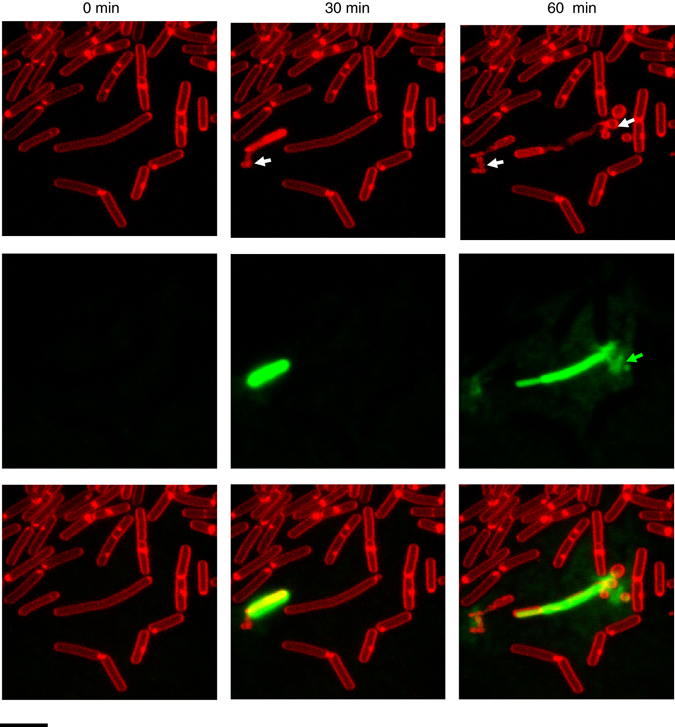
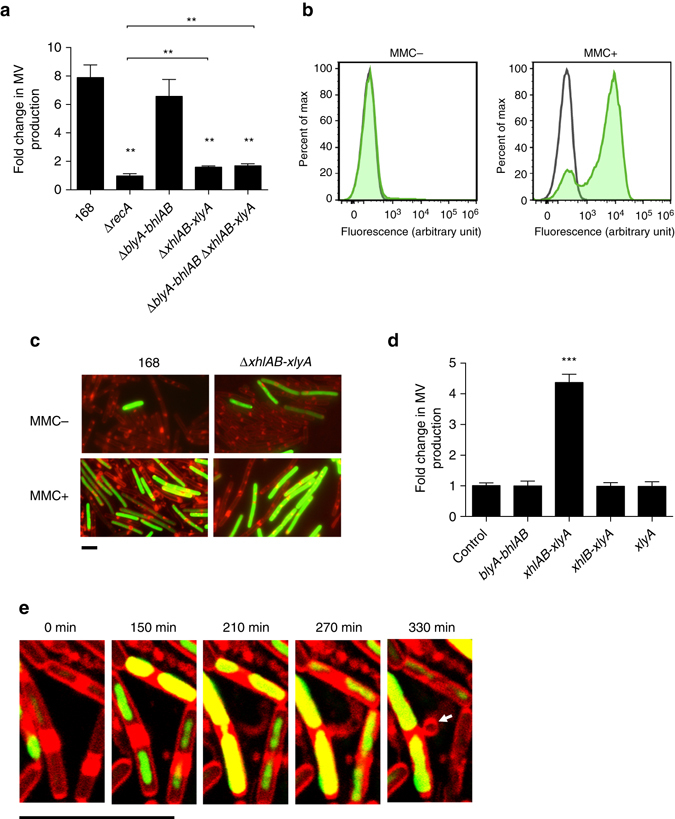
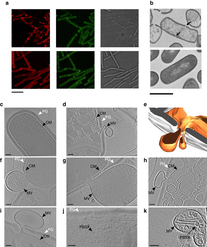
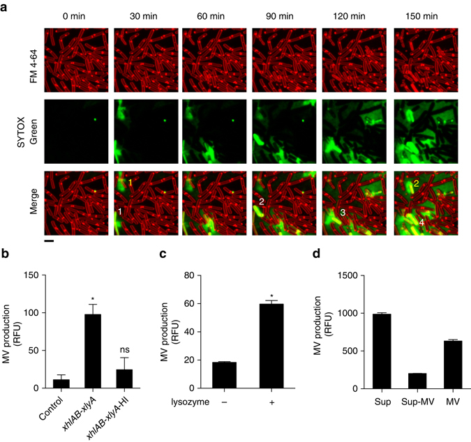
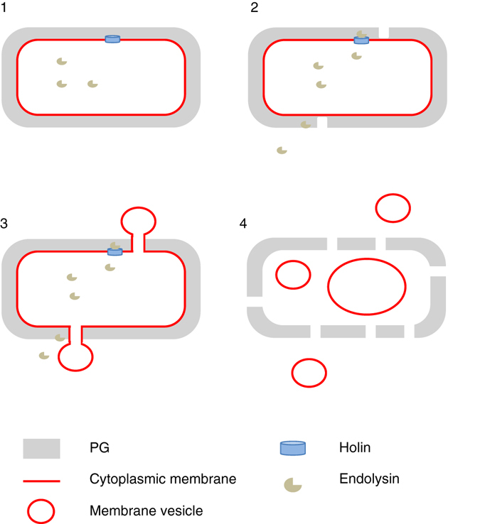
Bacteria release membrane vesicles (MVs) that play important roles in various biological processes. However, the mechanisms of MV formation in Gram-positive bacteria are unclear, as these cells possess a single cytoplasmic membrane that is surrounded by a thick cell wall. Here we use live cell imaging and electron cryo-tomography to describe a mechanism for MV formation in Bacillus subtilis. We show that the expression of a prophage-encoded endolysin in a sub-population of cells generates holes in the peptidoglycan cell wall. Through these openings, cytoplasmic membrane material protrudes into the extracellular space and is released as MVs. Due to the loss of membrane integrity, the induced cells eventually die. The vesicle-producing cells induce MV formation in neighboring cells by the enzymatic action of the released endolysin. Our results support the idea that endolysins may be important for MV formation in bacteria, and this mechanism may potentially be useful for the production of MVs for applications in biomedicine and nanotechnology.It is unclear how Gram-positive bacteria, with a thick cell wall, can release membrane vesicles. Here, Toyofuku et al. show that a prophage-encoded endolysin can generate holes in the cell wall through which cytoplasmic membrane material protrudes and is released as vesicles.


Conflict of interest statement

The authors declare no competing financial interests.

Figures

Fig. 1
Fig. 1
MVs are released by B. subtilis 168 at the onset of cell death. MV formation was followed at 30 min intervals at room temperature. Membranes were stained with the red fluorescent dye FM4-64 and SYTOX green was used to visualize dead cells and extracellular DNA (green). White arrows indicate cells releasing MVs and green arrows indicate extracellular DNA. Scale bar, 5 μm
Fig. 2
Fig. 2
Genotoxic stress induces MV formation through activation of the holin–endolysin system. a Fold-change of MV production of MMC-treated cells relative to non-treated cells. n = 3; mean ± s.d. **P < 0.01 (unpaired t-test with Welch’s correction). b Expression of the PBSX holin–endolysin gene cluster is induced by MMC. A transcriptional fusion of the PL promoter, which drives transcription of the PBSX late operon (including the holin–endolysin genes), to zsGreen was quantified by flow cytometry. Green curve shows activity of the PL promoter; black curve shows activity of the vector control. c Promoter (PL) activities of the PBSX holin–endolysin gene cluster under MMC non-inducing and inducing conditions. The image shows FM4-64 (red) merged with ZsGreen (green). Scale bar, 5 μm. Phase contrast images along with the images of control experiments are shown in Supplementary Fig. 2a. d Fold-change of MV production by the expression of holin–endolysin genes relative to the control strain 168 (PxylA). ***P < 0.001 (unpaired t-test with Welch’s correction). e Live cell imaging of MV formation in B. subtilis 168 (PxylA -xhlAB-xlyA) cells. Arrows indicate MVs. Cells were incubated on LB agarose pads containing 0.1% xylose, FM4-64 (red) and SYTOX green (green). Scale bar, 5 μm
Fig. 3
Fig. 3
The activity of the PBSX endolysin creates holes in the bacterial cell wall. a Staining of the PG with FDL in cells expressing the holin–endolysin genes and in uninduced control cells. Upper panel shows strain 168 (PxylA -xhlAB-xlyA), in which the holin–endolysin genes were expressed; the lower panel shows the control strain 168 (PxylA). Membranes were stained with FM4-64 (red, left) and the PG was stained with FDL (green, central). Corresponding bright field images are shown in the right panel. Scale bar, 5 μm. b Thin section of induced B. subtilis 168 (PxylA -xhlAB-xlyA) cells (upper panel) and of the control strain 168 (PxylA) (lower panel). Arrows indicate holes in the bacterial cell wall. Scale bar, 1 μm. ck ECT images of different Bacillus ΔponA strains. c Uninduced B. subtilis ΔponA (PxylA -xhlAB-xlyA) cells have an intact peptidoglycan (PG) layer and cytoplasmic membrane (CM) and a dense cytoplasm. dh Induced cells extrude MVs through holes in the PG layer. We also observed cells with lowered cytoplasmic densities that harbored intracellular vesicles (h). ik Likewise, cells of a MMC-induced B. subtilis ΔponA culture showed MV formation (i) in addition to phage particles (PBSX) inside cells (j) and sometimes within extracellular MVs (k). Shown are 1.38 nm (c, f, h, k), 6.9 nm (g), 11.86 nm (i), 29.64 nm (j) thick slices through cryotomograms and a model (e) of the cell shown in d. White, peptidoglycan; orange, cytoplasmic membrane. Scale bars, 100 nm
Fig. 4
Fig. 4
Endolysin triggers MV release in neighboring cells. a Cell death induces MV release in neighboring cells. Numbers depict the progression of cell death in the population. Scale bar, 5 μm. Membranes are stained with the red fluorescence dye FM4-64. Dead cells and extracellular DNA are visualized with SYTOX green. b MV production is increased in the presence of PBSX endolysin. Cells were stained with FM4-64FX, fixed and incubated with supernatants of the control strain 168 (PxylA) or with induced 168 (PxylA -xhlAB-xlyA) supernatants (untreated; xhlAB-xlyA and heat-inactivated; xhlAB-xlyA-HI). n = 6; mean ± s.d. ****P < 0.0001. c MV production in strain 168 is stimulated by treatment of exponentially grown cells with lysozyme. n = 3; mean ± s.d. ****P < 0.0001, or d in the presence of purified MVs. Cells were stained with FM1-43FX, fixed and incubated with the supernatant (Sup), the supernatant without MVs (Sup-MV) and purified MVs of induced 168 (PxylA -xhlAB-xlyA) cultures. n = 3
Fig. 5
Fig. 5
Schematic model of MV production by the holin–endolysin pathway in B. subtilis. Step 1, the holin forms pores in the cytoplasmic membrane to allow the endolysin to access the PG. Step 2, the endolysin degrades the PG and creates holes. The PG can also be degraded from outside, e.g., by endolysins released from neighboring cells. Step 3, the cytoplasmic membrane is extruded through the holes in the PG, presumably due to high cell turgor. Step 4, the MVs are released. We also observed that intracellular membrane fragments can vesicularize and may eventually be released from the dying cell

References

    1. Deatherage BL, Cookson BT. Membrane vesicle release in bacteria, eukaryotes, and archaea: a conserved yet underappreciated aspect of microbial life. Infect. Immun. 2012;80:1948–1957. doi: 10.1128/IAI.06014-11. - DOI - PMC - PubMed
    1. Schwechheimer C, Kuehn MJ. Outer-membrane vesicles from Gram-negative bacteria: biogenesis and functions. Nat. Rev. Microbiol. 2015;13:605–619. doi: 10.1038/nrmicro3525. - DOI - PMC - PubMed
    1. Brown L, Wolf JM, Prados-Rosales R, Casadevall A. Through the wall: extracellular vesicles in Gram-positive bacteria, mycobacteria and fungi. Nat. Rev. Microbiol. 2015;13:620–630. doi: 10.1038/nrmicro3480. - DOI - PMC - PubMed
    1. Mashburn LM, Whiteley M. Membrane vesicles traffic signals and facilitate group activities in a prokaryote. Nature. 2005;437:422–425. doi: 10.1038/nature03925. - DOI - PubMed
    1. Toyofuku M, et al. Membrane vesicle-mediated bacterial communication. ISME J. 2017;11:1504–1509. doi: 10.1038/ismej.2017.13. - DOI - PMC - PubMed

Publication types

MeSH terms

LinkOut - more resources