. 2017 Sep 7;8(1):473.
doi: 10.1038/s41467-017-00618-0.

Tau exacerbates excitotoxic brain damage in an animal model of stroke

Affiliations

Tau exacerbates excitotoxic brain damage in an animal model of stroke

Mian Bi et al. Nat Commun. .

Abstract

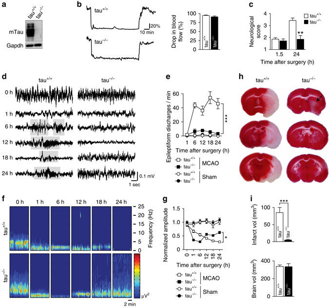
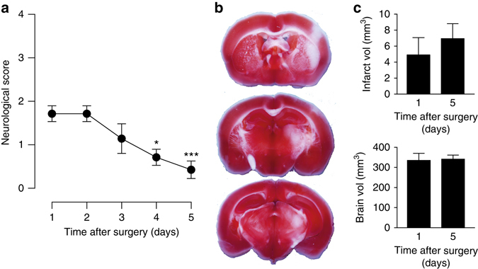
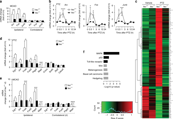
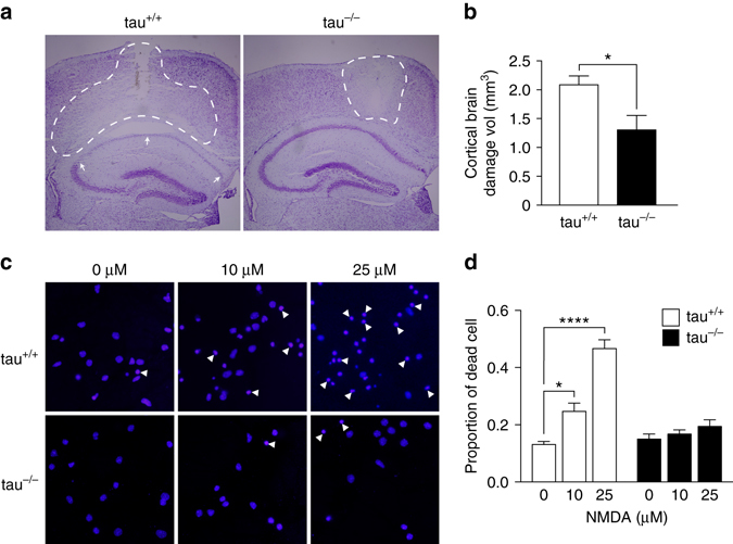
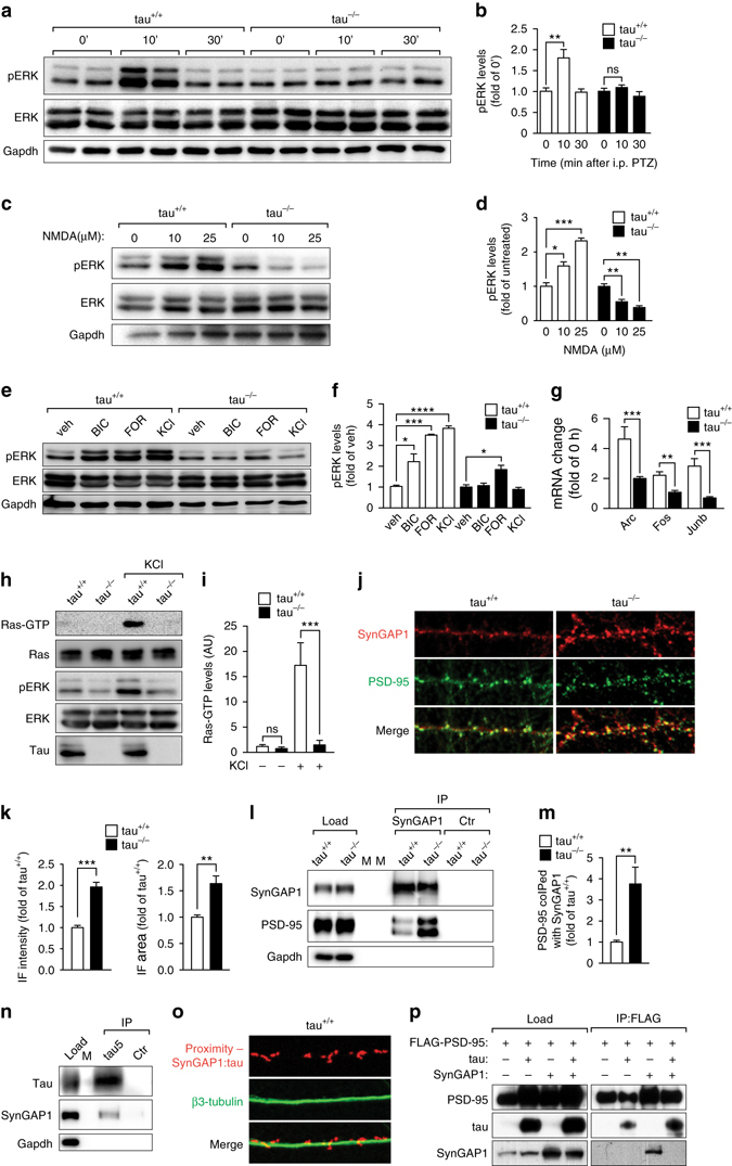
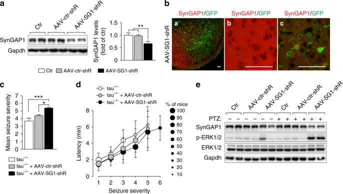
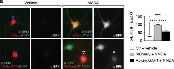
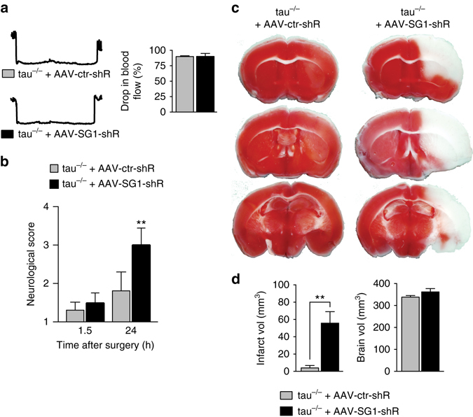
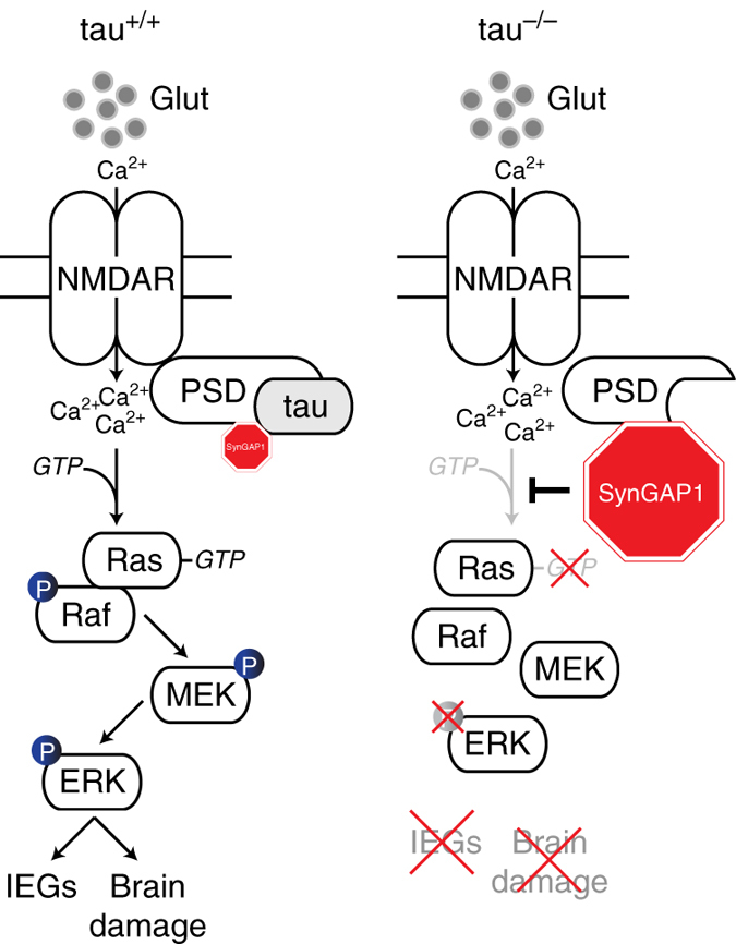
Neuronal excitotoxicity induced by aberrant excitation of glutamatergic receptors contributes to brain damage in stroke. Here we show that tau-deficient (tau-/-) mice are profoundly protected from excitotoxic brain damage and neurological deficits following experimental stroke, using a middle cerebral artery occlusion with reperfusion model. Mechanistically, we show that this protection is due to site-specific inhibition of glutamate-induced and Ras/ERK-mediated toxicity by accumulation of Ras-inhibiting SynGAP1, which resides in a post-synaptic complex with tau. Accordingly, reducing SynGAP1 levels in tau-/- mice abolished the protection from pharmacologically induced excitotoxicity and middle cerebral artery occlusion-induced brain damage. Conversely, over-expression of SynGAP1 prevented excitotoxic ERK activation in wild-type neurons. Our findings suggest that tau mediates excitotoxic Ras/ERK signaling by controlling post-synaptic compartmentalization of SynGAP1.Excitotoxicity contributes to neuronal injury following stroke. Here the authors show that tau promotes excitotoxicity by a post-synaptic mechanism, involving site-specific control of ERK activation, in a mouse model of stroke.


Conflict of interest statement

The authors declare no competing financial interests.

Figures

Fig. 1
Fig. 1
Tau−/− mice are protected from neurological deficits, aberrant hyperexcitation and extensive brain damage after transient MCAO. a Western blotting for murine tau (mTau) in brain extracts from tau+/+ and tau−/− mice. GAPDH confirmed equal loading. b Ischemic stroke was induced by middle cerebral artery occlusion (MCAO) for 1.5 h with subsequent reperfusion. Drop in blood flow in the MCA was the same in tau−/− and tau+/+ mice during MCAO, as determined by laser Doppler flowmetry (not significant; N = 12; Student’s t-test). Unit, % of baseline flow. c Neurological scoring (with higher numbers indicating more severe impairments) revealed similar deficits directly after MCAO in tau−/− and tau+/+ mice. Only tau+/+ mice showed a worsening of deficits at 24 h (**P < 0.01; N = 12; 2-way ANOVA (Sidak post hoc)). d Representative electroencephalography (EEG) recordings in tau−/− and tau+/+ mice at baseline (0 h) and indicated times after transient MCAO. Suppressed EEG signals recovered after 12 h following MCAO in tau−/− mice, while tau+/+ mice presented with epileptiform discharges (gray boxed) after 6 h. e Quantification revealed persistently high numbers of epileptiform discharges 6 h after transient MCAO (***p < 0.001 vs. tau−/− MCAO; N = 5; two-way ANOVA (Bonferroni post hoc)). A small increase in epileptiform spike trains was transient in tau−/− mice, and reached levels of sham operated tau−/− and tau+/+ mice 24 h after transient MCAO. f EEG frequency power spectrum (0–25 Hz) tau+/+ and tau−/− mice at baseline (0 h) indicated times following transient MCAO, with recovery only in tau−/− animals. g Quantification of EEG recordings showed a progressive amplitude decline in tau+/+ mice that partial recovered tau−/− animals (*p < 0.05; N = 5; two-way ANOVA (Bonferroni post hoc)). h TTC-stained serial brain sections of tau+/+ and tau−/− mice 24 h after MCAO (viable tissue stains red). Note the large infarcted area (white) in tau+/+, while only minimal brain damage was present in tau−/− mice (arrow). i Volumetric quantification of infarct and brain volumes in tau+/+ and tau−/− mice 24 h after transient MCAO (***p < 0.001; N = 12; Student’s t-test). All error bars are s.e.m
Fig. 2
Fig. 2
Improvement of functional deficits over 5 days after 90 min of transient MCAO in tau−/− mice. a Neurological impairments of tau−/− mice after 90 min of transient MCAO progressively improved over 5 days after the procedure (*p < 0.05; ***p < 0.001; N = 8; one-way ANOVA (Turkey post hoc)). b Example of a maximum infarct area 5 days after transient MCAO in tau−/− mice. TTC stains viable brain tissue in serial sections red. c Volumetric analysis showed similar infarct volumes 24 h and 5 days after MCAO (not significant; N = 12 (day 1), N = 8 (day 5); Student’s t-test). All error bars are s.e.m
Fig. 3
Fig. 3
Accelerated recovery of tau−/− mice from transient MCAO. a For longer-term follow up of tau−/− and tau+/+ mice after stroke, animals were exposed to 30 min of transient MCAO. Representative laser Doppler flowmetry of the MCAO procedure. Unit, % of baseline flow. b Significantly less neurological deficits with faster recovery in tau−/− compared to tau+/+ mice following to 30 min of transient MCAO (*p < 0.05; **p < 0.01; ****p < 0.0001; N = 8; two-way ANOVA (Sidak post-hoc)). c Significantly less body weight loss with faster recovery in tau−/− compared to tau+/+ mice following to 30 min of transient MCAO (*p < 0.05; **p < 0.01; ***p < 0.001; N = 8; two-way ANOVA (Sidak post hoc)). d Tau−/− mice performed significantly better on the accelerative mode Rota-Rod than tau+/+ animals (***p < 0.001; N = 8; two-way ANOVA (Sidak post hoc)). e TTC-stained serial brain sections of tau+/+ and tau−/− mice 14 days after 30 min of transient MCAO (viable tissue stains red). Note the larger infarcted brain area (white) in tau+/+, compared to minimal brain damage in tau−/− mice. f Quantification of infarct and brains volumes in tau−/− and tau+/+ mice (**P < 0.001; N = 8; Student’s t-test). All error bars are s.e.m
Fig. 4
Fig. 4
Excitotoxic seizures and transient MCAO cause similar differential gene regulation in tau−/− mice. a Immediate early gene (IEG) activation for Arc, Fos and Junb in the affected hemisphere (ipsilateral) 1 h after transient MCAO in tau+/+ and tau−/− mice (**p < 0.01; N = 6; Student’s t-test). No gene induction occurred on the contralateral side. b Time course of IEG mRNA induction of Arc, Fos and Junb after PTZ administration in tau+/+ but little to none in tau−/− mice (*p < 0.05; ***p < 0.001; ****p < 0.0001; N = 6 per time point; two-way ANOVA (Holm-Sidak post hoc)). mRNA levels were determined by real-time PCR (rtPCR) using gene-specific primers listed in Supplementary Table 3. c Whole transcriptome sequencing of tau−/− and tau+/+ mice treated with vehicle or PTZ revealed pronounced lack of gene induction/suppression 1 h after treatment in tau−/− mice. Red indicates up- and green down-regulation. Only genes with significant differential regulation are displayed. The displayed gene names are listed in order in Supplementary Data 1. d Quantitative rtPCR confirms differential regulation of selected transcripts from (see c) tau−/− mice (*p < 0.05; ***p < 0.0001; N = 6; Student’s t-test), as well as similar regulation of others (selected from Supplementary Fig. 6 and Supplementary Data 2) in tau−/− and tau+/+ mice. e Quantitative rtPCR showed a similar pattern of differentially regulated genes in the ipsilateral hemisphere after transient MCAO compared to after PTZ (see d) (*p < 0.05; ***p < 0.0001; N = 6; Student’s t-test), while there was no deregulation contralaterally in tau−/− and tau+/+ mice. f DAVID pathway analysis of genes that lacked response to PTZ administration in tau−/− mice (see c) identified pathways that were up- (black) and down-regulated (gray) in tau+/+ mice. All error bars are s.e.m
Fig. 5
Fig. 5
Tau-depletion reduced neurotoxicity of NMDA. a Representative Nissl staining of tau+/+ and tau−/− brains 24 h after infusion of 0.2 µl of 50 mM NMDA into the cortex with pale appearance of damaged brain area (broken lines). Note that the damaged area extends profoundly into the CA1 region of the hippocampus (arrows) in tau+/+ but not tau−/− brains, suggesting progressive expansion of excitotoxicity only in tau+/+ brains. b Quantification confirmed less total brain damage in tau−/− compared to tau+/+ mice (*p < 0.05, N = 4; Student’s t-test). c Primary cortical neurons treated with 0, 10 and 25 µM NMDA showed less cell death (determined by nuclear condensation (some indicated by arrowheads), DAPI) in tau−/− compared with tau+/+ cells. d Quantification of neuron death induced by NMDA in tau−/− and tau+/+ cells (*p < 0.05; ****p < 0.0001; N = 12; two-way ANOVA (Sidak post hoc)). All error bars are s.e.m
Fig. 6
Fig. 6
SynGAP1 accumulation blocks Ras/ERK signaling at the post-synapse in tau−/− mice. a Western blotting of brain extracts; PTZ induced transient ERK phosphorylation 10 min after administration in tau+/+ but not tau−/− mice, b as confirmed by quantification of independent blots (**p < 0.01; N = 6; one-way ANOVA (Tukey post hoc)). c Primary tau+/+ but not tau−/− neurons showed ERK phosphorylation when challenged with 10 and 25 µM NMDA, d as confirmed by quantification of independent blots (*p < 0.05; **p < 0.01; ***p < 0.001; N = 3; one-way ANOVA (Tukey post hoc)). e Primary tau+/+ neurons showed ERK phosphorylation when treated with bicuculline (BIC), forskolin (FOR) and KCl. In contrast, primary tau−/− neurons showed only increased ERK phosphorylation upon treatment with FOR, though to a lower degree. f Quantification of independent blots confirmed BIC-, FOR- and KCl-induced ERK phosphorylation in tau+/+ neurons, while only FOR significantly activated ERK in tau−/− cells (*p < 0.05; ***p < 0.001; ****p < 0.0001; N = 6; one-way ANOVA (Tukey post hoc)). g BIC treatment of primary neurons resulted in upregulation of Arc, Fos and Junb mRNA levels in tau+/+, but little to none in tau−/− cells (**p < 0.01; ***p < 0.001; N = 6; Student’s t-test). h Activated Ras (Ras-GTP) was pulled down from KCl treated tau+/+, but not tau−/− brain slices. Total Ras levels, however, were comparable tau−/− and tau+/+ mice. Concomitant ERK activation was only seen in KCl treated tau+/+ slices. i Quantification of independent blots confirmed that Ras activation occurred only in tau+/+ mice (***p < 0.001; N = 6; one-way ANOVA (Tukey post hoc)). j Confocal imaging of primary neurons co-stained with SynGAP1 (green) and PSD-95 (red) showed more intensive labeling of spines for SynGAP1 that co-localized with PSD-95 in tau−/− than in tau+/+ neurons. k Both, immunofluorescence intensity and cluster size of SynGAP1 in spines were significantly increased in tau−/− compared to tau+/+ neurons (**p < 0.01; ***p < 0.001; N = 5; Student’s t-test). l Immunoprecipitation (IP) with a SynGAP1-specific antibody co-purified 3.8-fold more PSD-95 from tau−/− brain extracts than from tau+/+ lysates. Control (ctr) precipitations were done without primary antibodies. Load represents brain extracts used for immunoprecipitation. M, marker. m Quantification of 6 independent experiments showed a significant increase in SynGAP1/PSD-95 interaction in tau−/− compared to tau+/+ mice (**p < 0.01; N = 6; Student’s t-test). n SynGAP1 interacts with tau in brain extracts from wild-type mice, as revealed by co-IP using a tau-specific antibodies (tau5). Control (ctr) precipitations were done without primary antibodies. M, marker lanes. o SynGAP1 and tau are in a complex within dendritic spines, as determined by a < 40 nm proximity ligation assay (SynGAP1:tau; red) using SynGAP1- and tau-specific antibodies. Dendritic shafts were counter-stained with β3-tubulin (green). p IP of FLAG-PSD95 co-precipitated SynGAP1 in the absence but not in the presence of tau from transiently transfected cells (load) (N = 3). All error bars are s.e.m
Fig. 7
Fig. 7
Reducing SynGAP1 levels in tau−/− mice reinstates susceptibility to seizures and ERK activation. a Neonatal (P1-3) wild-type C57Bl/6 mice were injected intracranially with AAVs expressing either shRNA to knockdown SynGAP1 (AAV-SG1-shR) or control shRNA (AAV-ctr-shR). Western blotting of cortical extracts from 2-month-old naive (ctr) and AAV-injected mice showed marked reduction of SynGAP1 levels in AAV-SG1-shR-injected mice. Quantification of band intensities confirmed significant SynGAP1 reduction by AAV-SG1-shR (**p < 0.01; N = 9; one-way ANOVA (Tukey post hoc)). b Example of GFP reporter expression in neurons throughout the cortex of AAV-SG1-shR-injected mice. Double labeling with a SynGAP1 antibody (red) revealed SynGAP1 expression in areas with no GFP expression and marked reduction of SynGAP1 in GFP-positive areas, suggesting efficient knockdown. Higher magnification of areas with neuronal and synaptic SynGAP1, but no GFP expression (middle), or neuronal GFP reporter expression with reduced SynGAP1 (right). Scale bars, 100 µm. c AAV-SG1-shR, but not AAV-ctr-shR injection increased the mean seizure severity in tau−/− mice after administration of 50 mg/kg PTZ (*p < 0.05; ***p < 0.001; N = 11 (tau−/−), N = 13 (AAV injected tau−/−); one-way ANOVA (Tukey post hoc)). d More tau−/− mice injected with AAV-SG1-shR developed higher degree seizures after administration of 50 mg/kg PTZ, compared with naive (ctr) or AAV-ctr-shR-injected tau−/− mice. The latency of seizure progression was comparable. e Strong ERK1/2 phosphorylation 10 min after 50 mg/kg PTZ administration in AAV-SG1-shR-injected, but not AAV-ctr-shR-injected or naive (ctr) tau−/− mice, together with reduction of SynGAP1 levels. Detection of ERK1/2 and Gapdh confirmed equal loading. All error bars are s.e.m
Fig. 8
Fig. 8
Expression of SynGAP1 mitigated NMDA-induced ERK activation in primary neurons. a Primary neurons transiently over-expressing V5-SynGAP1 (red) showed less ERK phosphorylation (p-ERK, green) in response to NMDA exposure, compared to control cells expressing mCherry (red) (or untransfected cells). No p-ERK was detected prior to NMDA treatments. b Quantification of fluorescence intensity of p-ERK signals in transfected neurons showed a significant reduction of ERK activation after NMDA in V5-SynGAP1− compared to mCherry-expressing neurons. Controls (ctr) comprised both mCherry, SynGAP1 or untransfected cells (***p < 0.001; ****p < 0.0001; N = 3; one-way ANOVA (Tukey post hoc)). All error bars are s.e.m
Fig. 9
Fig. 9
Reducing SynGAP1 levels in tau−/− mice confers susceptibility to transient MCAO. a Blood flow was reduced to the same extend during transient MCAO in 2 month-old tau−/− injected with either AAV-SG1-shR or AAV-ctr-shR at P1-3 (not significant; N = 5; Student’s t-test). b Neurological scoring (with higher numbers indicating more severe impairments) revealed similar deficits directly after transient MCAO in AAV-SG1-shR- and AAV-ctr-shR-injected tau−/− mice, but thereafter, neurological deficits in AAV-SG1-shR-injected tau−/− mice progressed more severely than in AAV-ctr-shR-injected tau−/− mice (**p < 0.01; N = 5; two-way ANOVA (Tukey post hoc)). c Representative TTC-stained brain sections of tau−/− mice injected with AAV-SG1-shR or AAV-ctr-shR 24 h after transient MCAO (viable tissue stains red). Note the large infarcted area (white) in AAV-SG1-shR-injected tau−/− mice, while only minimal brain damage was present in AAV-ctr-shR-injected tau−/− mice. d Infarct and brain volume quantification of serial TTC-stained sections from AAV-SG1-shR-injected compared to AAV-ctr-shR-injected tau−/− mice (**p < 0.01; N = 5; Student’s t-test). All error bars are s.e.m
Fig. 10
Fig. 10
Schematic of excitotoxic, NMDAR-mediated Ras/ERK signaling in tau+/+ and tau−/− mice. In wild-type mice (tau+/+), excessive synaptic glutamate (glut) levels trigger increased calcium (Ca2+) influx via NMDARs at the post-synapse. This triggers Ras activation by GTP binding, and eventually activation (phosphorylation; P) of ERK via the Ras/Raf/MEK/ERK cascade. To allow for Ras activation, levels of the site-specific inhibitor SynGAP1 (red) that resides in a complex with PSD-95 and tau need to be controlled (a process we found to be dependent directly or indirectly on tau). In contrast, our data revealed absence of excitotoxic Ras activation and hence downstream ERK phosphorylation in tau−/− mice. In the absence of tau, SynGAP1 is increasingly bound to PSD-95 and completely suppresses site-specific NMDAR-mediate activation of Ras at the post-synapse. As a consequence, neuronal damage and activation of immediate-early genes (IEGs) after stroke and excitotoxic seizures are suppressed

Comment in

References

    1. Donnan GA, Fisher M, Macleod M, Davis SM. Stroke. Lancet. 2008;371:1612–1623. doi: 10.1016/S0140-6736(08)60694-7. - DOI - PubMed
    1. Sanderson TH, Reynolds CA, Kumar R, Przyklenk K, Huttemann M. Molecular mechanisms of ischemia-reperfusion injury in brain: pivotal role of the mitochondrial membrane potential in reactive oxygen species generation. Mol. Neurobiol. 2013;47:9–23. doi: 10.1007/s12035-012-8344-z. - DOI - PMC - PubMed
    1. Hacke W, et al. Thrombolysis with alteplase 3 to 4.5 hours after acute ischemic stroke. N. Engl. J. Med. 2008;359:1317–1329. doi: 10.1056/NEJMoa0804656. - DOI - PubMed
    1. Kidwell CS, et al. A trial of imaging selection and endovascular treatment for ischemic stroke. N. Engl. J. Med. 2013;368:914–923. doi: 10.1056/NEJMoa1212793. - DOI - PMC - PubMed
    1. Campbell BC, et al. Endovascular therapy for ischemic stroke with perfusion-imaging selection. N. Engl. J. Med. 2015;372:1009–1018. doi: 10.1056/NEJMoa1414792. - DOI - PubMed

Publication types

MeSH terms