. 2017 Sep 7;7(1):10868.
doi: 10.1038/s41598-017-11455-y.

Techniques and long-term effects of transjugular intrahepatic portosystemic shunt on liver cirrhosis-related thrombotic total occlusion of main portal vein

Affiliations

Techniques and long-term effects of transjugular intrahepatic portosystemic shunt on liver cirrhosis-related thrombotic total occlusion of main portal vein

Lei Wang et al. Sci Rep. .

Abstract

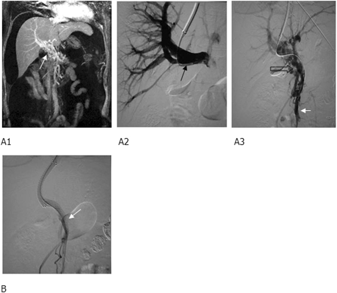
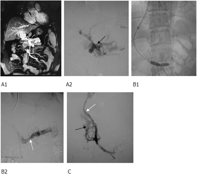
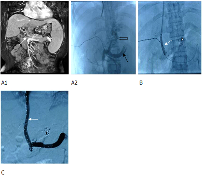
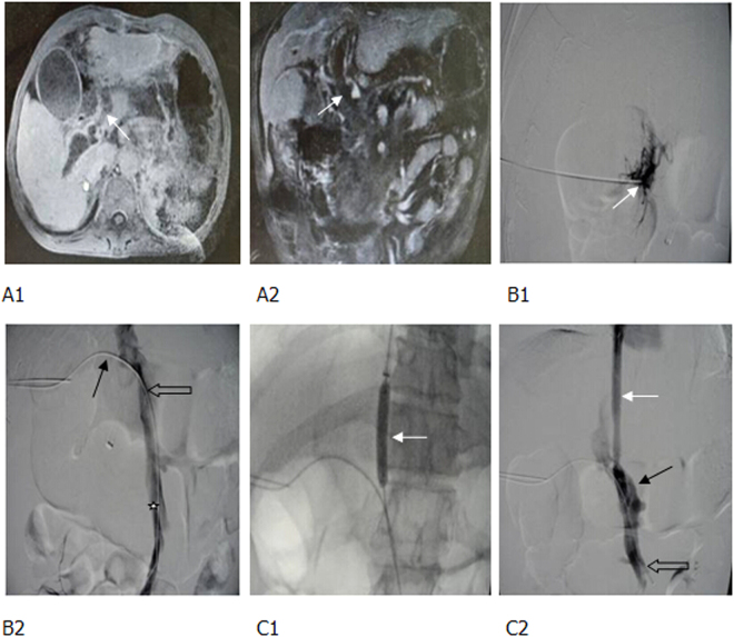
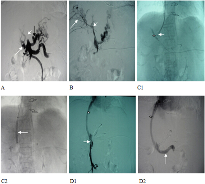
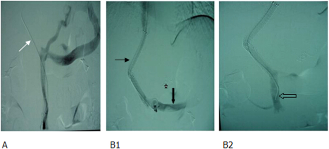
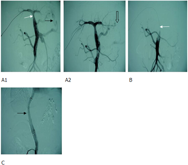
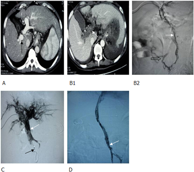
Portal vein hypertension (PVH) in liver cirrhosis complicated with portal venous thrombosis (PVT) has been mainly treated with transjugular intrahepatic portosystemic shunt (TIPS). The clinical effects of TIPS have been confirmed, however, no large-scale studies have been focused on technical analyses and a long-term follow-up, especially on thrombotic total occlusion of main portal vein (MPV). To demonstrate critical techniques and clinical outcome of TIPS on liver cirrhosis-related thrombotic total occlusion of MPV, 98 patients diagnosed with liver cirrhosis related thrombotic total occlusion of MPV and treated with TIPS from January 2000 to January 2010 were retrospectively analyzed. Twenty-three (23.5%) patients had MPV (single site) thrombosis, 55 (56.1%) had multiple site-thrombosis (MPV and other), 17 (17.3%) had cavernous transformation of portal vein, and 3 (3.1%) had post-transplant thrombosis. The successful rate of TIPS was 90.7%, without any procedure-related deaths or severe complications. Mean portal pressure was dropped from 33.08 ± 1.38 mmHg preoperatively to 20.18 ± 0.83 mmHg postoperatively (p < 0.001). Collectively, TIPS is safe and effective in treating liver cirrhosis-related thrombotic total occlusion of MPV. This complex procedure requires combination of indirect portography and percutaneous transhepatic portal techniques to increase the rate of success.


Conflict of interest statement

The authors declare that they have no competing interests.

Figures

Figure 1
Figure 1
MRPV (Magnetic Resonance of portal vein) and TIPS (transjugular intrahepatic portosystemic shunt) portography showed (A1) cavernous transformation of portal vein (white arrow), right portal branch (black arrow), (A2) occluded trunk of superior mesenteric vein (hollow arrow), and (A3) a relatively large branch (short and white arrow). (B) Two BMS (bare metal stent) were implanted between a hepatic vein and a relatively large branch of superior mesenteric vein to form a shunt, and the shunting blood flow was smooth, with the disapperance of a collateral vessel (white arrow).
Figure 2
Figure 2
MRPV and percutaneous transhepatic portography showed (A1) cavernous transformation of portal vein, numerous collateral vessels (black arrow) and (A2) occlusion at the openings of superior mesenteric vein (white thick arrow) and splenic vein (white thin arrow). (B1) After balloon dilatation of the thrombi in the splenic vein (white star), the splenic vein was opened and the collateral vessels were markedly reduced, (B2) however, an obvious stenosis (white arrow) remained there. (C) The superior mesenteric vein was re-opened and 2 BMS were implanted to form a shunt of smooth blood flow (white arrow), and some collateral vessels were observed (black arrow).
Figure 3
Figure 3
MRPV and percutaneous transhepatic portography showed (A1,A2) the thrombotic total occlusion of MPV (main portal vein) (white arrow), catheter penetrating through the thrombus to the splenic vein (black arrow), a significant varicose gastric coronary vein (hollow arrow), and countercurrent contrast agents in the superior mesenteric vein. (B) TIPS sheath was inserted into the MPV, a balloon was used to dilate the thrombus; shunt (white arrow) and coils were applied to embolize the gastric coronary vein (white star). (C) Two BMS were used to create a shunt. The blood flow in the shunt was smooth (white arrow) and the varicose vein disappeared (black star).
Figure 4
Figure 4
Thrombotic total occlusion of portal vein following splenectomy, gastrointestinal bleeding. (A1, A2) Enhanced MR and MRPV identified a totally occlusive thrombus in the MPV (white arrow). (B1, B2) The puncture needle was inserted into the thrombus following one-step procedure (white arrow), but it failed to pass through the thrombus. Therefore, a 18 G puncture needle was used, and the guide wire and catheter (black arrow) passed through the thrombus. Percutaneous transhepatic portography showed a totally occlusive thrombus in the MPV (hollow arrow) and partial thrombosis of superior mesenteric vein (white star). (C1, C2) The balloon-dilated thrombus and a shunt (white arrow). After implanting one covered stent to form a shunt, both the shunt (white arrow) and portal vein (black arrow) were unblocked. A few thrombi remained in the superior mesenteric vein (hollow arrow).
Figure 5
Figure 5
Thrombotic total occlusion of MPV (short and white arrow). (A) small amount of collateral formation was observed (long and white arrow), and gastric coronary vein presented with obvious varicoses (long and black arrow). (B) Splenic venous catheter highly selective for a branch of portal vein. Portography identified intrahepatic portal vein thrombosis (short and white arrow) and small branches (long and white arrow). (C1, C2). A balloon (6 × 40 mm) was used to dilate the thrombus (white star). The outer sheath of RUPS-100 (short and white arrow) alignment balloon punctured intrahepatic portal vein, a balloon (8 × 40 mm) was applied to dilate distributary channel (long and white arrow). (D1, D2) Portography identified the smooth blood flow in the shunt (white star) and superior mesenteric vein, where opening residual thrombosis (short and white arrow) had been opened. (long and white arrow) was unblocked without thrombus.
Figure 6
Figure 6
Acute total portal venous thrombosis in patients with liver cirrhosis and gastrointestinal bleeding. (A) Portography showed that the contrast agents was passing through the superior mesenteric vein to gastric coronary vein and retrograding to splenic vein, whlie the thrombus totally occluded portal vein (white arrow). (B1, B2) Portography showed that two BMS had formeda shunt (thin black arrow) while coils had embolized gastric coronary vein (white star). The shunt, superior mesenteric vein (hollow arrow) and splenic vein (thick black arrow) were unblocked.
Figure 7
Figure 7
After cirrhotic liver transplantation, gastrointestinal bleeding, and total thrombosis of portal vein occurred. (A1, A2) Percutaneous transhepatic portography identified stenosis of the distal end of MPV (white arrow) and significant varicoses (black arrow). After embolization, varicoses (hollow arrow) disappeared. (B) At 2 months following varicocele embolization, total thrombus was observed in portal vein (white arrow). (C) During TIPS, 2 covered stents were implanted to creat a shunt (black arrow).
Figure 8
Figure 8
Cirrhotic portal hypertension and total thrombosis in portal vein 5 days after splenectomy, percutaneous transhepatic embolectomy and thrombectomy; 2 days after local thrombolysis and anticoagulant therapy, complicated with gastrointestinal bleeding. (A) Before splenectomy, enhanced CT scan demonstrated that portal vein was completely unblocked (black arrow), without any thrombus. (B1, B2) At the third and fifth day following splenectomy, enhanced CT scan and percutaneous transhepatic portography identified total thrombi in portal vein and superior mesenteric vein (white arrow). A few collateral vessels of superior mesenteric vein were present (hollow arrow). (C) After embolectomy, thrombectomy was conducted with a balloon, local thrombolysis, and anticoagulant therapy. Thus, thrombi in portal vein were decreased (white arrow), however, thrombi in superior mesenteric vein were not significantly changed (black arrow), complicated with gastrointestinal bleeding. (D) After establishing a shunt using 2 covered stents during TIPS, portography demonstrated the shunt, as well as unblocked MPV and superior mesenteric vein. However, some thrombi remained in portal vein (white arrow). CT, computed tomography.

References

    1. Okuda K, et al. Incidence of portal vein thrombosis in liver cirrhosis. An angiographic study in 708 patients. Gastroenterology. 1985;89:279–286. doi: 10.1016/0016-5085(85)90327-0. - DOI - PubMed
    1. Janssen HL, et al. Extrahepatic portal vein thrombosis: aetiology and determinants of survival. Gut. 2001;49:720–724. doi: 10.1136/gut.49.5.720. - DOI - PMC - PubMed
    1. Ogren M, et al. Portal vein thrombosis: prevalence, patient characteristics and lifetime risk: a population study based on 23,796 consecutive autopsies. World journal of gastroenterology: WJG. 2006;12:2115–2119. doi: 10.3748/wjg.v12.i13.2115. - DOI - PMC - PubMed
    1. Belli L, Romani F, Sansalone CV, Aseni P, Rondinara G. Portal thrombosis in cirrhotics. A retrospective analysis. Annals of surgery. 1986;203:286–291. doi: 10.1097/00000658-198603000-00012. - DOI - PMC - PubMed
    1. Nonami T, Yokoyama I, Iwatsuki S, Starzl TE. The incidence of portal vein thrombosis at liver transplantation. Hepatology. 1992;16:1195–1198. doi: 10.1002/hep.1840160515. - DOI - PMC - PubMed

MeSH terms