. 2017 Sep 7;7(1):10801.
doi: 10.1038/s41598-017-09790-1.

Global fibroblast activation throughout the left ventricle but localized fibrosis after myocardial infarction

Affiliations

Global fibroblast activation throughout the left ventricle but localized fibrosis after myocardial infarction

Chandan K Nagaraju et al. Sci Rep. .

Abstract

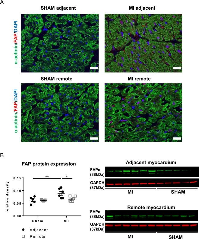
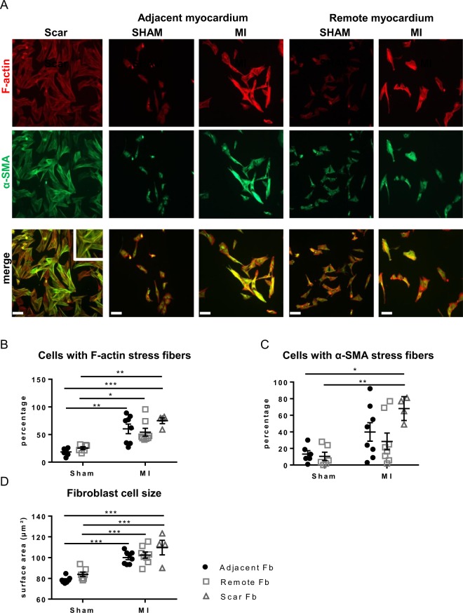
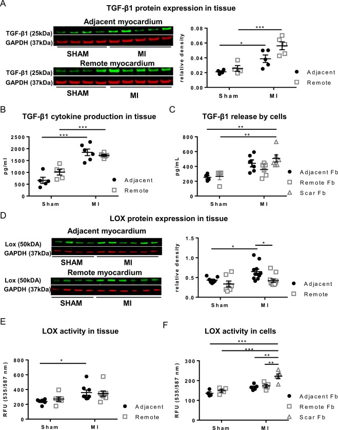
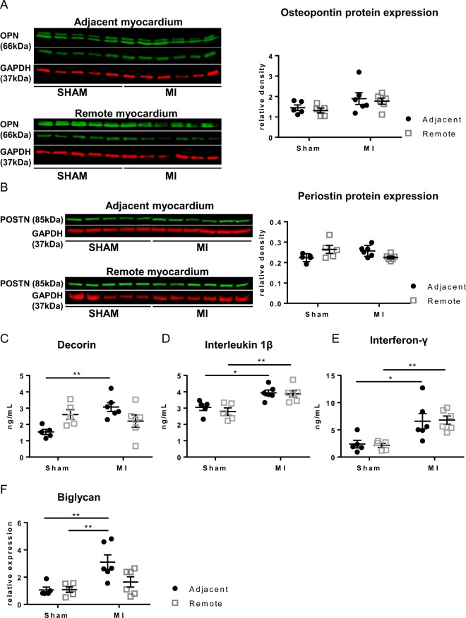
Fibroblast (Fb) differentiation and interstitial fibrosis contribute to cardiac remodeling and loss of function after myocardial infarction (MI). We investigated regional presence and regulation of fibrosis in a pig MI model. In vivo analysis of regional function and perfusion defined three regions: the scar, the myocardium adjacent to the scar (MIadjacent, reduced function, reduced perfusion reserve), and the remote myocardium (MIremote, minimal functional deficit, maintained perfusion). Interstitial and perivascular fibrosis, and increase of collagen type I, was only observed in the MIadjacent. Fb activated protein-alpha (FAP-α) was enriched in MIadjacent compared to MIremote. TGF-β1, which triggers Fb differentiation, was upregulated in both MIadjacent and MIremote, whereas lysyl oxidase, a regulator of collagen cross-linking, and the proteoglycans decorin and biglycan were only increased in the MIadjacent. Fb isolated and cultured for 4 days had myoFb characteristics with little difference between MIremote and MIadjacent, although RNA sequencing revealed differences in gene expression profiles. Fbs from all regions maintained proliferative capacity, and induced contraction of 3-D collagen matrices but scar myoFb was more effective. These data suggest that after MI, signaling through TGF-β1, possibly related to increased mechanical load, drives Fb activation throughout the left ventricle while regional signaling determines further maturation and extracellular matrix remodeling after MI.


Conflict of interest statement

The authors declare that they have no competing interests.

Figures

Figure 1
Figure 1
Interstitial fibrosis in different regions of the LV. (A) Representative images of Sirius red stained sections and polarized light microscopy from the adjacent, remote and scar tissue of SHAM and MI. (B) Analysis of interstitial fibrosis, and in polarized light collagen type I (red-yellow) and collagen type III (green) in SHAM and MI. Scale bars represent 50 µm. (C) Collagen type I and III mRNA expression in the adjacent and remote myocardium of SHAM and MI. (**p < 0.01: ***p < 0.001) (2-way ANOVA with Bonferroni post hoc test).
Figure 2
Figure 2
Perivascular fibrosis in the adjacent myocardium. (A) Representative images of Sirius red stained arterioles from the adjacent and remote myocardium of SHAM and MI acquired via light and polarization microscopy. (B) Quantification of perivascular fibrosis and collagen isoforms within the perivascular area. Scale bars represent 20 µm. (**p < 0.01) (2-way ANOVA with Bonferroni post hoc test).
Figure 3
Figure 3
Myofibroblasts in situ in the adjacent myocardium. (A) Immunofluorescence staining of Fibroblast Activation Protein α, FAP-α, (red), α-actinin (green) and DAPI (blue) in SHAM and MI tissue sections. Scale bars represent 30 µm. (B) Western blotting of FAP-α (88-kDa) in tissue from the adjacent and remote myocardium of SHAM and MI. (*p < 0.05: **p < 0.01). (2-way ANOVA with Bonferroni post hoc test).
Figure 4
Figure 4
Differentiation of fibroblasts derived from scar, adjacent and remote myocardium. (A) Fluorescent images of fibroblastic cells stained for α-SMA (green) and F-actin (red) and the corresponding overlay after 4 days in culture. (B) Quantification of Fb positive for immuno-stained F-actin as percentage of all cells. (C) Quantification of α-SMA-positive Fb as percentage of all cells. (D) Cell size of Fb from SHAM and MI adjacent and remote myocardium. (*p < 0.05: **p < 0.01: *** < 0.001). (1-way ANOVA with Bonferroni post hoc test).
Figure 5
Figure 5
Proliferative and contractile properties of fibroblasts from the adjacent and remote myocardium. (A) Proliferation capacity of Fb, expressed as cell number after 3 days in culture with 5000 cells seeded at d0. (B) Percentage of Fb positive for Ki-67 marker. (C) Representative images of 3-DCM contraction by fibroblastic cells. (D) Analysis of the collagen gel diameter after 3 days in culture. (*p < 0.05: **p < 0.01: *** < 0.001). (1-way ANOVA with Bonferroni post hoc test).
Figure 6
Figure 6
TGF-β1 and LOX expression during post-MI remodeling. (A) Protein expression by Western blotting of TGF-β1. (B) TGF-β1 concentration measured in cytokine array. (C) TGF-β1 secretion by cultured Fb cells derived from SHAM and MI. (D) LOX expression in tissue from the adjacent and remote myocardium of SHAM and MI. (E) LOX activity in tissue, (F) Lox activity in cultured Fb cells derived from SHAM and MI. *p < 0.05, **p < 0.01. (*p < 0.05: **p < 0.01: *** < 0.001). (2-way ANOVA with Bonferroni post hoc test for 6A, B, D, E;1-way ANOVA with Bonferroni post hoc test for 6 C, F)
Figure 7
Figure 7
Cytokines in the adjacent and remote myocardium of SHAM and MI. (A,B) Cytokines assessed with Western blotting for osteopontin and periostin in tissue from the adjacent and remote myocardium. (CE) Assessment in cytokine array of secretion of decorin (C), interleukin 1β (D), interferon-γ (E). (F) mRNA expression of biglycan (*p < 0.05: **p < 0.01). (2-way ANOVA with Bonferroni post hoc test).

References

    1. Pinto AR, et al. Revisiting Cardiac Cellular Composition. Circ. Res. 2016;118:400–409. doi: 10.1161/CIRCRESAHA.115.307778. - DOI - PMC - PubMed
    1. Krenning G, Zeisberg EM, Kalluri R. The origin of fibroblasts and mechanism of cardiac fibrosis. J. Cell. Physiol. 2010;225:631–637. doi: 10.1002/jcp.22322. - DOI - PMC - PubMed
    1. Ieda M, et al. Cardiac fibroblasts regulate myocardial proliferation through beta1 integrin signaling. Dev. Cell. 2009;16:233–244. doi: 10.1016/j.devcel.2008.12.007. - DOI - PMC - PubMed
    1. Takeda N, et al. Cardiac fibroblasts are essential for the adaptive response of the murine heart to pressure overload. J. Clin. Invest. 2010;120:254–265. doi: 10.1172/JCI40295. - DOI - PMC - PubMed
    1. Ali SR, et al. Developmental heterogeneity of cardiac fibroblasts does not predict pathological proliferation and activation. Circ. Res. 2014;115:625–635. doi: 10.1161/CIRCRESAHA.115.303794. - DOI - PubMed

Publication types

MeSH terms