. 2017 Sep 7;7(1):10787.
doi: 10.1038/s41598-017-11289-8.

Evidence that endogenous formaldehyde produces immunogenic and atherogenic adduct epitopes

Affiliations

Evidence that endogenous formaldehyde produces immunogenic and atherogenic adduct epitopes

Jun Nakamura et al. Sci Rep. .

Abstract

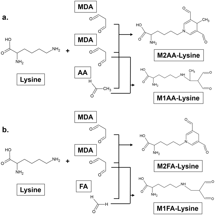
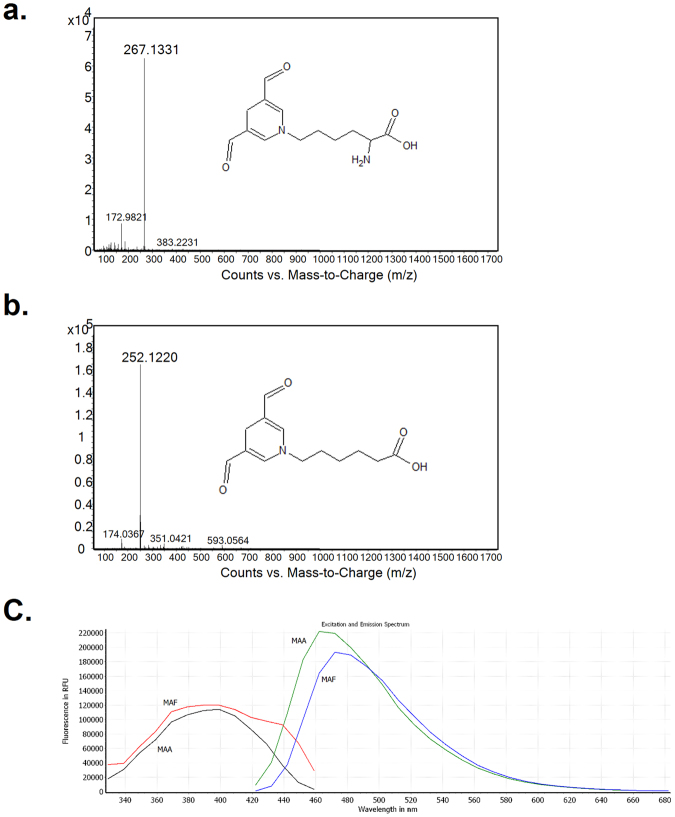
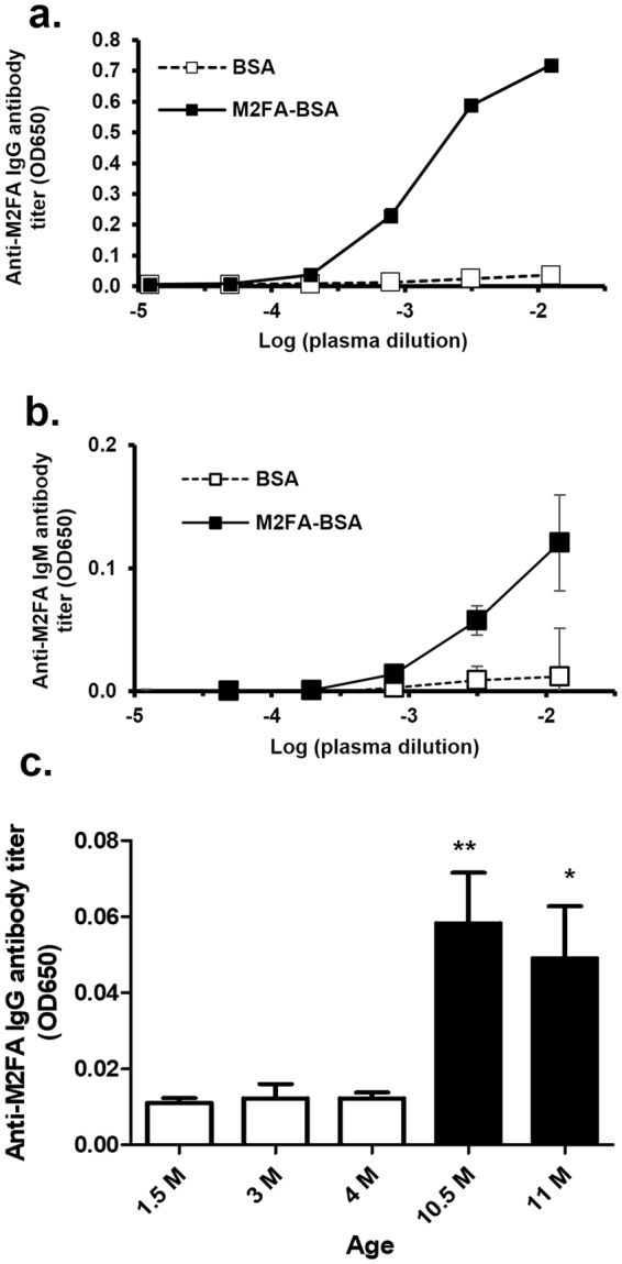
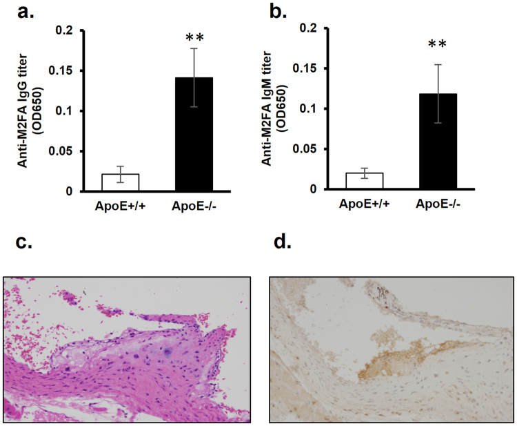
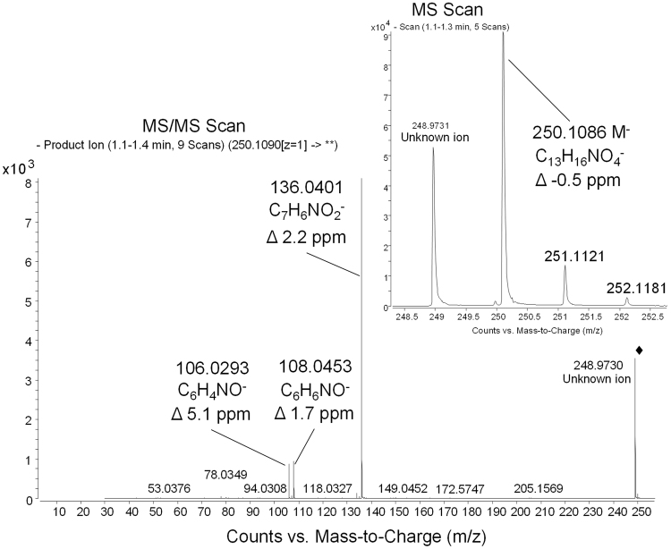
Endogenous formaldehyde is abundantly present in our bodies, at around 100 µM under normal conditions. While such high steady state levels of formaldehyde may be derived by enzymatic reactions including oxidative demethylation/deamination and myeloperoxidation, it is unclear whether endogenous formaldehyde can initiate and/or promote diseases in humans. Here, we show that fluorescent malondialdehyde-formaldehyde (M2FA)-lysine adducts are immunogenic without adjuvants in mice. Natural antibody titers against M2FA are elevated in atherosclerosis-prone mice. Staining with an antibody against M2FA demonstrated that M2FA is present in plaque found on the aortic valve of ApoE -/- mice. To mimic inflammation during atherogenesis, human myeloperoxidase was incubated with glycine, H2O2, malondialdehyde, and a lysine analog in PBS at a physiological temperature, which resulted in M2FA generation. These results strongly suggest that the 1,4-dihydropyridine-type of lysine adducts observed in atherosclerosis lesions are likely produced by endogenous formaldehyde and malondialdehyde with lysine. These highly fluorescent M2FA adducts may play important roles in human inflammatory and degenerative diseases.


Conflict of interest statement

The authors declare that they have no competing interests.

Figures

Figure 1
Figure 1
Structure of the M2AA-lysine, M1AA-lysine, M2FA-lysine, and M1FA-lysine adducts. (a) 1,4-dihydropyridine-type M2AA-Lys adduct is formed by a reaction between AA and two equivalents of MDA with a primary amine, usually at the ε-position amino moiety of a Lys residue on the target protein. M1AA-lysine adduct is produced by a reaction between AA and MDA in the presence of Lys (b) M2FA-Lys adduct is formed by a reaction between FA and two equivalents of MDA with Lys. M1FA-lysine adduct may be produced by a reaction between MA and MDA in the presence of Lys.
Figure 2
Figure 2
The full scan mass spectra of M2FA-lysine and M2FA-6ACA and fluorescence spectrum of M2FA- and M2AA-6ACA. (a) The background-subtracted full scan mass spectrum shows the protonated molecular ion for M2AA-lysine at m/z 267.1331. (b) The background-subtracted full scan mass spectrum shows the protonated molecular ion of M2AA-6ACA at m/z 252.1220. (c) Fluorescence spectrum of M2FA- and M2AA-6ACA.
Figure 3
Figure 3
Competitive inhibition of M2FA- and M2AA-MB in competitive ELISA. The relative ability of M2FA- and M2AA MB to inhibit antibody. Purified anti-M2AA and M2FA antibodies (a) moMono-1F83-M2AA Ab; (b) rabPoly-M2AA Ab; (c) rabPoly-M2FA Ab) were preincubated with the indicated competitors and controls prior to detection of IgG binding to plated M2FA-6ACA-BSA. Data are shown as B/B0 of triplicates for a representative experiment.
Figure 4
Figure 4
The immunogenicity of M2FA-lysine-BSA in the absence of adjuvant and anti-M2FA antibody titers in intact mice. C57BL/6 mice were injected i.p. with M2FA-lysine-BSA or BSA in the absence of adjuvant. The antibody titers of IgG (a) and IgM (b) against M2FA-lysine were detected using MFA-6ACA-KLH-coated plates. The anti-M2AA antibody titers were clearly increased in M2FA-lysine-BSA-immunized mice compared to the controls (BSA-treated mice). Values are mean and SD. (c) Intact female C57BL/6 mice (n = 4 or 5 per group) with different ages showed significantly different anti-M2FA IgG titers. Values are mean and SD. (*p < 0.05; between 1.5 M/3 M vs 11 M; **p < 0.01: 1.5 M/3 M/4 M vs 10.5 M).
Figure 5
Figure 5
Serum anti-M2FA IgG and IgM antibody levels in wild-type and ApoE −/− mice and immunohistochemical detection of M2FA-epitopes in heart valve of ApoE −/− mice. The anti-M2FA IgG (a) and IgM (b) antibody levels showed significant differences between wild-type and ApoE −/− mice with the M2FA-6ACA-BSA ELISAs (**p < 0.01). Representative H&E (c) and M2FA immunohistochemistry (d) sections of plaques in heart valve of ApoE −/− mice at 5 month old fed with normal diet. M2FA-lysine in the plaque was stained with rabPoly-M2FA Ab.
Figure 6
Figure 6
Liquid chromatography−mass spectrometry analyses demonstrating M2FA formation through the MPO-H2O2-halide system. Glycine, human MPO, H2O2 were incubated in PBS for 1 hour at 37 °C followed by incubation with MDA and 6-ACA (Lys analog) for 3 days at 37 °C. The reactant was applied to LC-MS analysis. Full scan and ms/ms spectra of M2FA-6ACA were obtained on an Agilent 6520 Accurate Mass Q-TOF in negative mode. Spectra show the intact de-protonated molecular ion at m/z 250.1086 (Δ −0.5 ppm) and major fragments at m/z 136.0401 (Δ 2.2 ppm), 108.0453 (Δ 1.7 ppm), and 106.0293 (Δ 5.1 ppm). Chemical formulas are proposed for the fragments, and deviations of measured masses to calculated masses for these formulas are all 5 ppm or less. The product ion spectrum was obtained using m/z 250.1090 as the precursor and 20 eV for collision energy.

References

    1. Pizzimenti, S. et al. Interaction of aldehydes derived from lipid peroxidation and membrane proteins. Frontiers in Physiology 4 SEP (2013). - PMC - PubMed
    1. Weismann D, et al. Complement factor H binds malondialdehyde epitopes and protects from oxidative stress. Nature. 2011;478:76–81. doi: 10.1038/nature10449. - DOI - PMC - PubMed
    1. Romero FJ, et al. Lipid peroxidation products and antioxidants in human disease. Environ. Health Perspect. 1998;106(Suppl):1229–34. doi: 10.1289/ehp.98106s51229. - DOI - PMC - PubMed
    1. Kikugawa K, Machida Y, Kida M, Kurechi T. Studies on Peroxidized Lipids. III. Fluorescent Pigments derived from the Reaction of Malonaldehyde and Amino AcidsNo Title. Chem. Pharm. Bull. (Tokyo). 1981;29:3003–3011. doi: 10.1248/cpb.29.3003. - DOI
    1. Summerfield FW, Tappel AL. Detection and measurement by high-performance liquid chromatography of malondialdehyde crosslinks in DNA. Anal. Biochem. 1984;143:265–271. doi: 10.1016/0003-2697(84)90662-6. - DOI - PubMed

Publication types