

Role of MRI in Diagnosis of Ruptured Intracranial Dermoid Cyst
- PMID: 28883682
- PMCID: PMC5544451
- DOI: 10.5455/aim.2017.25.141-144
Role of MRI in Diagnosis of Ruptured Intracranial Dermoid Cyst
Abstract
Introduction: Intracranial dermoid cystic tumors account for <1% of all intracranial masses.
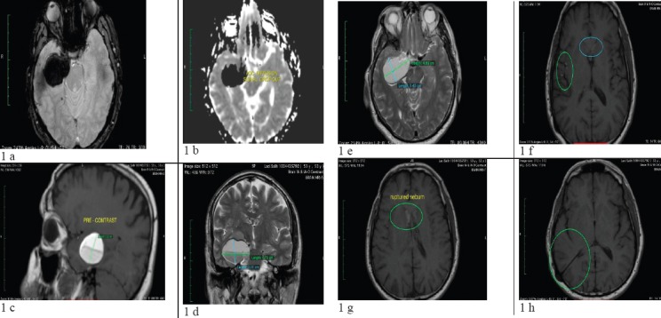
Case report: A 52-year-old male, having headaches, nausea and is presented with a history of 2 episodes of new onset seizures. On presentation, the patient had a normal physical exam, including a complete neurological and cranial nerve exam.
Methods: Precontrast MRI; TSE/T2Wsequence in axial/coronal planes; 3D - HI-resolution T1W sagittal; FLAIR/T2W axial; FLAIR/T2W, Flash/T2W oblique coronal plane, GRE/T2W axial. Post-contrast TSE/T1W sequence in axial, coronal and sagittal planes. Diffusion weighted and ADC mapping, postcontrast: TSE/T1W sequence in axial, coronal and sagittal planes.
Results: Subsequent MRI of the brain revealed an oval and lobulated 47x34x30mm (TRxAPxCC) non-enhancing T1-hyperintense mass in right cavernous sinus, with compression of surrounding mesial temporal lobe and right anterolateral aspect of mesencephalon. Findings are consistent with ruptured dermoid cyst, given the evacuated sebum content at its lower half. Sebum particles in millimetric sizes are seen within right Sylvian fissure, anterior horns of lateral ventricles and to a lesser extent within left Sylvian fissure, right parietal sulci, cerebral aqueduct, and basal cisterns. No restricted diffusion is seen, eliminating the possibility of epidermoid. A shunt catheter is evident traversing between right lateral ventricle and right parietal bone; besides, slit-like right lateral ventricle is noted (likely secondary to over-draining shunt catheter).
Conclusion: Intracranial dermoid cysts are benign rare slow-growing tumors that upon rupture, however, widespread presence of T1 hyperintense droplets and leptomeningeal enhancement can be noted-making MRI the best imaging modality for diagnosis of this rare entity.
Keywords: Intracranial dermoid cyst; Kosova; MRI; Pristine; UCCK; ruptured.
Conflict of interest statement
• Conflict of interest: none declared
Figures
References
-
- Rubin G, Scienza R, Pasqualin A, Rosta L, Da Pian R. Craniocerebral epidermoids and dermoids. A review of 44 cases. Acta Neurochir (Wien) 1989;97(1–2):1–16. - PubMed
-
- Osborn AG, Preece MT. Intracranial cysts: radiologic-pathologic correlation and imaging approach. Radiology. 2006;239(3):650–64. - PubMed
-
- Kim KS, Weinberg PE. Dermoid tumor. Surg Neurol. 1981;15(5):375–6. - PubMed
-
- Graham DV, Tampieri D, Villemure JG. Intramedullary dermoid tumor diagnosed with the assistance of magnetic resonance imaging. Neurosurgery. 1988;23(6):7657. - PubMed
-
- Cha JG, Paik SH, Park JS, Park SJ, Kim DH, Lee HK. Ruptured spinal dermoid cyst with disseminated intracranial fat droplets. Br J Radiol. 2006;79(938):167–9. - PubMed
Publication types
LinkOut - more resources
Full Text Sources
Other Literature Sources