

Schistosoma japonicum attenuates dextran sodium sulfate-induced colitis in mice via reduction of endoplasmic reticulum stress
- PMID: 28883695
- PMCID: PMC5569284
- DOI: 10.3748/wjg.v23.i31.5700
Schistosoma japonicum attenuates dextran sodium sulfate-induced colitis in mice via reduction of endoplasmic reticulum stress
Abstract
Aim: To elucidate the impact of Schistosoma (S.) japonicum infection on inflammatory bowel disease by studying the effects of exposure to S. japonicum cercariae on dextran sodium sulfate (DSS)-induced colitis.
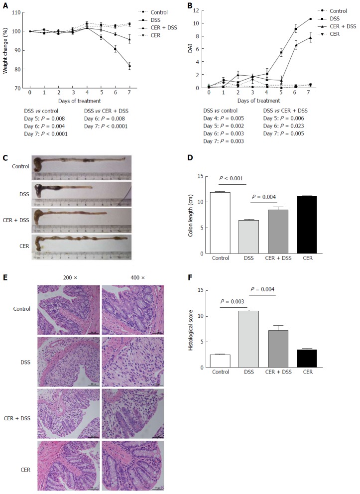
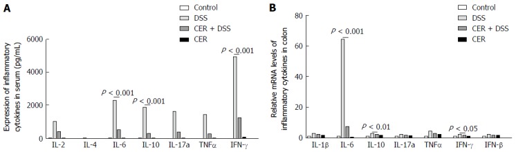
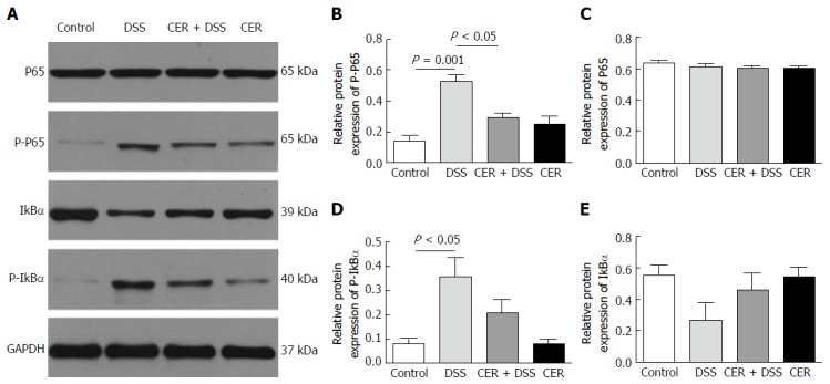
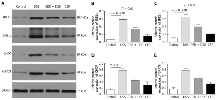
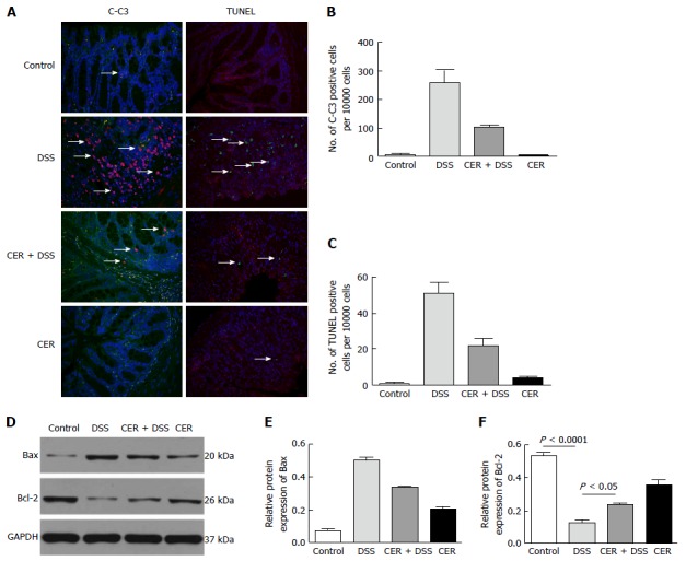
Methods: Infection was percutaneously established with 20 ± 2 cercariae of S. japonicum, and colitis was induced by administration of 3% DSS at 4 wk post infection. Weight change, colon length, histological score (HS) and disease activity index (DAI) were evaluated. Inflammatory cytokines, such as IL-2, IL-10 and IFN-γ, were tested by a cytometric bead array and real-time quantitative polymerase chain reaction (RT-PCR). Protein and mRNA levels of IRE1α, IRE1β, GRP78, CHOP, P65, P-P65, P-IκBα and IκBα in colon tissues were examined by Western blot and RT-PCR, respectively. Terminal deoxynucleotidyl transferase-mediated dUTP nick-end labeling positive cells, cleaved-caspase 3 expression and Bcl2/Bax were investigated to assess the apoptosis in colon tissues.
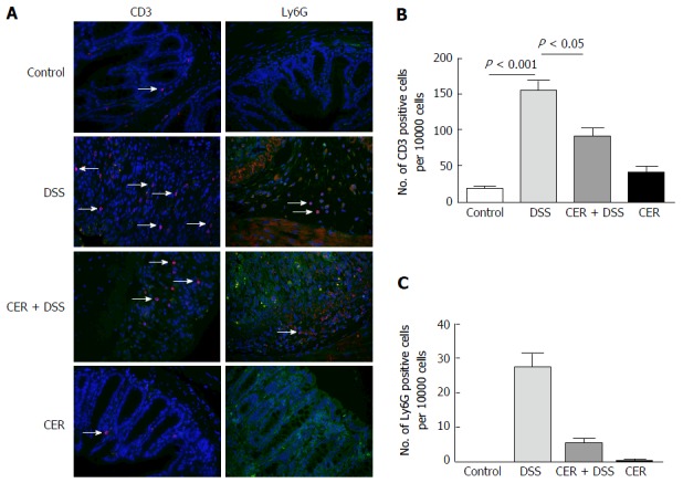
Results: Mice infected with S. japonicum cercariae were less susceptible to DSS. Mice infected with S. japonicum cercariae and treated with DSS showed decreased weight loss, longer colon, and lower HS and DAI compared with mice treated with DSS alone. A substantial decrease in Th1/Th2/Th17 response was observed after infection with S. japonicum. Endoplasmic reticulum (ER) stress and the nuclear factor-kappa B (NF-κB) pathway were reduced in mice infected with S. japonicum cercariae and treated with DSS, along with ameliorated celluar apoptosis, in contrast to mice treated with DSS alone.
Conclusion: Exposure to S. japonicum attenuated inflammatory response in a DSS-induced colitis model. In addition to the Th1/Th2/Th17 pathway and NF-κB pathway, ER stress was shown to be involved in mitigating inflammation and decreasing apoptosis. Thus, ER stress is a new aspect in elucidating the relationship between helminth infection and inflammatory bowel disease (IBD), which may offer new therapeutic methods for IBD.
Keywords: Colitis; Endoplasmic reticulum stress; Schistosoma japonicum.
Conflict of interest statement
Conflict-of-interest statement: To the best of our knowledge, no conflict of interest exists.
Figures
Similar articles
-
Anti-inflammatory effects of Brucea javanica oil emulsion by suppressing NF-κB activation on dextran sulfate sodium-induced ulcerative colitis in mice.J Ethnopharmacol. 2017 Feb 23;198:389-398. doi: 10.1016/j.jep.2017.01.042. Epub 2017 Jan 22. J Ethnopharmacol. 2017. PMID: 28119098
-
Schistosoma japonicum peptide SJMHE1 inhibits acute and chronic colitis induced by dextran sulfate sodium in mice.Parasit Vectors. 2021 Sep 6;14(1):455. doi: 10.1186/s13071-021-04977-y. Parasit Vectors. 2021. PMID: 34488863 Free PMC article.
-
Sirtuin 1 alleviates endoplasmic reticulum stress-mediated apoptosis of intestinal epithelial cells in ulcerative colitis.World J Gastroenterol. 2019 Oct 14;25(38):5800-5813. doi: 10.3748/wjg.v25.i38.5800. World J Gastroenterol. 2019. PMID: 31636473 Free PMC article.
-
Infection characteristics of Schistosoma japonicum in mice and relevance to the assessment of schistosome vaccines.Adv Parasitol. 1991;30:167-200. doi: 10.1016/s0065-308x(08)60308-5. Adv Parasitol. 1991. PMID: 1906234 Review. No abstract available.
-
Schistosoma japonicum Associated Colorectal Cancer and Its Management.Acta Parasitol. 2023 Dec;68(4):723-734. doi: 10.1007/s11686-023-00707-9. Epub 2023 Aug 18. Acta Parasitol. 2023. PMID: 37594685 Review.
Cited by
-
The Inhibitory Effect of Artesunate on Excessive Endoplasmic Reticulum Stress Alleviates Experimental Colitis in Mice.Front Pharmacol. 2021 Mar 9;12:629798. doi: 10.3389/fphar.2021.629798. eCollection 2021. Front Pharmacol. 2021. PMID: 33767628 Free PMC article.
-
Single-sex schistosomiasis: a mini review.Front Immunol. 2023 Apr 19;14:1158805. doi: 10.3389/fimmu.2023.1158805. eCollection 2023. Front Immunol. 2023. PMID: 37153566 Free PMC article. Review.
-
Gα12 and endoplasmic reticulum stress-mediated pyroptosis in a single cycle of dextran sulfate-induced mouse colitis.Sci Rep. 2024 Mar 15;14(1):6335. doi: 10.1038/s41598-024-56685-z. Sci Rep. 2024. PMID: 38491049 Free PMC article.
-
Recombinant protein Schistosoma japonicum-derived molecule attenuates dextran sulfate sodium-induced colitis by inhibiting miRNA-217-5p to alleviate apoptosis.World J Gastroenterol. 2021 Dec 14;27(46):7982-7994. doi: 10.3748/wjg.v27.i46.7982. World J Gastroenterol. 2021. PMID: 35046625 Free PMC article.
-
Painong San, a Traditional Chinese Compound Herbal Medicine, Restores Colon Barrier Function on DSS-Induced Colitis in Mice.Evid Based Complement Alternat Med. 2021 Dec 20;2021:2810915. doi: 10.1155/2021/2810915. eCollection 2021. Evid Based Complement Alternat Med. 2021. PMID: 34966434 Free PMC article.
References
-
- Xavier RJ, Podolsky DK. Unravelling the pathogenesis of inflammatory bowel disease. Nature. 2007;448:427–434. - PubMed
-
- Molodecky NA, Soon IS, Rabi DM, Ghali WA, Ferris M, Chernoff G, Benchimol EI, Panaccione R, Ghosh S, Barkema HW, et al. Increasing incidence and prevalence of the inflammatory bowel diseases with time, based on systematic review. Gastroenterology. 2012;142:46–54.e42; quiz e30. - PubMed
-
- Prideaux L, Kamm MA, De Cruz PP, Chan FK, Ng SC. Inflammatory bowel disease in Asia: a systematic review. J Gastroenterol Hepatol. 2012;27:1266–1280. - PubMed
MeSH terms
Substances
LinkOut - more resources
Full Text Sources
Other Literature Sources
Medical
Research Materials
Miscellaneous
