. 2017 Sep;21(5):509-518.
doi: 10.4196/kjpp.2017.21.5.509. Epub 2017 Aug 22.

Effect of glucose level on chemical hypoxia- and hydrogen peroxide-induced chemokine expression in human glioblastoma cell lines

Affiliations

Effect of glucose level on chemical hypoxia- and hydrogen peroxide-induced chemokine expression in human glioblastoma cell lines

Yieun Jung et al. Korean J Physiol Pharmacol. 2017 Sep.

Abstract

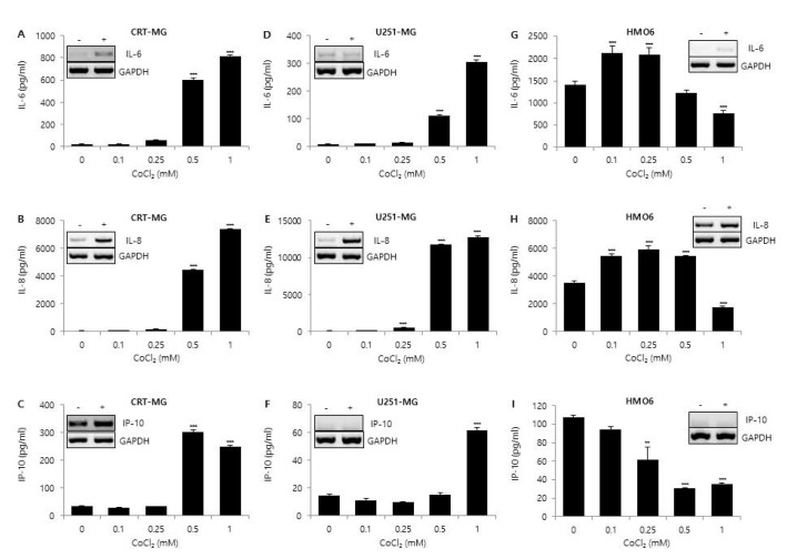
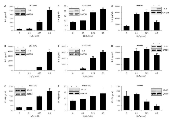
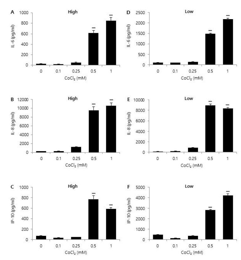
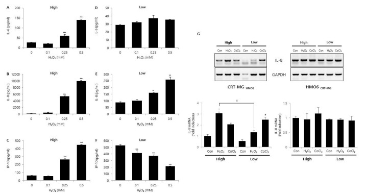
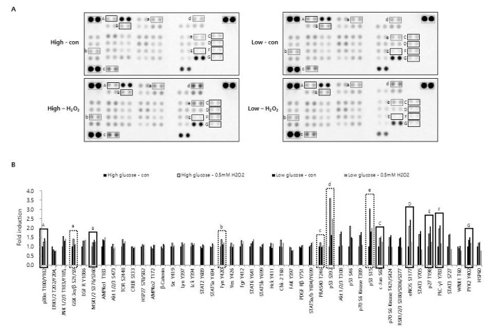
Glioblastoma multiforme (GBM) is the most common primary intracranial tumor in adults and has poor prognosis. The GBM-specific tumor microenvironment (TME) plays a crucial role in tumor progression, immune escape, local invasion, and metastasis of GBM. Here, we demonstrate that hypoxia, reactive oxygen species (ROS), and differential concentration of glucose influence the expression of cytokines and chemokines, such as IL-6, IL-8, and IP-10, in human glial cell lines. Treatment with cobalt chloride (CoCl2) and hydrogen peroxide (H2O2) significantly increased the expression levels of IL-6, IL-8, and IP-10 in a dose-dependent manner in CRT-MG and U251-MG astroglioma cells, but not in microglia cells. However, we found strikingly different patterns of expression of cytokines and chemokines between H2O2-treated CRT-MG cells cultured in low- and high-glucose medium. These results suggest that astroglioma and microglia cells exhibit distinct patterns of cytokine and chemokine expression in response to CoCl2 and H2O2 treatment, and different concentrations of glucose influence this expression under either hypoxic or oxidant-enriched conditions.

Keywords: Chemokines; Cytokines; Glioblastoma; Tumor microenvironment.


Conflict of interest statement

CONFLICTS OF INTEREST: The authors declare no conflicts of interest.

Figures

Fig. 1
Fig. 1. Effect of CoCl2 on IL-6, IL-8, and IP-10 expression.
CRT-MG and U251-MG astroglioma cell lines and HMO6 microglia cells were either treated with the indicated concentrations of CoCl2 or not treated for 24 h. After incubation, supernatants were collected, and the protein levels of chemokines from the culture supernatants were analyzed via ELISA for IL-6 (A, D, G), IL-8 (B, E, H), and IP-10 (C, F, I). Absorbance was measured at 450 nm. (Inset) RT-PCR of CRT-MG, U251-MG and HMO6 cells treated with or without 0.5 mM CoCl2. Data shown are representative of at least three independent experiments. *p<0.05, **p<0.01, ***p<0.001 vs. untreated control.
Fig. 2
Fig. 2. Effect of H2O2 on IL-6, IL-8, and IP-10 expression.
CRT-MG, U251 MG, and HMO6 cells were either treated with the indicated concentrations of H2O2 or not treated for 24 h. After incubation, supernatants were collected, and the protein levels of chemokines from the culture supernatants were analyzed via ELISA for IL-6 (A, D, G), IL-8 (B, E, H), and IP-10 (C, F, I). Absorbance was measured at 450 nm. (Inset) RT-PCR of CRT-MG, U251-MG and HMO6 cells treated with or without 0.5 mM H2O2. Data shown are representative of at least three independent experiments. *p<0.05, **p<0.01, ***p<0.001 vs. untreated control.
Fig. 3
Fig. 3. Effect of glucose concentration on CoCl2-induced chemokine expression.
CRT-MG astroglioma cells were cultured in either low (1,000 mg/L) or high (4,500 mg/L) glucose medium with the indicated concentration of CoCl2 for 24 h. After incubation, supernatants were collected and the level of IL-6 (A, D), IL-8 (B, E), and IP-10 (C, F) protein was measured via ELISA. Absorbance was measured at 450 nm. Data shown are representative of at least three experiments. *p<0.05, **p<0.01, ***p<0.001 vs. untreated control.
Fig. 4
Fig. 4. Effect of glucose concentration on H2O2-induced chemokine expression.
CRT-MG cells were cultured in either low or high glucose medium with the indicated concentration of H2O2 for 24 h. After incubation, supernatants were collected, and the level of IL-6 (A, B), IL-8 (C, D), and IP-10 (E, F) protein was measured using ELISA. Absorbance was measured at 450 nm. (G) Indirect co-culture assay. CRT-MG astroglioma cells were placed on the 24 well-plate, and HMO6 microglia cells were seeded onto the microporous membranes of the transwell insert, and then cells were treated with either 0.5 mM H2O2 or 0.5 mM CoCl2 for 24 h under low and high glucose conditions. After incubation, total RNA from CRT-MG and HMO6 cells were extracted and analyzed by RT-PCR for IL-8. Data shown are representative of at least three experiments. *p<0.05, **p<0.01, ***p<0.001 vs. untreated control.
Fig. 5
Fig. 5. Effect of H2O2 and glucose concentration on phosphorylation of protein kinases.
CRT-MG cells cultured in low (1,000 mg/L) and high (4,500 mg/L) glucose medium were either treated with 0.5 mM of H2O2 or not treated for 24 h. After incubation, cells were lysed on ice for 30 min in lysis buffer, and 430 mg of cell lysate was analyzed for the phosphokinase assay as described in the Methods. (A) The duplicate spots corresponding to reduced phosphorylation of protein kinase in response to H2O2 plus low glucose compared to H2O2 plus high glucose or low glucose alone are highlighted in the solid box (A~G). Increased phosphorylation in response to H2O2 plus high glucose compared to high glucose alone, low glucose alone, or H2O2 plus low glucose is highlighted in the dashed box (a~e). The gray box indicates reference spots. (B) The intensity of spots corresponding to solid and dashed boxes is shown in A. The positive control was quantified using Image J software and subtracted from the background, then expressed as a ratio to the positive control. The ratio of each spot from untreated control cells, cultured in high glucose medium was assigned a value of one.

Similar articles

Cited by

References

    1. Friedmann-Morvinski D, Narasimamurthy R, Xia Y, Myskiw C, Soda Y, Verma IM. Targeting NF-κB in glioblastoma: a therapeutic approach. Sci Adv. 2016;2:e1501292. - PMC - PubMed
    1. Kupp R, Shtayer L, Tien AC, Szeto E, Sanai N, Rowitch DH, Mehta S. Lineage-restricted OLIG2-RTK signaling governs the molecular subtype of glioma stem-like cells. Cell Rep. 2016;16:2838–2845. - PMC - PubMed
    1. McLendon RE, Halperin EC. Is the long-term survival of patients with intracranial glioblastoma multiforme overstated? Cancer. 2003;98:1745–1748. - PubMed
    1. Puli S, Lai JC, Bhushan A. Inhibition of matrix degrading enzymes and invasion in human glioblastoma (U87MG) cells by isoflavones. J Neurooncol. 2006;79:135–142. - PubMed
    1. Kesari S. Understanding glioblastoma tumor biology: the potential to improve current diagnosis and treatments. Semin Oncol. 2011;38(Suppl 4):S2–S10. - PubMed