

Rosmarinic Acid Alleviates the Endothelial Dysfunction Induced by Hydrogen Peroxide in Rat Aortic Rings via Activation of AMPK
- PMID: 28883905
- PMCID: PMC5572610
- DOI: 10.1155/2017/7091904
Rosmarinic Acid Alleviates the Endothelial Dysfunction Induced by Hydrogen Peroxide in Rat Aortic Rings via Activation of AMPK
Abstract
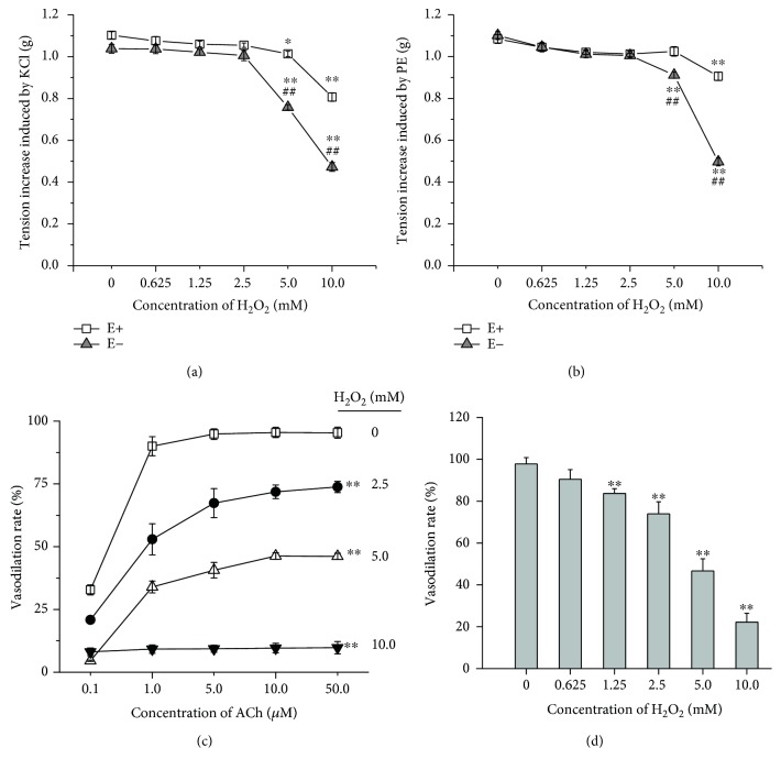
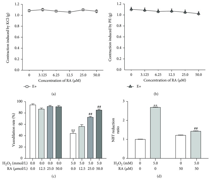
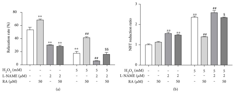
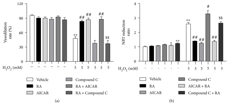
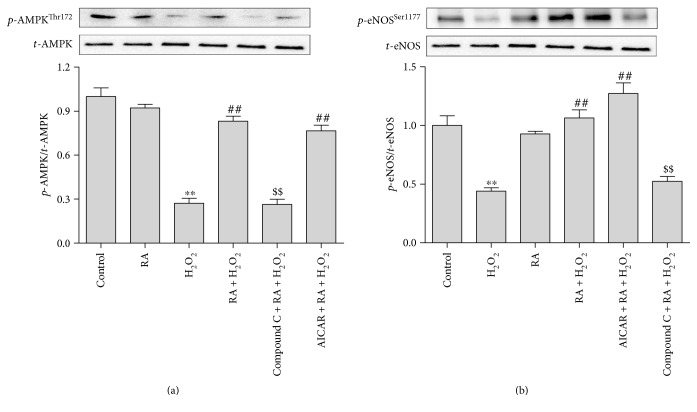
Endothelial dysfunction is the key player in the development and progression of vascular events. Oxidative stress is involved in endothelial injury. Rosmarinic acid (RA) is a natural polyphenol with antioxidative, antiapoptotic, and anti-inflammatory properties. The present study investigates the protective effect of RA on endothelial dysfunction induced by hydrogen peroxide (H2O2). Compared with endothelium-denuded aortic rings, the endothelium significantly alleviated the decrease of vasoconstrictive reactivity to PE and KCl induced by H2O2. H2O2 pretreatment significantly injured the vasodilative reactivity to ACh in endothelium-intact aortic rings in a concentration-dependent manner. RA individual pretreatment had no obvious effect on the vasoconstrictive reaction to PE and KCl, while its cotreatment obviously mitigated the endothelium-dependent relaxation impairments and the oxidative stress induced by H2O2. The RA cotreatment reversed the downregulation of AMPK and eNOS phosphorylation induced by H2O2 in HAEC cells. The pretreatment with the inhibitors of AMPK (compound C) and eNOS (L-NAME) wiped off RA's beneficial effects. All these results demonstrated that RA attenuated the endothelial dysfunction induced by oxidative stress by activating the AMPK/eNOS pathway.
Figures
Similar articles
-
Rosmarinic acid administration attenuates diabetes-induced vascular dysfunction of the rat aorta.J Pharm Pharmacol. 2013 May;65(5):713-23. doi: 10.1111/jphp.12037. Epub 2013 Feb 27. J Pharm Pharmacol. 2013. PMID: 23600389
-
Contribution of oxidative stress and prostanoids in endothelial dysfunction induced by chronic fluoxetine treatment.Vascul Pharmacol. 2015 Oct;73:124-37. doi: 10.1016/j.vph.2015.06.015. Epub 2015 Jun 30. Vascul Pharmacol. 2015. PMID: 26141931
-
Protective effects of rosmarinic acid against hydrogen peroxide‑induced cellular senescence and the inflammatory response in normal human dermal fibroblasts.Mol Med Rep. 2017 Dec;16(6):9763-9769. doi: 10.3892/mmr.2017.7804. Epub 2017 Oct 17. Mol Med Rep. 2017. PMID: 29039587
-
[Novel antioxidant therapeutic strategies for cardiovascular dysfunction associated with ageing].Orv Hetil. 2008 Dec 14;149(50):2377-85. doi: 10.1556/OH.2008.28502. Orv Hetil. 2008. PMID: 19073445 Hungarian.
-
Ethanol induces vascular relaxation via redox-sensitive and nitric oxide-dependent pathways.Vascul Pharmacol. 2012 Jan-Feb;56(1-2):74-83. doi: 10.1016/j.vph.2011.11.006. Epub 2011 Dec 3. Vascul Pharmacol. 2012. PMID: 22155162
Cited by
-
Ginsenoside Rb1 reduces H2O2‑induced HUVEC dysfunction by stimulating the sirtuin‑1/AMP‑activated protein kinase pathway.Mol Med Rep. 2020 Jul;22(1):247-256. doi: 10.3892/mmr.2020.11096. Epub 2020 Apr 28. Mol Med Rep. 2020. PMID: 32377712 Free PMC article.
-
Natural Compounds Rosmarinic Acid and Carvacrol Counteract Aluminium-Induced Oxidative Stress.Molecules. 2020 Apr 15;25(8):1807. doi: 10.3390/molecules25081807. Molecules. 2020. PMID: 32326410 Free PMC article.
-
Vasorelaxant Mechanism of Herbal Extracts from Mentha suaveolens, Conyza canadensis, Teucrium polium and Salvia verbenaca in the Aorta of Wistar Rats.Molecules. 2022 Dec 9;27(24):8752. doi: 10.3390/molecules27248752. Molecules. 2022. PMID: 36557886 Free PMC article.
-
Role of Polyphenols and Carotenoids in Endothelial Dysfunction: An Overview from Classic to Innovative Biomarkers.Oxid Med Cell Longev. 2020 Oct 19;2020:6381380. doi: 10.1155/2020/6381380. eCollection 2020. Oxid Med Cell Longev. 2020. PMID: 33133348 Free PMC article. Review.
-
Preliminary Characterization of the Vasorelaxant Effect of Thymus atlanticus (Ball) Roussine using Optical Methods.Curr Drug Discov Technol. 2025;22(3):e15701638309612. doi: 10.2174/0115701638309612240726060844. Curr Drug Discov Technol. 2025. PMID: 39501951
References
-
- McIntyre M., Bohr D. F., Dominiczak A. F. Endothelial function in hypertension. Hypertension. 1999;34(4):539–545. - PubMed
MeSH terms
Substances
LinkOut - more resources
Full Text Sources
Other Literature Sources
