Case Reports
. 2017:2017:5972940.
doi: 10.1155/2017/5972940. Epub 2017 Aug 14.

New Insights about Pulmonary Epithelioid Hemangioendothelioma: Review of the Literature and Two Case Reports

Affiliations
Case Reports

New Insights about Pulmonary Epithelioid Hemangioendothelioma: Review of the Literature and Two Case Reports

Romeu Duarte Mesquita et al. Case Rep Radiol. 2017.

Abstract

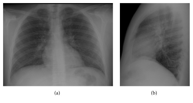
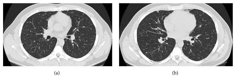
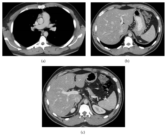
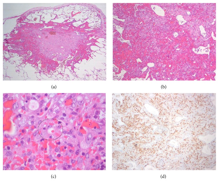
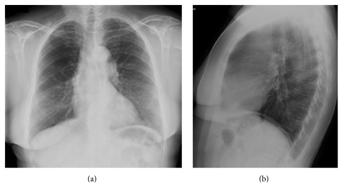
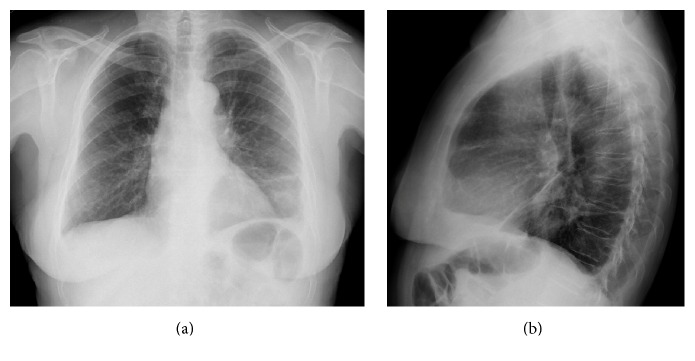
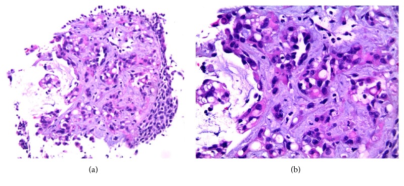
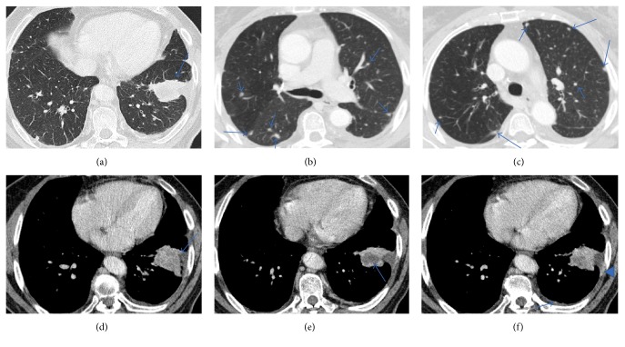
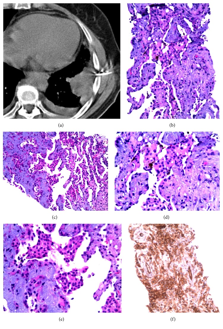
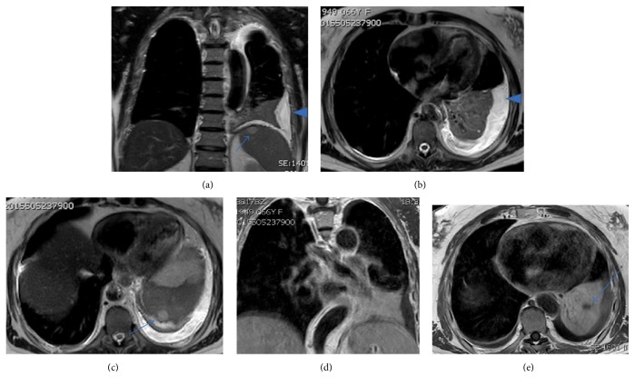
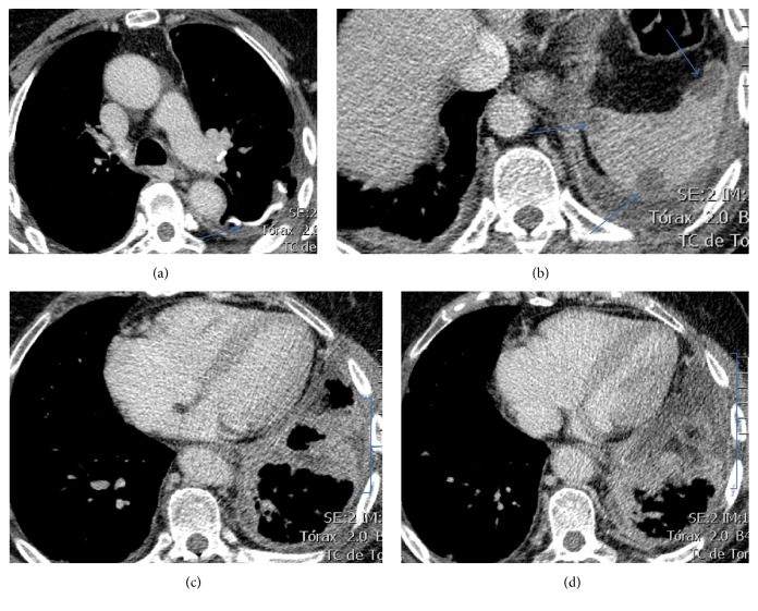
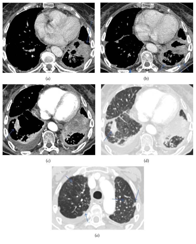
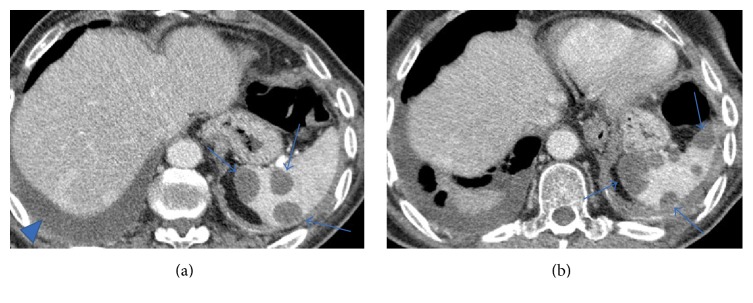
Pulmonary epithelioid hemangioendothelioma (PEH) is a rare neoplasm of vascular origin. There are three different major imaging patterns identified in thoracic manifestation of epithelioid hemangioendothelioma: (1) multiple pulmonary nodules; (2) multiple pulmonary reticulonodular opacities; and (3) diffuse infiltrative pleural thickening. Radiographically, presence of bilateral multiple nodules is the most common pattern of presentation. The diagnosis is made on the basis of histopathological findings and confirmed by positive immunohistochemistry staining. Although the prognostic factors for PEH have not yet been well established, a better prognosis is usually associated with the multinodular pattern. We report two different imagological presentations of this rare disease, based on two institutional experiences, along with a review of the relevant literature.


Figures

Figure 1
Figure 1
Posteroanterior (a) and lateral (b) chest X-rays. The chest radiography showed a diffuse nodular pulmonary pattern in both lungs.
Figure 2
Figure 2
Chest CT (a and b). Multislice chest CT-images on lung window showed innumerable, well-defined, small, round, and noncalcified pulmonary nodular opacities, scattered in both lungs. These nodules were present in all pulmonary lobes and adjacent to bronchioles and medium/small vessels.
Figure 3
Figure 3
Contrast-enhanced multislice CT of the chest (a) on mediastinal window showed no thoracic lymphadenopathy. Multislice abdominopelvic CT scans (b and c) with intravenous contrast administration showed no significant findings, excluding focal liver or splenic lesions.
Figure 4
Figure 4
Histopathologic findings of hemangioendothelioma. (a) Low magnification of the specimen obtained from pulmonary wedge resection reveals a subpleural nodule that extends to adjacent alveoli, with vascular proliferation in the peripheral zone and areas of recent and old hemorrhage. (b) Neoplastic nodule showing tumor cells with an eosinophilic stroma at the periphery. The lesion contains blood filled spaces and respiratory epithelium “trapped” within the lesion is also shown. (c) Higher magnification of the tumor reveals vacuolation of some of the tumor cells, representing primitive angiogenesis. One of these epithelioid cells can be observed with intracytoplasmic lumen and an erythrocyte inside, indicating their vascular nature. (d) Immunostaining for CD34 revealed positivity of the neoplastic cells (brown colour), confirming the endothelial lineage of the tumor.
Figure 5
Figure 5
Posteroanterior (a) and lateral (b) chest X-rays. In a routine chest radiography 2 years before presentation in the ER it was difficult to detect the lesion in the left lower lung.
Figure 6
Figure 6
Posteroanterior (a) and lateral (b) chest X-rays. The chest radiography showed a peripheral lesion in the left lower lung and a small volume of pleural effusion on the same side.
Figure 7
Figure 7
Transbronchial biopsy of a pulmonary mass (H-E). (a) 10x. (b) 20x. Vascular proliferation and endothelial cells, frequently with citoplasmatic vacuolization, in a fibromyxoid stroma, with bronchial surface epithelial lining.
Figure 8
Figure 8
Chest CT. Multislice chest CT-images on lung window (a, b, and c) showed a lung mass with irregular borders (a); they also showed multiple small, round, and noncalcified pulmonary nodular opacities, scattered in both lungs (b and c). On mediastinal window (d, e, and f) it is possible to better identify the pleural invasion (d) and the necrotic areas in lesion (e), retrospectively, and valorization is hard when the exam was performed, and we can recognize areas of enhancement in the left pleura, more nodular (arrow) or linear (arrowhead) (f).
Figure 9
Figure 9
20 G Tru-Cut biopsy of the lung mass (a). H-E (b–e) images show vascular proliferation with epithelioid cells in a fibromyxoid stroma and presence of haemosiderophages ((b, c) 10x; (d) 20x). Some of the epithelioid cells show vacuolation (e). Immunostaining for CD31 revealed positivity of the neoplastic cells (brown colour), confirming the endothelial lineage of the tumor.
Figure 10
Figure 10
Thoracic MR images. Fluid sensitive sequences (a–c) show the presence of left pleural effusion (arrowhead) and also the nodular lesions in the spleen, which do not correspond to cysts (arrows). T1 weighted images with contrast (d and e) show the pulmonary mass and hypodense zones representing necrotic areas (arrow).
Figure 11
Figure 11
Chest CT. Multislice chest axial CT-images on mediastinal window show (a) signs of previous atypical wedge resection in the left lower lobe (arrow); (b) more nodular lesions in the spleen (arrows); (c and d) more pronounced and diffuse pleural invasion (brackets).
Figure 12
Figure 12
Chest CT. Multislice chest axial CT-images on mediastinal window (a, b, and c) show increase in size of the main lesion (a) and pleural metastases in the left side (arrows) and in contralateral side (arrowhead); a new lesion in the right lung is observed (arrow in (c)). Multislice chest axial CT-images on lung window (d and e) show the new contralateral lesion (arrow in (d)) and the parenchymal pulmonary nodules did not vary significantly in size from the previous studies (arrows in (e)).
Figure 13
Figure 13
Multislice chest CT. Axial images on mediastinal window at the pulmonary bases (a and b) show increase in size and number of the nodular spleen lesions (arrows). A pleural effusion in the right side is also apparent (arrowhead).

Similar articles

Cited by

References

    1. Weiss S. W., Ishak K. G., Dail D. H., Sweet D. E., Enzinger F. M. Epithelioid hemangioendothelioma and related lesions. Semin Diagn Pathol. 1986;3(4):259–287. http://www.ncbi.nlm.nih.gov/pubmed/3303234. - PubMed
    1. Dail D. H., Liebow A. A., Gmelich J. T., et al. Intravascular, bronchiolar, and alveolar tumor of the lung (IVBAT): An analysis of twenty cases of a peculiar sclerosing endothelial tumor. Cancer. 1983;51(3):452–464. doi: 10.1002/1097-0142(19830201)51:3<452::AID-CNCR2820510317>3.0.CO;2-M. doi: 10.1002/1097-0142(19830201)51:3<452::AID-CNCR2820510317>3.0.CO;2-M. - DOI - DOI - PubMed
    1. de Albuquerque A. K. A. C., Romano S. d. O., Eisenberg A. L. A. Epithelioid hemangioendothelioma: 15 years at the National Cancer Institute. Literature review. Brazilian Journal of Pathology and Laboratory Medicine. 2013;49(2):119–125. http://www.scielo.br/scielo.php?script=sci_arttext&amp;pid=S1676-2444201....
    1. Fletcher C. D. M., Bridge J. A., Hogendoorn PCW M. F. WHO Classification of Tumours of Soft Tissue and Bone. 4th. Vol. 5. WHO/IARC Classification of Tumours; 2013. http://publications.iarc.fr/Book-And-Report-Series/Who-Iarc-Classificati....
    1. Corrin B., Manners B., Millard M., Weaver L. Histogenesis of the so‐called “intravascular bronchioloalveolar tumour”. The Journal of Pathology. 1979;128(3):163–167. doi: 10.1002/path.1711280308. - DOI - PubMed

Publication types

LinkOut - more resources