Review
. 2017 Aug 24:16:461-467.
doi: 10.1016/j.nicl.2017.08.018. eCollection 2017.

Connectome-based lesion-symptom mapping (CLSM): A novel approach to map neurological function

Affiliations
Review

Connectome-based lesion-symptom mapping (CLSM): A novel approach to map neurological function

Ezequiel Gleichgerrcht et al. Neuroimage Clin. .

Abstract

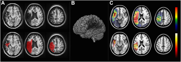
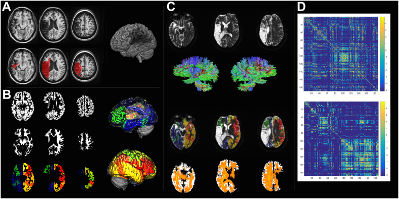
Lesion-symptom mapping is a key tool in understanding the relationship between structure and function in neuroscience as it can provide objective evidence about which regions are crucial for a given process. Initial limitations with this approach were largely overcome by voxel-based lesion-symptom mapping (VLSM), a method introduced in the early 2000s, which allows for a whole-brain approach to study the association between damaged areas and behavioral impairment by applying an independent statistical test at every voxel. By doing so, this technique eliminated the need to predefine regions of interest or classify patients into groups based on arbitrary cutoff scores. VLSM has nonetheless its own limitations; chiefly, a bias towards recognizing cortical necrosis/gliosis but with poor sensitivity for detecting injury along long white matter tracts, thus ignoring cortical disconnection, which can per se lead to behavioral impairment. Here, we propose a complementary method that, instead, establishes a statistical relationship between the strength of connections between all brain regions of the brain (as defined by a standard brain atlas) and the array of behavioral performance seen in patients with brain injury: connectome-based lesion-symptom mapping (CLSM). Whole-brain CLSM therefore has the potential to identify key connections for behavior independently of a priori assumptions with applicability across a broad spectrum of neurological and psychiatric diseases. We propose that this approach can further our understanding of brain-structure relationships and is worth exploring in clinical and theoretical contexts.

Keywords: Connectome-based lesion-symptom mapping; Connectomics; Diffusion tensor imaging; Voxel-based lesion-symptom mapping.


Figures

Fig. 1
Fig. 1
Voxel-based lesion-symptom mapping (VLSM). VLSM is performed by first defining the location of the post-stroke necrotic/gliotic tissue. Panel A demonstrates axial T1-weighted slices of one representative patient with a chronic post-stroke lesion (hypointense areas on top row slices), which is demarcated in red on the bottom row slices. Panel B is a 3D rendering that illustrates the magnitude of brain damage. Lesions from multiple individuals are then transformed into the stereotaxic MNI space and, for each voxel, a statistical analysis is performed by assessing whether there is a difference in a given behavioral measure (e.g. test score) in the group of subjects with a lesion in that voxel, versus the group of subjects without the lesion in that voxel. The results are then corrected for multiple comparisons based on the number of tested voxels. The top row in Panel C demonstrates the overlay of multiple lesions (red indicating areas with higher overlap) and the bottom row demonstrates an example of a voxel-wise statistical analyses (white-yellow voxels more strongly associated with behavioral measures).
Fig. 2
Fig. 2
The methodological steps involved in the calculation of the connectome share similarities with VLSM. First, the necrotic/gliotic image is defined on T1 or T2 weighted images as shown in Panel A. Again, here we see a 3D render of an individual patient's brain with a lesion. Subsequently, an iterative segmentation and cost-function normalization approach is employed to define probabilistic maps of gray (Panel B, top row) and white matter (Panel B, middle row). The transformation matrix between T1 to MNI space is used to transfer an anatomical atlas to T1-weighted space and segment the probabilistic gray matter into regions of interest (Panel C, bottom row). Panel C also shows the 3D renders of segmentation into regions of interest (left and right lateral views with different colors for different regions). Tractography is performed in diffusion space, so the white matter mask and the segmented gray matter maps are transferred to B0 space (Panel C) and tractography is used to assess the number of streamlines linking each possible pairs of regions. Care is taken to ensure that tractography is performed being guided by the white matter probabilistic map, excluding the lesion site. The bottom row of Panel C shows a fiber density image in orange. Finally, a 2D matrix is generated where each entry represents the connection weight between the region in the row and column. The top matrix in Panel D shows the connectome, which is then arranged anatomically (Panel D, bottom matrix) to demonstrate the difference in the number of fibers in the left hemisphere (left upper matrix quadrant) versus the right hemisphere (right lower matrix quadrant).

Similar articles

Cited by

References

    1. Agosta F., Henry R.G., Migliaccio R. Language networks in semantic dementia. Brain J. Neurol. 2010;133(Pt 1):286–299. (published Online First: Epub Date) - PMC - PubMed
    1. Assaf Y., Johansen-Berg H., Thiebaut de Schotten M. The role of diffusion MRI in neuroscience. NMR Biomed. 2017 (published Online First: Epub Date) - PubMed
    1. Basilakos A., Rorden C., Bonilha L., Moser D., Fridriksson J. Patterns of poststroke brain damage that predict speech production errors in apraxia of speech and aphasia dissociate. Stroke. 2015;46(6):1561–1566. (published Online First: Epub Date) - PMC - PubMed
    1. Bates E., Wilson S.M., Saygin A.P. Voxel-based lesion-symptom mapping. Nat. Neurosci. 2003;6(5):448–450. (published Online First: Epub Date) - PubMed
    1. Bonilha L., Fridriksson J. Subcortical damage and white matter disconnection associated with non-fluent speech. Brain J. Neurol. 2009;132(Pt 6) (published Online First: Epub Date) - PMC - PubMed

LinkOut - more resources