. 2017 Nov;33(11):1917-1926.
doi: 10.1007/s00381-017-3588-6. Epub 2017 Sep 7.

Time-to-event analysis of surgically treated posthemorrhagic hydrocephalus in preterm infants: a single-institution retrospective study

Affiliations

Time-to-event analysis of surgically treated posthemorrhagic hydrocephalus in preterm infants: a single-institution retrospective study

Rowland H Han et al. Childs Nerv Syst. 2017 Nov.

Abstract

Purpose: The purpose of this study is to report time points relevant to the neurosurgical management of posthemorrhagic hydrocephalus (PHH).

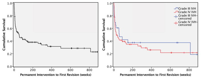
Methods: Data were collected retrospectively on 104 preterm infants with intraventricular hemorrhage (IVH) who received neurosurgical intervention for PHH at St. Louis Children's Hospital from 1994 to 2016. Kaplan-Meier curves were constructed for various endpoints.

Results: IVH grade on head ultrasound obtained through routine clinical care was II, III, and IV in 5 (4.8%), 33 (31.7%), and 66 (63.5%) of the patients, respectively. Neither IVH size nor location appeared to affect development of PHH. Days from birth to IVH, ventriculomegaly, temporizing neurosurgical procedure (TNP), and permanent neurosurgical intervention were 2.0 (95% CI 1.7-2.3), 3.0 (2.5-3.5), 24.0 (22.2-25.8), and 101.0 (90.4-111.6), respectively. Grades III and IV IVH did not differ in age at IVH diagnosis (Χ 2 (1 d.f.) = 1.32, p = 0.25), ventriculomegaly (Χ 2 = 0.73, p = 0.40), TNP (Χ 2 = 0.61, p = 0.43), or permanent intervention (Χ 2 = 2.48, p = 0.17). Ventricular reservoirs and ventriculosubgaleal shunts were used in 71 (68.3%) and 30 (28.8%), respectively. Eighty (76.9%) of the patients ultimately received a VPS. Five (4.8%) underwent a primary endoscopic third ventriculostomy (ETV), and two (1.9%) had ETV for a revision procedure. Four of the seven ETVs had choroid plexus cauterization.

Conclusions: Although most infants who develop IVH and ventriculomegaly will do so within a few days of birth, at-risk infants should be observed for at least 4 weeks with serial head ultrasounds to monitor for PHH requiring surgery.

Keywords: Intraventricular hemorrhage; Temporizing neurosurgical procedure; Ventriculomegaly; Ventriculoperitoneal shunt.


Figures

Fig. 1
Fig. 1
Coronal (left) and sagittal (right) head ultrasound screen captures from patients with grade II (top row), III (middle row), and IV (bottom row) IVH, respectively.
Fig. 2
Fig. 2
Boxplots of FOR measured at the time of each subject’s first ever imaging study, ventriculomegaly diagnosis, and TNP. Boxes indicate interquartile ranges; lines inside boxes represent medians; whiskers at 5 to 95 percentiles
Fig. 3
Fig. 3
KM survival curves showing time from birth to IVH diagnosis (top row), ventriculomegaly diagnosis (middle row), and TNP (bottom row). Data are shown for all 104 preterm infants (left column) and separated by IVH grade for 33 infants with grade III and 66 infants with grade IV IVH (right column). All subjects experienced the event, and none were censored
Fig. 4
Fig. 4
KM survival curves showing time from birth to permanent neurosurgical intervention. Data are shown for all 104 preterm infants (left) and separated by IVH grade for 33 infants with grade III and 66 infants with grade IV IVH (right). Tick marks indicate censoring due to end of study period or death (1 subject)
Fig. 5
Fig. 5
KM survival curves showing time from initial permanent intervention to first revision. Data are shown for all 80 preterm infants who required a VPS or ETV (left) and separated by IVH grade for 23 infants with grade III and 54 infants with grade IV IVH (right). Tick marks indicate censoring due to end of study period or death

References

    1. Adams-Chapman I, Hansen NI, Stoll BJ, Higgins R. Neurodevelopmental outcome of extremely low birth weight infants with posthemorrhagic hydrocephalus requiring shunt insertion. Pediatrics. 2008;121:e1167–77. doi: 10.1542/peds.2007-0423. - DOI - PMC - PubMed
    1. Al-Abdi SY, Al-Aamri MA. A Systematic Review and Meta-analysis of the Timing of Early Intraventricular Hemorrhage in Preterm Neonates: Clinical and Research Implications. J Clin Neonatol. 2014;3:76–88. doi: 10.4103/2249-4847.134674. - DOI - PMC - PubMed
    1. Badhiwala JH, Hong CJ, Nassiri F, Hong BY, Riva-Cambrin J, Kulkarni AV. Treatment of posthemorrhagic ventricular dilation in preterm infants: a systematic review and meta-analysis of outcomes and complications. J Neurosurg Pediatr. 2015;16:545–555. doi: 10.3171/2015.3.PEDS14630. - DOI - PubMed
    1. Beverley DW, Chance GW, Coates CF. Intraventricular haemorrhage--timing of occurrence and relationship to perinatal events. Br J Obstet Gynaecol. 1984;91:1007–1013. - PubMed
    1. Chittiboina P, Pasieka H, Sonig A, Bollam P, Notarianni C, Willis BK, Nanda A. Posthemorrhagic hydrocephalus and shunts: what are the predictors of multiple revision surgeries? J Neurosurg Pediatr. 2013;11:37–42. doi: 10.3171/2012.8.PEDS11296. - DOI - PubMed

LinkOut - more resources