. 2017 Oct 4;139(39):13692-13700.
doi: 10.1021/jacs.7b01380. Epub 2017 Sep 21.

A Small Molecule Causes a Population Shift in the Conformational Landscape of an Intrinsically Disordered Protein

Affiliations

A Small Molecule Causes a Population Shift in the Conformational Landscape of an Intrinsically Disordered Protein

David Ban et al. J Am Chem Soc. .

Abstract

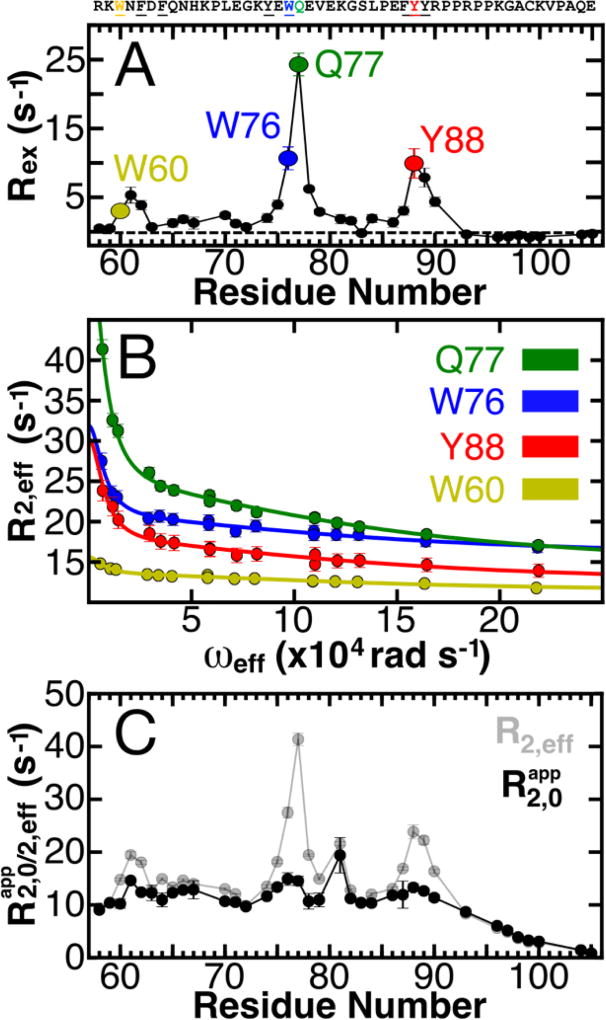
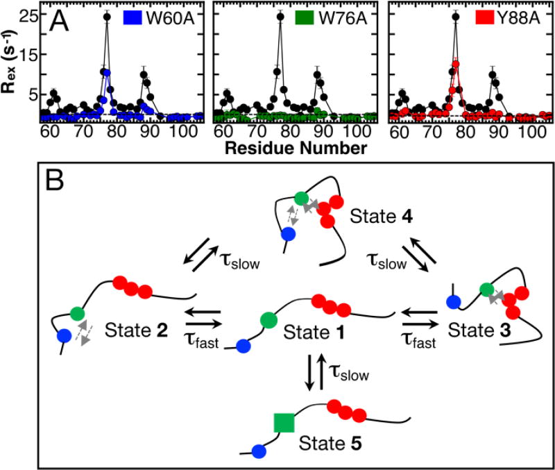
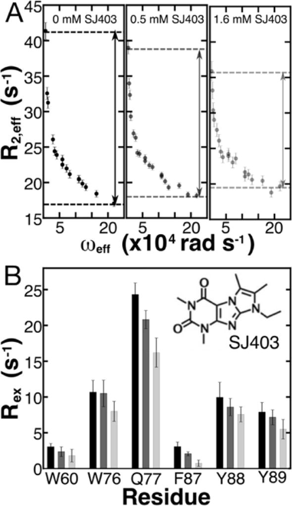
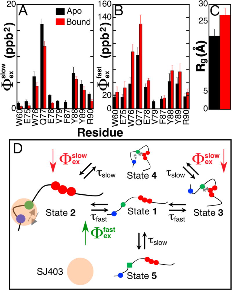
Intrinsically disordered proteins (IDPs) have roles in myriad biological processes and numerous human diseases. However, kinetic and amplitude information regarding their ground-state conformational fluctuations has remained elusive. We demonstrate using nuclear magnetic resonance (NMR)-based relaxation dispersion that the D2 domain of p27Kip1, a prototypical IDP, samples multiple discrete, rapidly exchanging conformational states. By combining NMR with mutagenesis and small-angle X-ray scattering (SAXS), we show that these states involve aromatic residue clustering through long-range hydrophobic interactions. Theoretical studies have proposed that small molecules bind promiscuously to IDPs, causing expansion of their conformational landscapes. However, on the basis of previous NMR-based screening results, we show here that compound binding only shifts the populations of states that existed within the ground state of apo p27-D2 without changing the barriers between states. Our results provide atomic resolution insight into how a small molecule binds an IDP and emphasize the need to examine motions on the low microsecond time scale when probing these types of interactions.


Figures

Figure 1
Figure 1. Defining the conformational landscape of p27-D2
(A) Large effective refocusing field strength (ωeff) and low temperature 1HN relaxation dispersion (RD) experiments reveal multi-state conformational exchange for the intrinsically disordered protein, p27-D2. Conformational exchange is sensed by many residues within p27-D2, which exhibit quantifiable exchange contributions ( Rex=R2,efflowωeffR2,effhighωeff; the dashed line indicates a Rex value of zero). The greatest observed conformational exchange is observed for residues in p27-D2 within three aromatic clusters, including W60 (yellow), W76 (blue), and Y88 (red); the 1HN displaying the largest amplitude of motion is Q77 (green). (B) The observed RD monitors the change in the effective transverse relaxation rate (R2,eff) as a function of ωeff and reports that some residues undergo conformational exchange that has two kinetic phases. Solid lines correspond to global fits of all RD data to a thermodynamic model that describes nuclei which sense two separate kinetic phases. Data presented in A & B were collected at 274 K. (C) Plot of R2,eff measured at low ωeff (grey points) where a maximal contribution of microsecond exchange is detected as compared to the transverse relaxation rate due to the apparent intrinsic fast nanosecond exchange ( R2,0app; black points) across all residues. R2,0app was determined from the fitted RD data and shows that large amplitude 1HN RD permits extensive sampling of Rex by using high ωeff which substantially quenches RD. The fast microsecond exchange detected in p27-D2 accounts for the elevated R2,eff values and once these are taken into account, a flat, featureless R2,0app profile is observed as expected for a prototypical disordered protein.
Figure 2
Figure 2. Mutagenesis of p27-D2 affect RD
(A) Contributions of Rex for different point mutants of p27-D2 in which critical aromatic residues were substituted with alanine. Mutational analysis showed that W76 (green) abrogates all microsecond exchange in p27-D2 whereas the other mutants, W60A (blue) and Y88A (red), exhibit little exchange near the site of mutation and reduced exchange at the two non-mutated sites. Points in black correspond to wild-type p27-D2 data and the dashed black line is drawn at zero which indicates no detected exchange. Analysis of residual RD within the p27-D2 mutants can be found in Figure S8 and Table S4. (B) Schematic of the proposed model for observed microsecond p27-D2 dynamics. The three critical hydrophobic residues are colored in blue, green, and red and correspond to W60, W76 in the central region W76, and F87-Y89, respectively. The grey arrows indicate transient hydrophobic interactions.
Figure 3
Figure 3. Binding of SJ403 to p27-D2 causes concentration-dependent reductions in Rex for 1HN nuclei
(A) Increasing concentration of SJ572403 (SJ403) reveals a concentration-dependent decrease in the observable contribution of conformational exchange. The black, grey and light grey points correspond to 1HN RD performed with 0 µM, 500 µM and 1600 µM, respectively, with a constant p27-D2 concentration of 400 µM at 274 K. The example shown in A is for residue Q77. The dependence of relaxation on compound concentration is readily observed in the RD data, as highlighted by the dashed lines in the three panels. The value of Rex decreased by 14% and 32%, respectively, at 0.5 and 1.6 mM SJ403 for the example plotted above. See Figure S12 in the Supporting Information for RD curves for other residues of p27-D2. The double headed arrows are used to guide the reader to see the change in Rex as a function of SJ403 concentration. (B) All residues that displayed large Rex and multi-state exchange processes show a significant decrease in Rex due to titration of SJ403. The RD performed with 500 µM and 1600 µM were statistically relevant across all values with a p-value of 1.2·10−3 and 7.16·10−14, respectively. The chemical structure of SJ403 is shown as an inset in B.
Figure 4
Figure 4. A small molecule, SJ403, induces a shift in the populations of the ground-state for p27-D2
(A & B). Comparison of conformational amplitudes (Φexslow/fast) for all residues that displayed multi-state exchange from fits of temperature dependent RD data for apo p27-D2 (black bars) and p27-D2 bound to SJ403 (red bars). The ratio of p27-D2 to SJ403 was 1:4. The conformational amplitudes associated with the slower kinetic phase (Φexslow) decrease when SJ403 is bound as compared with those associated with the faster kinetic process (Φexfast), which increase for all residues. Note that for the slow process, the value of Φexslow for V79 and F87 was zero. (C) Binding of SJ403 causes an increase in the Rg of p27-D2. Rg values were determined from a Guinier analysis of small angle X-ray scattering (SAXS) data. The increased Rg is also in agreement with the elevated Rg measured for p27-D2-Y88A mutant which is displaced due to its inability to engage in long-range hydrophobic interactions. The concentration of SJ403 during the SAXS measurement was 3 mM; therefore, based on the known dissociation constant, the bound population of p27-D2 was 55%. (D) The schematic illustrates populations changes associated with SJ403 binding to p27-D2; the population of State 2 increases while that of State 4 decreases. Population differences are represented by the relative sizes of the graphical icons for States 2 and 4; larger relative size indicates increased population. SJ403 preferentially binds to the W60 and W76 regions of p27-D2 increasing the population of State 2 with a concomitant decrease in that of State 4. This scenario is supported by the SAXS data which showed expansion of the Y88A p27-D2 mutant (Figure S15). See the Supporting Information for a further discussion of this conformational landscape model. The grey arrows indicate the hydrophobic interactions between residues.

References

    1. Csizmok V, Follis AV, Kriwacki RW, Forman-Kay JD. Chem. Rev. 2016;116:6424–6462. - PMC - PubMed
    1. Mitrea DM, Cika JA, Guy CS, Ban D, Banerjee PR, Stanley CB, Nourse A, Deniz AA, Kriwacki RW. eLIFE. 2016;5:e13571. - PMC - PubMed
    1. Dyson HJ, Wright PE. Nat. Rev. Mol. Cell Biol. 2005;6:197–208. - PubMed
    1. Heller GT, Pietro Sormanni, Vendruscolo M. Trends Biochem. Sci. 2015;40:491–496. - PubMed
    1. Jin F, Yu C, Lai L, Liu Z. PLoS Comput. Biol. 2013;9(10):e1003249. - PMC - PubMed

Publication types

MeSH terms

Substances

LinkOut - more resources