. 2019 Apr;39(4):704-717.
doi: 10.1177/0271678X17729280. Epub 2017 Sep 8.

Targeting the central projection of the dural trigeminovascular system for migraine prophylaxis

Affiliations

Targeting the central projection of the dural trigeminovascular system for migraine prophylaxis

Simon Akerman et al. J Cereb Blood Flow Metab. 2019 Apr.

Abstract

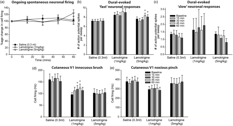
Migraine abortives likely target both peripheral-dural and central trigeminovascular mechanisms in mediating their therapeutic effects. However, in preclinical assays, many migraine preventives have little success at inhibiting similar trigeminovascular-mediated peripheral changes within the dural microenvironment. In addition, their effects on central trigeminovascular neuronal responses are largely unknown. Using a validated preclinical model of acute dural-intracranial (migraine-like) head pain, using Sprague Dawley rats, we tested whether migraine preventives suppress ongoing firing of central trigeminocervical neurons, and evoked responses to cranial neurovascular activation. Flunarizine, sodium valproate, propranolol, and amitriptyline, all dose-dependently inhibited ongoing spontaneous firing of dural trigeminovascular neurons, and differentially affected neuronal responses to intracranial-dural and extracranial-cutaneous somatosensory stimulation. Lamotrigine, only effective in the treatment of migraine aura, did not affect responses. These data provide a mechanistic rationale for the clinical effects of migraine preventives in the treatment of migraine, via the modulation of dural-responsive central trigeminovascular neurons. Also, given their limited effect on peripheral dural vasdilatory responses, these data also suggest that migraine preventives specifically target central, rather than peripheral, components of trigeminal neurovascular mechanisms involved in migraine pathophysiology, to mediate their preventive action. Finally, these data further validate this preclinical model of central trigeminovascular activation to screen migraine preventives.

Keywords: Migraine; central; peripheral; preventives; trigeminovascular system.


Figures

Figure 1.
Figure 1.
Experimental set-up for electrophysiological studies. (a). Neurons of the trigeminocervical complex were recorded in response to electrical stimulation of the trigeminal innervation of the dural meninges, and measurements of the cutaneous facial receptive field (shaded area). (b). Somatotopic representation of the trigeminal territories for receptive field characterization; V1, ophthalmic; V2, maxillary; V3, mandibular; 5Gn, trigeminal ganglion; C1-4, cervical regions. (c). Original tracing of a single sweep (stimulus) of a reproducible, dural-evoked multi-unit neuronal cluster classified as receiving Aδ-fiber input (<20 ms latency, ‘fast’ neuronal responses), but also with later unitary discharges that are classified as receiving C-fiber input (between 35 and 65 ms; slow neuronal responses), prior to any treatment (baseline – upper plot). As an example (lower plot), after administration of an effective migraine preventive, amitriptyline (5 mg/kg), the number of Aδ-fiber and C-fiber latency action potentials spikes is attenuated, indicative of inhibiting activation of these nociceptive nerve fibers. (d). Original example of the electrophysiological neuronal response to innocuous brush and noxious pinch of the cutaneous V1 receptive field. Top panel is original electrophysiological output, bottom panel is responses that cross the window discriminator.
Figure 2.
Figure 2.
Effects of flunarizine on dural-evoked trigeminocervical neuronal responses. (a) Time course changes in ongoing spontaneous trigeminocervical neuronal firing in response to flunarizine (1 and 5 mg/kg). Only 5 mg/kg caused a significant inhibition of ongoing spontaneous neuronal firing. The data have been normalized to represent the percentage change from baseline, and are expressed as mean ± SEM. Histograms of the time course changes in the average number of action potential spikes per sweep (mean ± SEM) of (b) intracranial dural-evoked multi-unit trigeminocervical neuronal clusters with inputs in the Aδ-fiber range (3–20 ms; ‘fast’ neuronal responses), and (c) unitary discharges within the C-fiber latency range (‘slow’ neuronal responses). Only 5 mg/kg flunarizine inhibited dural-evoked Aδ and C-fiber neuronal responses in the trigeminocervical complex (TCC). It also inhibited TCC neuronal responses to both (d) innocuous and (e) noxious somatosensory-evoked stimulation of the cutaneous facial receptive field. In all panels, vehicle control (hydroxypropyl-β-cyclodextrin; HBC) had no significant effects on neuronal responses. *P < 0.05 compared to baseline.
Figure 3.
Figure 3.
Effects of sodium valproate on dural-evoked trigeminocervical neuronal responses. (a) Time course changes in ongoing spontaneous trigeminocervical neuronal firing in response to sodium valproate (30 and 100 mg/kg). Only 100 mg/kg caused a significant inhibition of ongoing spontaneous neuronal firing. The data have been normalized to represent the percentage change from baseline, and are expressed as mean ± SEM. Histograms of the time course changes in the average number of action potential spikes per sweep (mean ± SEM) of (b) intracranial dural-evoked multi-unit trigeminocervical neuronal clusters with inputs in the Aδ-fiber range (3–20 ms; ‘fast’ neuronal responses), and (c) unitary discharges within the C-fiber (‘slow’ neuronal responses) latency range. Both 30 and 100 mg/kg sodium valproate inhibited dural-evoked Aδ and C-fiber neuronal responses in the trigeminocervical complex (TCC). Sodium valproate also inhibited TCC neuronal responses to (d) innocuous (100 mg/kg) and (e) noxious (30 and 100 mg/kg) somatosensory-evoked stimulation of the cutaneous facial receptive field. In all panels, vehicle control (saline) had no significant effects on neuronal responses. *P < 0.05 compared to baseline.
Figure 4.
Figure 4.
Effects of propranolol on dural-evoked trigeminocervical neuronal responses. (a) Time course changes in ongoing spontaneous trigeminocervical neuronal firing in response to propranolol (1 and 5 mg/kg). Both 1 and 5 mg/kg caused a significant inhibition of ongoing spontaneous neuronal firing. The data have been normalized to represent the percentage change from baseline, and are expressed as mean ± SEM. Histograms of the time course changes in the average number of action potential spikes per sweep (mean ± SEM) of (b) intracranial dural-evoked multi-unit trigeminocervical neuronal clusters with inputs in the Aδ-fiber and C-fiber range (3–30 ms; ‘fast’ neuronal responses), and (c) unitary discharges within the C-fiber (‘slow’ neuronal responses) latency range. Neither dose affected dural-evoked Aδ and C-fiber neuronal responses in the trigeminocervical complex (TCC). Similarly, neither dose affected TCC neuronal responses to (d) innocuous and (e) noxious somatosensory-evoked stimulation of the cutaneous facial receptive field. In all panels, vehicle control (saline) had no significant effects on neuronal responses. *P < 0.05 compared to baseline.
Figure 5.
Figure 5.
Effects of amitriptyline on dural-evoked trigeminocervical neuronal responses. (a) Time course changes in ongoing spontaneous trigeminocervical neuronal firing in response to amitriptyline (1 and 5 mg/kg). Both doses caused a significant inhibition of ongoing spontaneous neuronal firing. The data have been normalized to represent the percentage change from baseline, and are expressed as mean ± SEM. Histograms of the time course changes in the average number of action potential spikes per sweep (mean ± SEM) of (b) intracranial dural-evoked multi-unit trigeminocervical neuronal clusters with inputs in the Aδ-fiber range (3–25 ms; ‘fast’ neuronal responses), and (c) unitary discharges within the C-fiber (‘slow’ neuronal responses) latency range. Both 1 and 5 mg/kg amitriptyline significantly inhibited dural-evoked Aδ and C-fiber neuronal responses in the trigeminocervical complex (TCC). However, only 5 mg/kg inhibited TCC neuronal responses to (d) innocuous and (e) noxious somatosensory-evoked stimulation of the cutaneous facial receptive field. In all panels, vehicle control (saline) had no significant effects on neuronal responses. *P < 0.05 compared to baseline.
Figure 6.
Figure 6.
Effects of lamotrigine on dural-evoked trigeminocervical neuronal responses. (a) Time course changes in ongoing spontaneous trigeminocervical neuronal firing in response to lamotrigine (1 and 5 mg/kg). Neither dose had an effect on ongoing spontaneous neuronal firing. The data have been normalized to represent the percentage change from baseline, and are expressed as mean ± SEM. Histograms of the time course changes in the average number of action potential spikes per sweep (mean ± SEM) of (b) intracranial dural-evoked multi-unit trigeminocervical neuronal clusters with inputs in the Aδ-fiber range (3–25 ms; ‘fast’ neuronal responses), and (c) unitary discharges within the C-fiber (‘slow’ neuronal responses) latency range. Both 1 and 5 mg/kg lamotrigine significantly increased dural-evoked Aδ-fiber neuronal responses in the trigeminocervical complex (TCC), but neither dose had an effect on unitary C-fiber discharges. Only 1 mg/kg lamotrigine increased TCC neuronal responses to (d) innocuous somatosensory-evoked stimulation of the cutaneous facial receptive field. There was not effect of either dose on (e) noxious-evoked responses. In all panels, vehicle control (saline) had no significant effects on neuronal responses. *P < 0.05 compared to baseline.

References

    1. Noseda R, Burstein R. Migraine pathophysiology: anatomy of the trigeminovascular pathway and associated neurological symptoms, cortical spreading depression, sensitization, and modulation of pain. Pain 2013; 154(Suppl 1): S44–S53. - PubMed
    1. Goadsby PJ, Holland PR, Martins-Oliveira M, et al. Pathophysiology of migraine: a disorder of sensory processing. Physiol Rev 2017; 97: 553–622. - PMC - PubMed
    1. Burstein R, Yarnitsky D, Goor-Aryeh I, et al. An association between migraine and cutaneous allodynia. Ann Neurol 2000; 47: 614–624. - PubMed
    1. Lipton RB, Bigal ME, Ashina S, et al. Cutaneous allodynia in the migraine population. Ann Neurol 2008; 63: 148–158. - PMC - PubMed
    1. Penfield W, McNaughton F. Dural headache and innervation of the dura mater. Arch Neurol Psychiatry 1940; 44: 43–75.

Publication types

LinkOut - more resources