Review
. 2017 Sep 8;18(9):1932.
doi: 10.3390/ijms18091932.

The Effect of Sepsis on the Erythrocyte

Affiliations
Review

The Effect of Sepsis on the Erythrocyte

Ryon M Bateman et al. Int J Mol Sci. .

Abstract

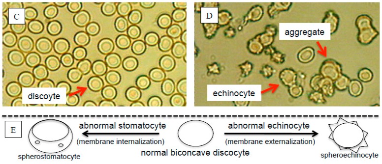
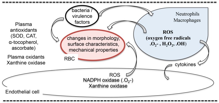
Sepsis induces a wide range of effects on the red blood cell (RBC). Some of the effects including altered metabolism and decreased 2,3-bisphosphoglycerate are preventable with appropriate treatment, whereas others, including decreased erythrocyte deformability and redistribution of membrane phospholipids, appear to be permanent, and factors in RBC clearance. Here, we review the effects of sepsis on the erythrocyte, including changes in RBC volume, metabolism and hemoglobin's affinity for oxygen, morphology, RBC deformability (an early indicator of sepsis), antioxidant status, intracellular Ca2+ homeostasis, membrane proteins, membrane phospholipid redistribution, clearance and RBC O₂-dependent adenosine triphosphate efflux (an RBC hypoxia signaling mechanism involved in microvascular autoregulation). We also consider the causes of these effects by host mediated oxidant stress and bacterial virulence factors. Additionally, we consider the altered erythrocyte microenvironment due to sepsis induced microvascular dysregulation and speculate on the possible effects of RBC autoxidation. In future, a better understanding of the mechanisms involved in sepsis induced erythrocyte pathophysiology and clearance may guide improved sepsis treatments. Evidence that small molecule antioxidants protect the erythrocyte from loss of deformability, and more importantly improve septic patient outcome suggest further research in this area is warranted. While not generally considered a critical factor in sepsis, erythrocytes (and especially a smaller subpopulation) appear to be highly susceptible to sepsis induced injury, provide an early warning signal of sepsis and are a factor in the microvascular dysfunction that has been associated with organ dysfunction.

Keywords: erythrocyte; morphology and microcirculation; rheology; sepsis.


Conflict of interest statement

The authors declare no conflict of interest.

Figures

Figure 1
Figure 1
Sepsis alters erythrocyte morphology and blood flow in capillary networks. (A,B) Intravital multiphoton images of the extensor digitorum longus (EDL) hindlimb skeletal muscle microcirculation of a control and septic mouse, 5 h after the onset of septic injury, respectively (Bateman et al. [34]). Solid lines indicate perfused capillaries, whereas broken lines indicate stopped-flow capillaries. Capillaries were pseudo colored for depth below the surface: brown (surface), green (75 µm), blue (150 µm) (Bateman et al. [35]). (C,D) Blood smears from a healthy volunteer with normal red blood cell (RBC) discocyte morphology and a septic shock patient with abnormal RBC morphology, respectively. Note the presence of echinocytes (also reported by de Oliveira et al. [4]), RBC aggregates and abnormal RBCs in the septic patient [8] (Copyright from John Wiley and Sons and Copyright Clearance Center). (E) The range of abnormal RBC morphologies, from spherostomatocyte to spheroechinocyte, described by Reinhart et al. [26], where damaged RBC membranes can either be internalized forming vacuoles or externalized forming spikes, respectively, as seen in the blood smear of septic RBCs.
Figure 1
Figure 1
Sepsis alters erythrocyte morphology and blood flow in capillary networks. (A,B) Intravital multiphoton images of the extensor digitorum longus (EDL) hindlimb skeletal muscle microcirculation of a control and septic mouse, 5 h after the onset of septic injury, respectively (Bateman et al. [34]). Solid lines indicate perfused capillaries, whereas broken lines indicate stopped-flow capillaries. Capillaries were pseudo colored for depth below the surface: brown (surface), green (75 µm), blue (150 µm) (Bateman et al. [35]). (C,D) Blood smears from a healthy volunteer with normal red blood cell (RBC) discocyte morphology and a septic shock patient with abnormal RBC morphology, respectively. Note the presence of echinocytes (also reported by de Oliveira et al. [4]), RBC aggregates and abnormal RBCs in the septic patient [8] (Copyright from John Wiley and Sons and Copyright Clearance Center). (E) The range of abnormal RBC morphologies, from spherostomatocyte to spheroechinocyte, described by Reinhart et al. [26], where damaged RBC membranes can either be internalized forming vacuoles or externalized forming spikes, respectively, as seen in the blood smear of septic RBCs.
Figure 2
Figure 2
Interactions between erythrocytes, bacterial virulence factors, phagocytes and endothelial cells and sources of red blood cell (RBC) exposure to reactive oxygen species (ROS). The schematic depicts interactions between bacteria and virulence factors with erythrocytes and phagocytes. Phagocytes generate oxygen free radicals, release reactive oxygen species and activate endothelial cells (EC) that also release ROS, exposing RBCs to exogenous sources of ROS. The plasma contains oxidant generating enzymes and various antioxidants that act to scavenge ROS. In addition to exposure from exogenous ROS, the RBC is also exposed to endogenous sources of ROS via hemoglobin autoxidation. Abbreviations: SOD (superoxide dismutase), CAT (catalase), .O2 (superoxide anion), H2O2 (hydrogen peroxide), .OH (hydroxyl radical).
Figure 3
Figure 3
Glycolysis is regulated at the RBC membrane via molecular switch (on/off) mechanisms involving phosphorylation, deoxyhemoglobin (Deoxy-Hb) and a complex of inactive glycolytic enzymes (GEs) [151,153] bound to the cytoplasmic domain of band 3 (cdb3). (A) phosphotyrosine phosphatase (PTP) maintains cdb3 in a dephosphorylated state [154]. (B) Hydrogen peroxide (H2O2) [155] and Ca2+ induce band 3 phosphorylation by disassociating PTP from band 3 [156] allowing a phosphotyrosine kinase (PTK) to then phosphorylate band 3. Tyrosine phosphorylation of band 3 leads to the displacement of GEs (broken arrow) and increased glycolysis [155]. (C) Similarly, deoxygenation of oxyhemoglobin (oxy-Hb) to deoxy-Hb (solid arrow) is associated with band 3 phosphorylation [157]. Moreover, deoxy-hemoglobin binding to cdb3 [158] is associated with release of GEs (broken arrow), glycolysis and increased release of adenosine triphosphate (ATP) [135]. Sepsis impairs this mechanism and RBC O2-dependent ATP efflux [13]. Abbreviations: PFK (phosphofructokinase), ALD (aldolase), GAPDH (glyceraldehyde 3-phosphate dehydrogenase), ADP (adenosine diphosphate), TYR (tyrosine), TYR-P (tyrosine-phosphate). (Modified from Bateman et al. [13], available online: https://creativecommons.org/licenses/by/4.0/).

References

    1. Singer M., Deutschman C.S., Seymour C.W., Shankar-Hari M., Annane D., Bauer M., Bellomo R., Bernard G.R., Chiche J.D., Coopersmith C.M., et al. The third international consensus definitions for sepsis and septic shock (sepsis-3) JAMA. 2016;315:801–810. doi: 10.1001/jama.2016.0287. - DOI - PMC - PubMed
    1. Baskurt O.K., Gelmont D., Meiselman H.J. Red blood cell deformability in sepsis. Am. J. Respir. Crit. Care Med. 1998;157:421–427. doi: 10.1164/ajrccm.157.2.9611103. - DOI - PubMed
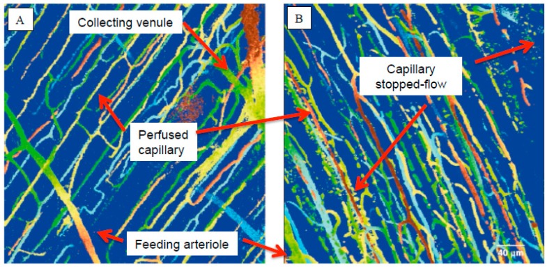
    1. Bateman R.M., Jagger J.E., Sharpe M.D., Ellsworth M.L., Mehta S., Ellis C.G. Erythrocyte deformability is a nitric oxide-mediated factor in decreased capillary density during sepsis. Am. J. Physiol. Heart Circ. Physiol. 2001;280:H2848–H2856. - PubMed
    1. De Oliveira Y.P.A., Pontes-de-Carvalho L.C., Couto R.D., Noronha-Dutra A.A. Oxidative stress in sepsis. Possible production of free radicals through an erythrocyte-mediated positive feedback mechanism. BJID. 2017;21:19–26. doi: 10.1016/j.bjid.2016.11.004. - DOI - PMC - PubMed
    1. Piagnerelli M., Boudjeltia K.Z., Brohee D., Piro P., Carlier E., Vincent J.L., Lejeune P., Vanhaeverbeek M. Alterations of red blood cell shape and sialic acid membrane content in septic patients. Crit. Care Med. 2003;31:2156–2162. doi: 10.1097/01.CCM.0000079608.00875.14. - DOI - PubMed

MeSH terms

LinkOut - more resources