. 2017 Sep 8;22(9):1498.
doi: 10.3390/molecules22091498.

Anti-HIV Activities and Mechanism of 12-O-Tricosanoylphorbol-20-acetate, a Novel Phorbol Ester from Ostodes katharinae

Affiliations

Anti-HIV Activities and Mechanism of 12-O-Tricosanoylphorbol-20-acetate, a Novel Phorbol Ester from Ostodes katharinae

Huan Chen et al. Molecules. .

Abstract

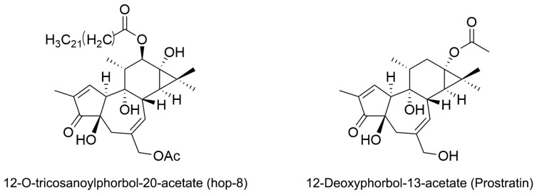
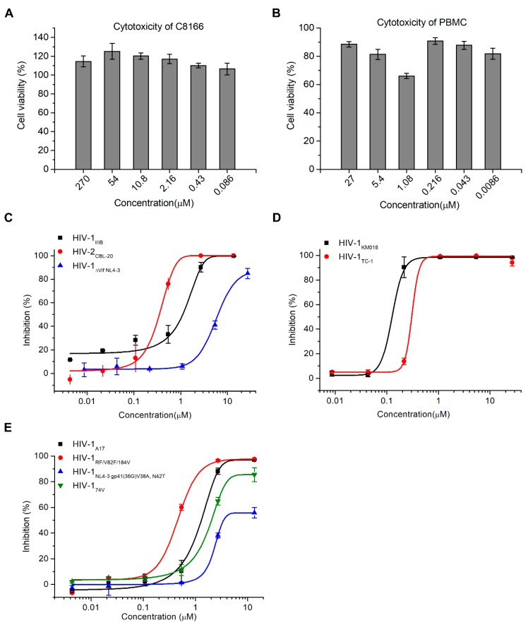
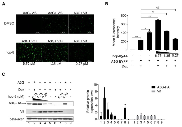
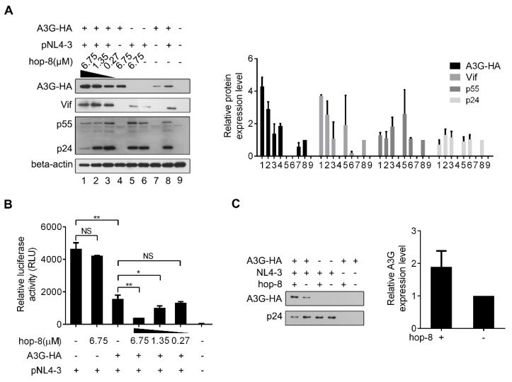
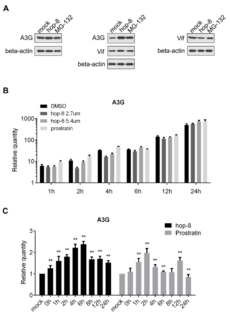
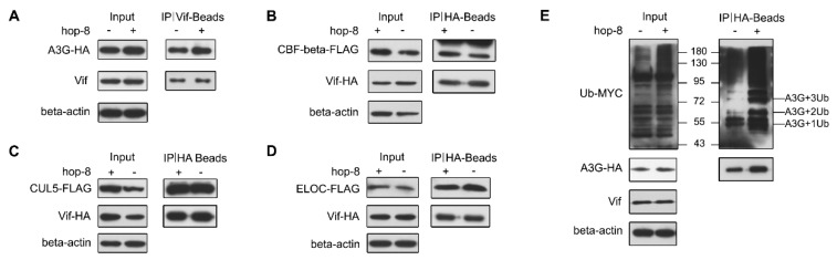
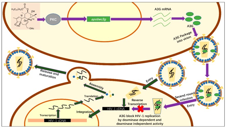
APOBEC3G is a member of the human cytidine deaminase family that restricts Vif-deficient viruses by being packaged with progeny virions and inducing the G to A mutation during the synthesis of HIV-1 viral DNA when the progeny virus infects new cells. HIV-1 Vif protein resists the activity of A3G by mediating A3G degradation. Phorbol esters are plant-derived organic compounds belonging to the tigliane family of diterpenes and could activate the PKC pathway. In this study, we identified an inhibitor 12-O-tricosanoylphorbol-20-acetate (hop-8), a novel ester of phorbol which was isolated from Ostodes katharinae of the family Euphorbiaceae, that inhibited the replication of wild-type HIV-1 and HIV-2 strains and drug-resistant strains broadly both in C8166 cells and PBMCs with low cytotoxicity and the EC50 values ranged from 0.106 μM to 7.987 μM. One of the main mechanisms of hop-8 is to stimulate A3G expressing in HIV-1 producing cells and upregulate the A3G level in progeny virions, which results in reducing the infectivity of the progeny virus. This novel mechanism of hop-8 inhibition of HIV replication might represents a promising approach for developing new therapeutics for HIV infection.

Keywords: 12-O-tricosanoylphorbol-20-acetate; APOBEC3G; HIV; Vif; antiviral agent; phorbol ester.


Conflict of interest statement

The authors declare no conflict of interest.

Figures

Figure 1
Figure 1
Structure of 12-O-tricosanoylphorbol-20-acetate (hop-8) and 12-Deoxyphorbol-13-acetate (prostratin).
Figure 2
Figure 2
Hop-8 inhibited HIV-1 and HIV-2 wild-type, clinical isolates and resistant strains with low cytotoxicity. (A) The cytotoxicity of hop-8 in C8166; (B) The cytotoxicity of hop-8 in peripheral blood mononuclear cells (PBMCs); (C) The antiviral activities of hop-8 against the clinical isolates HIV-1KM018, and HIV-1TC-1 were measured in PBMCs; (D) The antiviral activities of hop-8 against wild-type HIV-1IIIB, HIV-2CBL-20, and HIV-1ΔVif NL4-3 were measured in the C8166 cell line; (E) Antiviral activities of hop-8 against the HIV-1 drug resistant strains HIV-1A17, HIV-1NL4-3 gp41 (36G) V38A, N42T, HIV-1RF/V28F/184V, and HIV-174V. The levels of p24 in the cell culture supernatant were measured by ELISA. Each data point represents the mean percent inhibition (relative to the positive control) ± standard deviation (bars), n ≥ 2. Data were analyzed with Origin 8.5 (OriginLab, MA, USA).
Figure 3
Figure 3
Hop-8 restored A3G levels in cells undergoing Vif-mediated A3G degradation. (A) Fluorescence photographs were taken with a fluorescent microscope (Leica DMI4000B); (B) Cells were harvested, and EYFP expression in live cells was analyzed with a FACSVerse flow cytometer. Data are expressed as the means ± SD of at least three independent measurements. Statistical comparisons were performed between A3G-EYFP, the Vif positive group (column 2) and other groups with the pair-sample t-test with Origin 8.5. (p < 0.05, significant difference; *: p < 0.05; **: p < 0.01; NS: not significant); (C) pcDNA3.1-APOBEC3G-HA (0.5 μg) was transfected into TRex-hvif-15 cells. The compound (6.75, 1.35, and 0.27 μM) containing 0.1 μg/mL Dox was added 6 h post-transfection. The relative expression levels of A3G-HA and Vif were normalized by the levels of beta-actin. Values are presented as normalized intensities relative to the values of the Dox and A3G-HA positive groups (lane 5). Each data point represents the mean relative quantity ± standard deviation (bars), n ≥ 3.
Figure 4
Figure 4
Hop-8 enhanced the expression of A3G and reduced p24 expression in HIV-1 producing cells and reduced the infectivity of the produced virus. (A) Hop-8 increased the expression of A3G and Vif while reduced the p24 expression in HIV-1 producing cells. The plasmids pcDNA3.1-APOBEC3G-HA (0.125 μg) and pNL4-3 (0.375 μg) were transfected into 2 × 10⁵ 293T cells. The compound was diluted to 6.75, 1.35, and 0.27 μM with DMEM and added to the wells 6 h post-transfection. The relative expression levels of A3G-HA, Vif, p55, and p24 were normalized by the levels of beta-actin. Values are presented as normalized intensities relative to the values of the pNL4-3 and A3G-HA positive groups (lane 8). Each data point represents the mean relative quantity ± standard deviation (bars), n ≥ 3. (B) Hop-8 reduced the infectivity of the progeny virus. The supernatant of NL4-3 was collected and 4 × 10⁴ TZM-bl cells were infected with NL4-3 that contained 20 ng p24. The residual infection was determined using relative luciferase activity. All data represent the means ± standard deviation (bars), n ≥ 3. Data were analyzed by the unpair-sample t-test with Origin 8.5. (p < 0.05, significant difference; *: p < 0.05; **: p < 0.01; NS: not significant). (C) Hop-8 increased the incorporation of A3G in progeny virions. pcDNA-APOBEC3G-HA (1 μg) and pNL4-3 (1 μg) were transfected consecutively or co-transfected into 293T cells in 6-well cell culture plates. Cells were cultured with or without hop-8 (2.7 μM) for 48 h. The supernatant was collected and centrifuged at 2000× g and the cell debris was discarded. The viral particles were lysed with 0.5% Triton-X100. A3G and p24 levels in the supernatant were determined by western blot. The relative expression levels of A3G-HA were normalized by the levels of p24. Each data point represents the mean relative quantity ± standard deviation (bars), n ≥ 3.
Figure 5
Figure 5
Hop-8 upregulated both the protein and mRNA levels of A3G. (A) Hop-8 upregulated A3G protein level regardless of whether Vif was present or absent and had no influence on Vif expression. pcDNA3.1-APOBEC3G-HA and pcDNA3.1-Vif-HA were transfected consecutively or co-transfected into 293T cells and cultured with DMSO, 2.7 μM hop-8, or 10 μM of the proteasome inhibitor MG-132 for 24 h. A3G, Vif, and beta-actin were detected by western blot; (B) A3G mRNA levels of pcDNA3.1-APOBEC3G-HA transfected 293T cells in the presence of the 2.7 μM (dark gray) and 5.4 μM (gray) hop-8, prostratin (2.7 μM) (light gray), or DMSO (black). The cells collected at 4 h post transfection (0 h of compounds treatment) were used as a control. Each data point represents the mean relative quantity (relative to the mock) ± standard deviation (bars), n = 3; (C) A3G mRNA levels in PBMCs which treated with hop-8 (2 μM) (black) or prostratin (2 μM) (gray). Each data point represents the mean relative quantity (relative to the mock) ± standard deviation (bars), n ≥ 3. Data were analyzed by the Mann Whitney test with GraphPad Prism 6.0 (GraphPad Software, Inc., La Jolla, CA, USA). (p < 0.05, significant difference; *: p <0.05; **: p < 0.01).
Figure 6
Figure 6
Hop-8 did not interfere in the interaction of Vif with A3G, CBF-β, Elongin C, or Cullin 5. (A) Hop-8 did not interfere in Vif and A3G interaction. The plasmid pcDNA3.1-APOBEC3G-HA (3 μg) was transfected into 2 × 10⁶ Trex-hvif-15 cells and treated with 0.1 μg/mL Dox and 2.7 μM hop-8 or DMSO for 48 h. Cells were treated with 10 μM MG-132 for 16 h. Co-IP assays were performed with an anti-HIV1 Vif antibody; (BD) Hop-8 did not block the interaction between Vif and CBF-β (B), Elongin C (C), or Cullin 5 (D). The plasmids pcDNA3.1-Vif-HA (1.5 μg) and pcDNA3.1-CBF-β-FLAG (1.5 μg) or pcDNA3.1-Elogin C-FLAG or pcDNA3.1-Cullin 5-FLAG were co-transfected into 2 × 10⁶ 293T cells. Cells were treated with 2.7 μM hop-8 or DMSO. Co-IP assays were performed with an anti-HA antibody; (E) The influence of hop-8 on A3G ubiquitination. The plasmids pcDNA3.1-APOBEC3G-HA (1.5 μg) and pUb-MYC (1.5 μg) were co-transfected into 2 × 10⁶ Trex-hvif-15 cells. Cells were treated with 0.1 μg/mL Dox and 2.7 μM hop-8 or DMSO. The Co-IP assay was performed with a rabbit anti-HA antibody.
Figure 7
Figure 7
Schematic of the anti-HIV-1 activity mechanism of hop-8. The process shown with deep green arrows represents a simple schematic of HIV replication. Hop-8 stimulates A3G expression via the PKC pathway. A3G is packaged into the progeny virions and restricts the replication of the progeny virions (shown with purple arrows).

Similar articles

Cited by

References

    1. Spivak A.M., Planelles V. HIV-1 Eradication: Early Trials (and Tribulations) Trends Mol. Med. 2016;22:10–27. doi: 10.1016/j.molmed.2015.11.004. - DOI - PMC - PubMed
    1. Da Cunha J., Maselli L.M., Stern A.C., Spada C., Bydlowski S.P. Impact of antiretroviral therapy on lipid metabolism of human immunodeficiency virus-infected patients: Old and new drugs. World J. Virol. 2015;4:56–77. doi: 10.5501/wjv.v4.i2.56. - DOI - PMC - PubMed
    1. Hill A.M., Moecklinghoff C., DeMasi R. When can HIV clinical trials detect treatment effects on drug resistance? Int. J. STD AIDS. 2015;26:268–278. doi: 10.1177/0956462414536885. - DOI - PubMed
    1. Margolis A.M., Heverling H., Pham P.A., Stolbach A. A review of the toxicity of HIV medications. J. Med. Toxicol. 2014;10:26–39. doi: 10.1007/s13181-013-0325-8. - DOI - PMC - PubMed
    1. Stolbach A., Paziana K., Heverling H., Pham P. A Review of the Toxicity of HIV Medications II: Interactions with Drugs and Complementary and Alternative Medicine Products. J. Med. Toxicol. 2015;11:326–341. doi: 10.1007/s13181-015-0465-0. - DOI - PMC - PubMed

MeSH terms

LinkOut - more resources