. 2017 Sep 7;101(3):391-403.
doi: 10.1016/j.ajhg.2017.08.003.

CDK10 Mutations in Humans and Mice Cause Severe Growth Retardation, Spine Malformations, and Developmental Delays

Affiliations

CDK10 Mutations in Humans and Mice Cause Severe Growth Retardation, Spine Malformations, and Developmental Delays

Christian Windpassinger et al. Am J Hum Genet. .

Abstract

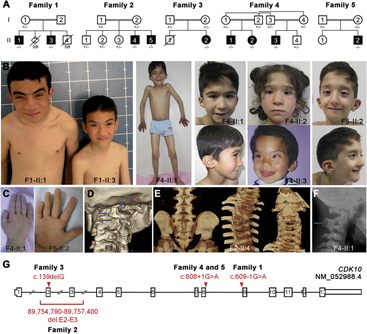
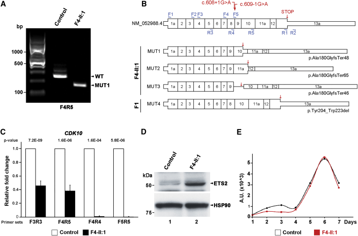
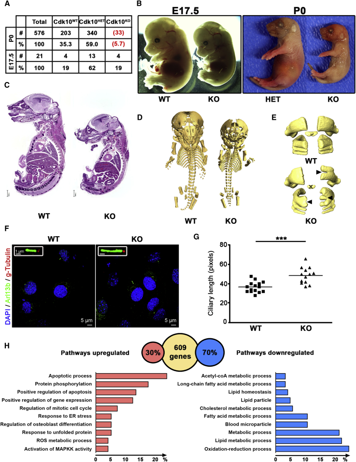
In five separate families, we identified nine individuals affected by a previously unidentified syndrome characterized by growth retardation, spine malformation, facial dysmorphisms, and developmental delays. Using homozygosity mapping, array CGH, and exome sequencing, we uncovered bi-allelic loss-of-function CDK10 mutations segregating with this disease. CDK10 is a protein kinase that partners with cyclin M to phosphorylate substrates such as ETS2 and PKN2 in order to modulate cellular growth. To validate and model the pathogenicity of these CDK10 germline mutations, we generated conditional-knockout mice. Homozygous Cdk10-knockout mice died postnatally with severe growth retardation, skeletal defects, and kidney and lung abnormalities, symptoms that partly resemble the disease's effect in humans. Fibroblasts derived from affected individuals and Cdk10-knockout mouse embryonic fibroblasts (MEFs) proliferated normally; however, Cdk10-knockout MEFs developed longer cilia. Comparative transcriptomic analysis of mutant and wild-type mouse organs revealed lipid metabolic changes consistent with growth impairment and altered ciliogenesis in the absence of CDK10. Our results document the CDK10 loss-of-function phenotype and point to a function for CDK10 in transducing signals received at the primary cilia to sustain embryonic and postnatal development.

Keywords: Al Kaissi syndrome knockout mice; CDK10; ETS2; cilia; congenital disorder; growth retardation; metabolism; spine malformation.


Figures

Figure 1
Figure 1
Biallelic Germline Mutations in CDK10 in Nine Individuals from Five Consanguineous Families (A) Pedigrees of families from Tunisia (1), Algeria (2), Saudi Arabia (3), and Turkey (4 and 5). The affected individuals carry homozygous germline mutations in CDK10. Open symbols represent unaffected individuals, and filled symbols represent affected individuals. (B) Photographs of affected individuals show facial dysmorphisms. (C) Hand anomalies with clinodactyly of the fifth finger and single transverse palmar crease. (D) Severe mal-segmentation of the cervical vertebrae in proband F1-II:1. (E) Lack of fusion of the posterior arches of S2–S5, right T4 hemivertebrae, and lack of fusion of the anterior arch of the atlas and partial fusion of C2 and C3 vertebrae in proband F2-II:4. (F) Fusion of the cervical vertebrae in proband F4-II:1. (G) Exon-intron structure of CDK10 on chromosome 16 shows the position of the four identified homozygous mutations.
Figure 2
Figure 2
CDK10 Splice-Site Mutations Reduce Endogenous mRNA Levels and Increase ETS2 Levels (A) RT-PCR performed on primary dermal fibroblasts derived from affected (F4-II:1) and control individuals indicates aberrant splicing of endogenous CDK10 mRNA. (B) Schematic representations of the wild-type CDK10 isoform and four mutant transcripts cloned from primary fibroblasts with intron 8 donor and acceptor splice-site mutations. (C) qRT-PCR analysis demonstrates lower endogenous CDK10 mRNA levels in cells derived from affected individuals than in control cells, suggestive of nonsense-mediated decay. (D) Whole-cell lysates of human fibroblasts from control and affected individuals were separated on SDS-PAGE, and immunoblots were stained with antibodies against ETS2 and HSP90. Cells with reduced or absent CDK10 kinase activity displayed increased levels of ETS2, which is normally degraded once it is phosphorylated by CDK10. (E) Primary human fibroblasts from control and affected individuals were grown in the presence of serum over 7 days and did not show any noticeable differences.
Figure 3
Figure 3
Mice Lacking Cdk10 as a Model for the Human Disease (A) Heterozygous Cdk10WT/KO mice were interbred, and the genotypes of the offspring were analyzed at embryonic day 17.5 (E17.5) and birth (P0). Numbers in red indicate that pups were dead at the moment of collection. (B) Control Cdk10WT/WT (WT), heterozygous Cdk10WT/KO (HET), and knockout Cdk10KO/KO (KO) embryos at E17.5 and pups at P0 were isolated and photographed. (C) Hematoxylin & eosin staining of sagittal sections of the entire Cdk10WT/KO and Cdk10KO/KO P0 pups. The scale bar represents 1 mm. (D) 3D μCT images of the mineralized part of the entire skeleton from Cdk10WT/WT and Cdk10KO/KO P0 pups. Note the shorter stature of the Cdk10KO pups and the defects in the vertebrae. (E) 3D μCT images of C1–C4 vertebrae from Cdk10WT/WT and Cdk10KO/KO P0 mice. Note the presence of bifidity (black arrow) and the absence of mineralized dens (cross) in the Cdk10KO/KO mice. (F) Cdk10KO and WT MEFs were starved for 4 days, and cilia were stained with Arl13b and gamma-tubulin antibodies. The inset shows magnified cilia. (G) Quantification of the maximal length of the cilia in WT and Cdk10KO MEFs. ∗∗∗p value < 0.001 (Student’s t test). (H) Pathway analysis from gene expression data of eight different tissues obtained from Cdk10WT/WT and Cdk10KO/KO P0 mice. From the entire dataset, 609 genes were differentially expressed in at least two tissues. In the absence of Cdk10, 30% of these genes were downregulated and 70% were upregulated. Pathway analysis for datasets of up- and downregulated genes is displayed as the percentage of genes annotated for the indicated pathway.

References

    1. Lim S., Kaldis P. Cdks, cyclins and CKIs: roles beyond cell cycle regulation. Development. 2013;140:3079–3093. - PubMed
    1. Malumbres M., Harlow E., Hunt T., Hunter T., Lahti J.M., Manning G., Morgan D.O., Tsai L.H., Wolgemuth D.J. Cyclin-dependent kinases: a family portrait. Nat. Cell Biol. 2009;11:1275–1276. - PMC - PubMed
    1. Morgan D.O. New Science Press Ltd; 2007. The cell cycle: principles of control.
    1. Brambilla R., Draetta G. Molecular cloning of PISSLRE, a novel putative member of the cdk family of protein serine/threonine kinases. Oncogene. 1994;9:3037–3041. - PubMed
    1. Graña X., Claudio P.P., De Luca A., Sang N., Giordano A. PISSLRE, a human novel CDC2-related protein kinase. Oncogene. 1994;9:2097–2103. - PubMed

MeSH terms