Review
. 2017 Sep 8:71:539-556.
doi: 10.1146/annurev-micro-090816-093754.

Outer Membrane Biogenesis

Affiliations
Review

Outer Membrane Biogenesis

Anna Konovalova et al. Annu Rev Microbiol. .

Abstract

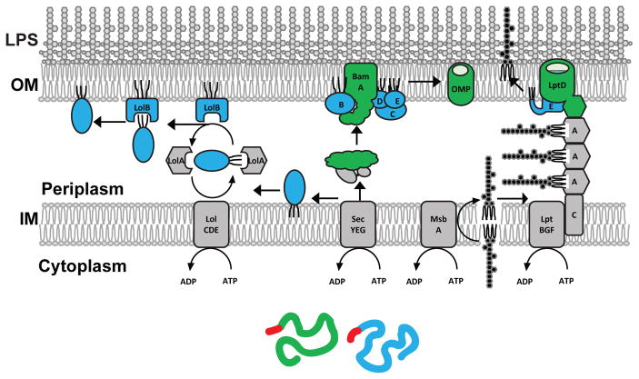
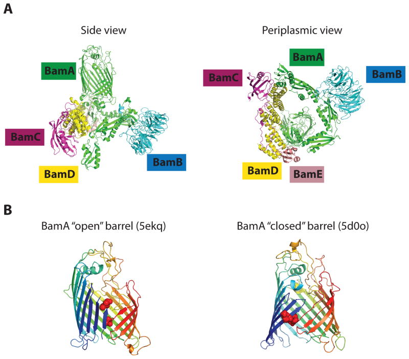
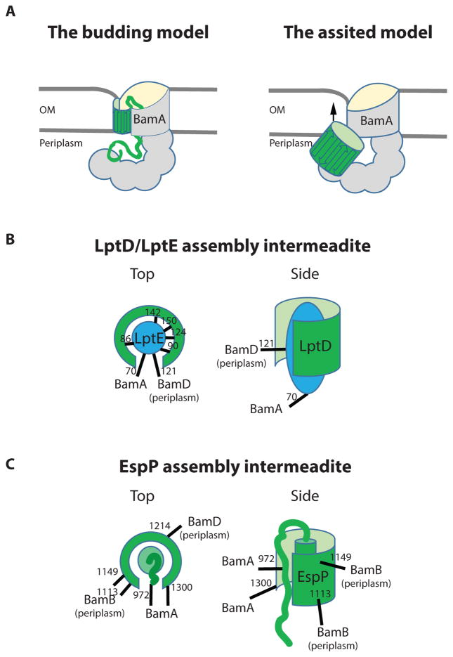
The hallmark of gram-negative bacteria and organelles such as mitochondria and chloroplasts is the presence of an outer membrane. In bacteria such as Escherichia coli, the outer membrane is a unique asymmetric lipid bilayer with lipopolysaccharide in the outer leaflet. Integral transmembrane proteins assume a β-barrel structure, and their assembly is catalyzed by the heteropentameric Bam complex containing the outer membrane protein BamA and four lipoproteins, BamB-E. How the Bam complex assembles a great diversity of outer membrane proteins into a membrane without an obvious energy source is a particularly challenging problem, because folding intermediates are predicted to be unstable in either an aqueous or a hydrophobic environment. Two models have been put forward: the budding model, based largely on structural data, and the BamA assisted model, based on genetic and biochemical studies. Here we offer a critical discussion of the pros and cons of each.

Keywords: LptD; envelope biogenesis; lateral gate; outer membrane protein; protein folding.


Figures

Figure 1
Figure 1. The structure of Gram-negative envelope and the pathways required for OM biogenesis
The envelope of Gram-negative bacteria contains two membranes. The OM in an asymmetric bilayer with LPS in its outer leaflet. LPS is synthesized on the cytoplasmic side of the IM and translocated across the IM by MsbA. LPS is extracted from the IM by LptBFG. LPS is then translocated across the periplasm by being pushes by the following LPS molecule through the periplasmic LptA bridge to the OM translocon, LptD/LptE, which inserts LPS directly into the outer leaflet (see the recent reviews (62; 85). OM proteins, such as β-barrel OMPs (green) or lipoproteins (blue) are synthesized on ribosomes and contain a signal sequence (red) that targets them to the Sec complex, which translocates them across the IM. Unfolded OMPs are bound by periplasmic chaperones which escort them to the Bam complex. The Bam complex assembles OMPs into the OM (see the recent review (73). Lipoproteins undergo lipid modifications and are extracted from the IM by the LolCDE and passed to the periplasmic chaperone LolA that delivers them to the OM acceptor protein LolB, which inserts lipoproteins into the inner leaflet of the OM (see the recent review (64).
Figure 2
Figure 2. The structure of the Bam complex
(A) The structure of the Bam complex by cryo-EM (5LJO) (37). The central component, BamA, contains a β-barrel and a periplasmic domain, consisting of five POTRA domains that scaffold lipoproteins BamB-E. POTRA domains together with lipoproteins form a ring-like structure with the cavity underneath the BamA barrel. Note, that the position of the ring relative to BamA barrel varies substantially in different structures of the Bam complex. (B) The architecture of the BamA barrel and the seam in two different crystal structures of BamA (2; 31). The residues of the seam used for a disulfide crosslinking are highlighted by the red spheres. These structures have been proposed to represent BamA in its “open” and “closed” conformations.
Figure 3
Figure 3. The proposed models for BamA function and experimental evidence for the folding in the periplasm
(A) The BamA budding model suggests that BamA opens laterally and templates the β-strands of an incoming OMP. As a result, a hybrid BamA/OMP barrel is formed. Once all OMP β-strand are templated, the OMP barrel buds off laterally into the OM (67). The BamA assisted model suggests that OMP folding is driven by their intrinsic thermodynamic properties and occurs largely at the periplasmic side of the OM. The function of BamA is to reduce the kinetic barrier for membrane integration by creating a membrane defect (illustrated by OM thinning) in the proximity to the BamA seam (24). (B) LptD/LptE assembly intermediate represented by the lptD4213 assembly-defective mutant (56). The LptD barrel (green) is largely formed because it established contacts LptE (blue), but not fully closed because LptE is still crosslinked to BamD. Sites of LptE crosslinking to the indicated proteins are shown. This LptD/E complex remains in the periplasm because it crosslinks to the periplasmic lipoprotein BamD. The N-terminal domains of LptD and LptE lipid moieties are not shown for clarity. (C) EspP assembly intermediate represented by espP_G1066A or G1081A assembly-defective mutants or WT under the conditions of slow growth (40; 71). EspP barrel largely formed because the α-helix of the passenger domain is buried inside the barrel. The barrel remains open to facilitate translocation of the passenger domain, which can still be crosslinked to BamA. Sites of EspP crosslinking to the indicated proteins are shown. This partially folded EspP barrel remains in the periplasm because it crosslinks to the periplasmic lipoproteins BamB and BamD, and cannot be crosslinked to LPS. Only the C-terminal part of the passenger domain is shown for clarity.

References

    1. Albrecht R, Schutz M, Oberhettinger P, Faulstich M, Bermejo I, et al. Structure of BamA, an essential factor in outer membrane protein biogenesis. Acta Crystallogr D Biol Crystallogr. 2014;70:1779–89. - PubMed
    1. Bakelar J, Buchanan SK, Noinaj N. The structure of the beta-barrel assembly machinery complex. Science. 2016;351:180–6. - PMC - PubMed
    1. Basle A, Rummel G, Storici P, Rosenbusch JP, Schirmer T. Crystal structure of osmoporin OmpC from E. coli at 2.0 A. J Mol Biol. 2006;362:933–42. - PubMed
    1. Bernstein HD. Looks can be deceiving: recent insights into the mechanism of protein secretion by the autotransporter pathway. Mol Microbiol. 2015;97:205–15. - PMC - PubMed
    1. Botos I, Majdalani N, Mayclin SJ, McCarthy JG, Lundquist K, et al. Structural and Functional Characterization of the LPS Transporter LptDE from Gram-Negative Pathogens. Structure. 2016;24:965–76. - PMC - PubMed

Publication types

MeSH terms