. 2017 Sep 8;18(1):67.
doi: 10.1186/s12868-017-0384-x.

Cerebral microvascular dysfunction in metabolic syndrome is exacerbated by ischemia-reperfusion injury

Affiliations

Cerebral microvascular dysfunction in metabolic syndrome is exacerbated by ischemia-reperfusion injury

Nathalie Obadia et al. BMC Neurosci. .

Abstract

Background: Metabolic syndrome (MetS) is associated with an increased risk of cerebrovascular diseases, including cerebral ischemia. Microvascular dysfunction is an important feature underlying the pathophysiology of cerebrovascular diseases. In this study, we aimed to investigate the impacts of ischemia and reperfusion (IR) injury on the cerebral microvascular function of rats with high-fat diet-induced MetS.

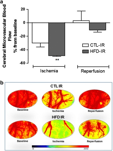
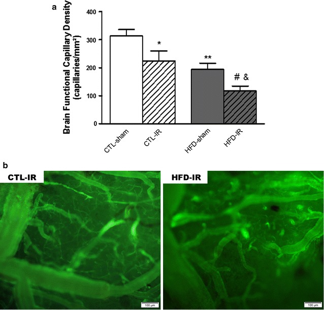
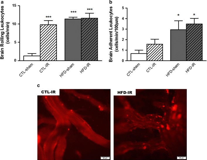
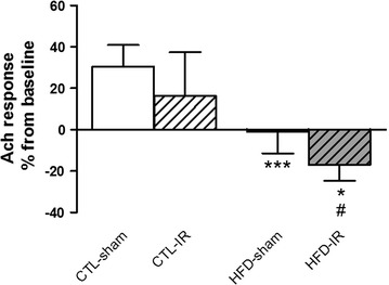
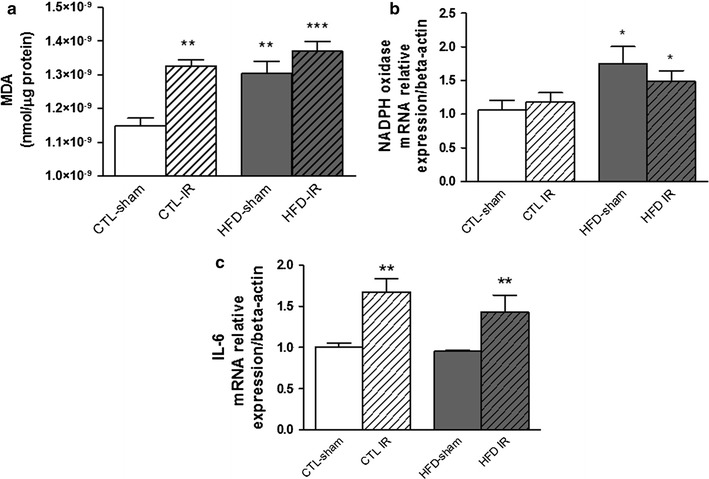
Results: We examined Wistar rats fed a high-fat diet (HFD) or normal diet (CTL) for 20 weeks underwent 30 min of bilateral carotid artery occlusion followed by 1 h of reperfusion (IR) or sham surgery. Microvascular blood flow was evaluated on the parietal cortex surface through a cranial window by laser speckle contrast imaging, functional capillary density, endothelial function and endothelial-leukocyte interactions by intravital videomicroscopy. Lipid peroxidation was assessed by TBARs analysis, the expression of oxidative enzymes and inflammatory markers in the brain tissue was analyzed by real-time PCR. The cerebral IR in MetS animals induced a functional capillary rarefaction (HFD IR 117 ± 17 vs. CTL IR 224 ± 35 capillary/mm2; p < 0.05), blunted the endothelial response to acetylcholine (HFD IR -16.93% vs. CTL IR 16.19% from baseline inner diameter p < 0.05) and increased the endothelial-leukocyte interactions in the venules in the brain. The impact of ischemia on the cerebral microvascular blood flow was worsened in MetS animals, with a marked reduction of cerebral blood flow, exposing brain tissue to a higher state of hypoxia.

Conclusions: Our results demonstrate that during ischemia and reperfusion, animals with MetS are more susceptible to alterations in the cerebral microcirculation involving endothelial dysfunction and oxidative stress events.

Keywords: Cerebral microvascular blood flow; Ischemia and reperfusion; Laser speckle contrast imaging (LSCI); Metabolic syndrome.


Figures

Fig. 1
Fig. 1
Cerebral microvascular blood flow assessed by laser speckle contrast imaging during cerebral ischemia and reperfusion. a Values represent the percentage of variation from basal values related to ischemia and reperfusion. b Representative images of laser speckle contrast imaging of the brain cortex of rats at baseline, at 30 min of ischemia and after 60 min of reperfusion in animals fed a control diet or high-fat diet and submitted to brain ischemia and reperfusion (CTL-IR and HFD-IR). Values represent the mean ± SEM, n = 5 per group. **p < 0.01 versus basal values
Fig. 2
Fig. 2
Brain microvascular functional capillary density in control or HFD animals after cerebral ischemia and reperfusion. a Brain functional capillary density in the cerebral microcirculation of rats with metabolic syndrome (HFD-sham) or control rats (CTL-sham) after ischemia and reperfusion (CTL-IR or HFD-IR). b Representative intravital fluorescence microscopic images of the effect of ischemia and reperfusion on the brain microcirculation of CTL-IR or HFD-IR rats after intravenous administration of FITC-labeled dextran (magnification ×100) Values represent the mean ± SEM. n = 8. *p < 0.05; **p < 0.01 versus CTL-sham; #p < 0.05 versus CTL-IR; & p < 0.05 versus HFD-sham
Fig. 3
Fig. 3
Leukocyte–endothelial interaction in brain microcirculation in control or HFD animals after cerebral ischemia and reperfusion. a Rolling and b adherent rhodamine 6G-labeled leukocytes evaluated by intravital fluorescence microscopy on the cerebral post-capillary venules of rats fed a normal or high-fat diet (CTL-sham, HFD-sham) after cerebral ischemia and reperfusion (CTL-IR or HFD-IR). c Representative images of the effects on the brain microcirculation in the cerebral post-capillary venules of rats after ischemia and reperfusion (CTL-IR or HFD-IR) (magnification ×200). Values represent the mean ± SEM. n = 8 per group. *p < 0.05; ***p < 0.001 versus CTL-sham
Fig. 4
Fig. 4
Evaluation of microvascular endothelial function by intravital fluorescence microscopy. Percentage of variation from the basal values of arteriolar inner diameter after topical acetylcholine (Ach, 10−6 μM) administration in the cranial window of rats fed a normal or high-fat diet (CTL-sham, HFD-sham) after cerebral ischemia and reperfusion (CTL-IR or HFD-IR). Values represent the mean ± SEM. n = 4 per group. *p < 0.05, ***p < 0.001 versus CTL-sham, #p < 0.05 versus CTL-IR
Fig. 5
Fig. 5
Oxidative stress, RNAm expression of NADPH and IL-6 in the brain of animals after cerebral ischemia and reperfusion. Malondialdehyde levels a and real-time PCR analyses of mRNA transcript levels of genes coding for the NADPH oxidase p47 subunit b and IL-6 c in the brains of rats fed a normal (CTL-sham) or high-fat diet (HFD-sham) or after cerebral ischemia and reperfusion (CTL-IR or HFD-IR). Values represent mean ± SEM, n = 6 per group *p < 0.05; ** p < 0.01, *** p < 0.001 versus CTL-sham

References

    1. Sacco RL, Kasner SE, Broderick JP, Caplan LR, Connors JJ, Culebras A, Elkind MS, George MG, Hamdan AD, Higashida RT, et al. An updated definition of stroke for the 21st century: a statement for healthcare professionals from the American Heart Association/American Stroke Association. Stroke. 2013;44(7):2064–2089. doi: 10.1161/STR.0b013e318296aeca. - DOI - PMC - PubMed
    1. Yang T, Sun Y, Lu Z, Leak RK, Zhang F. The impact of cerebrovascular aging on vascular cognitive impairment and dementia. Ageing Res Rev. 2016;34:15–29. doi: 10.1016/j.arr.2016.09.007. - DOI - PMC - PubMed
    1. Gao D, Kawai N, Tamiya T. The anti-inflammatory effects of D-allose contribute to attenuation of cerebral ischemia-reperfusion injury. Med Hypotheses. 2011;76(6):911–913. doi: 10.1016/j.mehy.2011.03.007. - DOI - PubMed
    1. Lam DW, LeRoith D. Metabolic syndrome. Endotext. In: De Groot LJ, Beck-Peccoz P, Chrousos G, Dungan K, Grossman A, Hershman JM, Koch C, McLachlan R, New M, Rebar R et al., editors. South Dartmouth MA: MDText.com, Inc.; 2000.
    1. Aoqui C, Chmielewski S, Scherer E, Eissler R, Sollinger D, Heid I, Braren R, Schmaderer C, Megens RT, Weber C, et al. Microvascular dysfunction in the course of metabolic syndrome induced by high-fat diet. Cardiovas Diabetol. 2014;13:31. doi: 10.1186/1475-2840-13-31. - DOI - PMC - PubMed

Publication types

MeSH terms