Review
. 2017 Oct;61(4):651-668.
doi: 10.1016/j.cden.2017.05.001.

The Tooth: Its Structure and Properties

Affiliations
Review

The Tooth: Its Structure and Properties

Dwayne D Arola et al. Dent Clin North Am. 2017 Oct.

Abstract

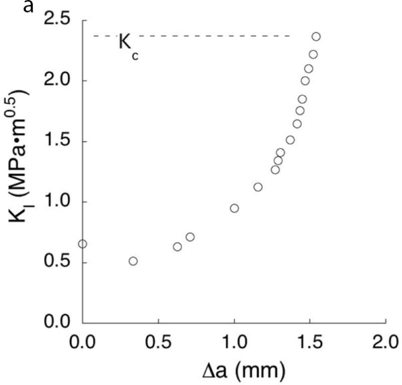
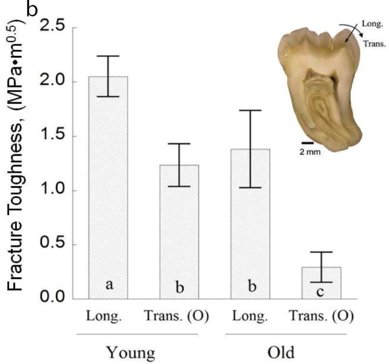
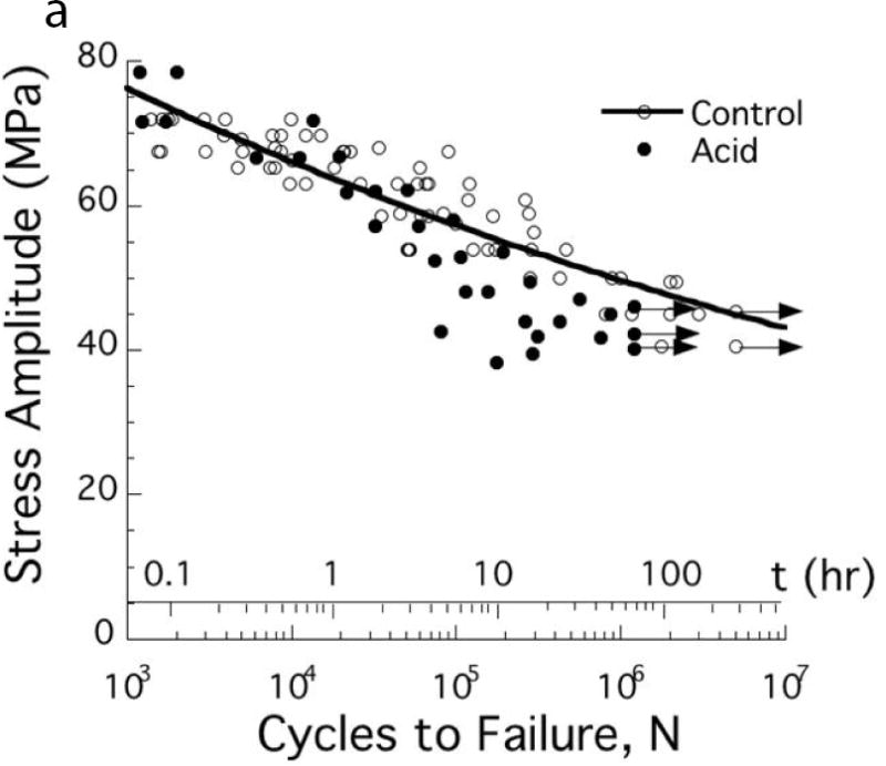
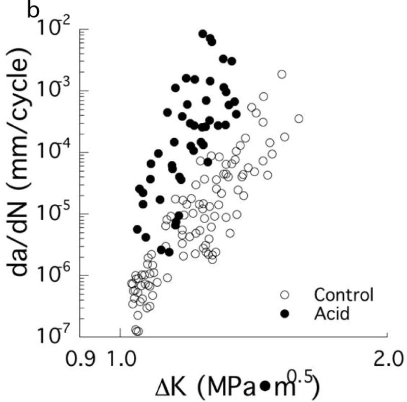
This article provides a brief review of recent investigations concerning the structure and properties of the tooth. The last decade has brought a greater emphasis on the durability of the tooth, an improved understanding of the fatigue and fracture behavior of the principal tissues, and their importance to tooth failures. The primary contributions to tooth durability are discussed, including the process of placing a restoration, the impact of aging, and challenges posed by the oral environment. The significance of these findings to the dental community and their importance to the pursuit of lifelong oral health are highlighted.

Keywords: Aging; Dentin; Durability; Enamel; Fatigue; Fracture; Tubules.


Figures

Fig. 1
Fig. 1
Importance of damage introduced during cavity preparations on the fatigue strength of coronal dentin. The “control” in these two diagrams consists of beams of coronal dentin prepared with diamond slicing wheels, and with average surface roughness (Ra) of less than 0.2 μm. (A) Comparison of the fatigue strength after cutting treatments with medium diamond abrasive and water spray irrigation. Results are shown for cutting parallel (//) and perpendicular (□) to the length of the beam, which is important to the orientation of damage. Data points with arrows represent beams that did not fail and the test was discontinued. (B) Comparison of the fatigue strength distribution of the control with dentin beams subjected to a bur cutting treatment followed by a 15 second etch with 37.5% gel. Cutting was performed with a 6-flute tungsten carbide straight fissure bur and commercial air turbine with water spray irrigation. A: Adapted from Majd B, Majd H, Porter JA, et al. Degradation in the fatigue strength of dentin by diamond bur preparations: Importance of cutting direction. J Biomed Mater Res B Appl Biomater 2016;104(1):39–49; with permission. B: Adapted from Lee HH, Majd H, Orrego S, et al. Degradation in the fatigue strength of dentin by cutting, etching and adhesive bonding. Dent Mater 2014;30(9):1061–72; with permission.
Fig. 1
Fig. 1
Importance of damage introduced during cavity preparations on the fatigue strength of coronal dentin. The “control” in these two diagrams consists of beams of coronal dentin prepared with diamond slicing wheels, and with average surface roughness (Ra) of less than 0.2 μm. (A) Comparison of the fatigue strength after cutting treatments with medium diamond abrasive and water spray irrigation. Results are shown for cutting parallel (//) and perpendicular (□) to the length of the beam, which is important to the orientation of damage. Data points with arrows represent beams that did not fail and the test was discontinued. (B) Comparison of the fatigue strength distribution of the control with dentin beams subjected to a bur cutting treatment followed by a 15 second etch with 37.5% gel. Cutting was performed with a 6-flute tungsten carbide straight fissure bur and commercial air turbine with water spray irrigation. A: Adapted from Majd B, Majd H, Porter JA, et al. Degradation in the fatigue strength of dentin by diamond bur preparations: Importance of cutting direction. J Biomed Mater Res B Appl Biomater 2016;104(1):39–49; with permission. B: Adapted from Lee HH, Majd H, Orrego S, et al. Degradation in the fatigue strength of dentin by cutting, etching and adhesive bonding. Dent Mater 2014;30(9):1061–72; with permission.
Fig. 2
Fig. 2
Contact damage of enamel resulting from cyclic loading. (A) Load-life diagram for cyclic contact of cuspal enamel described in terms of the maximum contact load and the number of cycles to failure. Failure was defined by an increase in maximum indentation depth that exceeded that in the first cycle by 15 μm. (B) Typical contact damage pattern resulting from cyclic loading. Note the large number of circumferential cracks inside the contact zone, and the radial cracks extending from the contact periphery. This particular damage pattern resulted from cyclic contact with maximum load of 400N and after a total of 160k cycles. A and B: Adapted from Gao SS, An BB, Yahyazadehfar M, et al. Contact fatigue of human enamel: Experiments, mechanisms and modeling. J Mech Behav Biomed Mater 2016;60:438–50; with permission.
Fig. 2
Fig. 2
Contact damage of enamel resulting from cyclic loading. (A) Load-life diagram for cyclic contact of cuspal enamel described in terms of the maximum contact load and the number of cycles to failure. Failure was defined by an increase in maximum indentation depth that exceeded that in the first cycle by 15 μm. (B) Typical contact damage pattern resulting from cyclic loading. Note the large number of circumferential cracks inside the contact zone, and the radial cracks extending from the contact periphery. This particular damage pattern resulted from cyclic contact with maximum load of 400N and after a total of 160k cycles. A and B: Adapted from Gao SS, An BB, Yahyazadehfar M, et al. Contact fatigue of human enamel: Experiments, mechanisms and modeling. J Mech Behav Biomed Mater 2016;60:438–50; with permission.
Figure 3
Figure 3
A comparison of the structure of coronal dentin obtained from donor teeth of residents from Colombia (CO) and the United States (US). The two groups consist of age matched young donors with 18≤age≤35 yrs. (A) Micrographs obtained from scanning electron microscopy of peripheral dentin obtained from representative donor teeth of residents from the US and CO. (B) The distribution of tubule lumen diameter of coronal dentin in the deep, central and peripheral regions. The column height represents the average and the caps indicate the standard deviation. Columns with different letters are significantly different (p≤0.05). Note the consistent diameter amongst the three locations of the Colombian teeth. A: Adapted from Ivancik J, Naranjo M, Correa S, et al. Differences in the microstructure and fatigue properties of dentin between residents of North and South America. Arch Oral Biol 2014;59(10):1001–1012; with permission. B: From Ivancik J, Naranjo M, Correa S, et al. Differences in the microstructure and fatigue properties of dentin between residents of North and South America. Arch Oral Biol 2014;59(10):1001–1012; with permission.
Figure 3
Figure 3
A comparison of the structure of coronal dentin obtained from donor teeth of residents from Colombia (CO) and the United States (US). The two groups consist of age matched young donors with 18≤age≤35 yrs. (A) Micrographs obtained from scanning electron microscopy of peripheral dentin obtained from representative donor teeth of residents from the US and CO. (B) The distribution of tubule lumen diameter of coronal dentin in the deep, central and peripheral regions. The column height represents the average and the caps indicate the standard deviation. Columns with different letters are significantly different (p≤0.05). Note the consistent diameter amongst the three locations of the Colombian teeth. A: Adapted from Ivancik J, Naranjo M, Correa S, et al. Differences in the microstructure and fatigue properties of dentin between residents of North and South America. Arch Oral Biol 2014;59(10):1001–1012; with permission. B: From Ivancik J, Naranjo M, Correa S, et al. Differences in the microstructure and fatigue properties of dentin between residents of North and South America. Arch Oral Biol 2014;59(10):1001–1012; with permission.
Fig. 4
Fig. 4
The importance of location and age on the fatigue crack growth resistance of coronal dentin. (A) Responses obtained for cyclic crack growth occurring “in-plane” with the dentin tubules. These responses are for young dentin (age ≤ 30 yrs) and stratified according to depth, including close to the pulp (inner), the mid–coronal region (central) and close to the DEJ (peripheral). (B) Comparison of cyclic crack growth in the dentin of teeth from young (age ≤ 30 yrs) and old (55 yrs ≤ age) donors. These responses are for cyclic crack growth occurring perpendicular to the dentin tubules. A: Adapted from Ivancik J, Neerchal NK, Romberg E, et al. The reduction in fatigue crack growth resistance of dentin with depth. J Dent Res 2011;90(8):1031–1036; with permission. B: Adapted from Ivancik J, Majd H, Bajaj D, et al. Contributions of aging to the fatigue crack growth resistance of human dentin. Acta Biomater 2012;8(7):2737–2746; with permission.
Fig. 4
Fig. 4
The importance of location and age on the fatigue crack growth resistance of coronal dentin. (A) Responses obtained for cyclic crack growth occurring “in-plane” with the dentin tubules. These responses are for young dentin (age ≤ 30 yrs) and stratified according to depth, including close to the pulp (inner), the mid–coronal region (central) and close to the DEJ (peripheral). (B) Comparison of cyclic crack growth in the dentin of teeth from young (age ≤ 30 yrs) and old (55 yrs ≤ age) donors. These responses are for cyclic crack growth occurring perpendicular to the dentin tubules. A: Adapted from Ivancik J, Neerchal NK, Romberg E, et al. The reduction in fatigue crack growth resistance of dentin with depth. J Dent Res 2011;90(8):1031–1036; with permission. B: Adapted from Ivancik J, Majd H, Bajaj D, et al. Contributions of aging to the fatigue crack growth resistance of human dentin. Acta Biomater 2012;8(7):2737–2746; with permission.
Fig. 5
Fig. 5
Characteristics of the crack growth resistance of enamel. (A) The increase in resistance to fracture of enamel with crack extension. This data was obtained from crack growth in the longitudinal direction of cuspal enamel, i.e. from the occlusal surface towards the DEJ. The “fracture toughness” of the specimen (Kc), was identified from the last point of stable crack extension preceding bulk fracture. (B) The fracture toughness of the enamel from 3rd molars for cracks extending from the occlusal surface to the DEJ (longitudinal) vs growth with buccal-lingual orientation (transverse). The columns represent the average values with standard deviations. Columns with different letters are significantly different. A: Data from Bajaj D, Arola D. Role of prism decussation on fatigue crack growth and fracture of human enamel. Acta Biomater 2009;5(8):3045–56. B: Adapted from Yahyazadehfar M, Zhang D, Arola D. On the importance of aging to the crack growth resistance of human enamel. Acta Biomater 2016;32:269; and Bajaj D, Arola D. Role of prism decussation on fatigue crack growth and fracture of human enamel. Acta Biomater 2009;5(8):3046; with permission.
Fig. 5
Fig. 5
Characteristics of the crack growth resistance of enamel. (A) The increase in resistance to fracture of enamel with crack extension. This data was obtained from crack growth in the longitudinal direction of cuspal enamel, i.e. from the occlusal surface towards the DEJ. The “fracture toughness” of the specimen (Kc), was identified from the last point of stable crack extension preceding bulk fracture. (B) The fracture toughness of the enamel from 3rd molars for cracks extending from the occlusal surface to the DEJ (longitudinal) vs growth with buccal-lingual orientation (transverse). The columns represent the average values with standard deviations. Columns with different letters are significantly different. A: Data from Bajaj D, Arola D. Role of prism decussation on fatigue crack growth and fracture of human enamel. Acta Biomater 2009;5(8):3045–56. B: Adapted from Yahyazadehfar M, Zhang D, Arola D. On the importance of aging to the crack growth resistance of human enamel. Acta Biomater 2016;32:269; and Bajaj D, Arola D. Role of prism decussation on fatigue crack growth and fracture of human enamel. Acta Biomater 2009;5(8):3046; with permission.
Fig. 6
Fig. 6
Degradation in the fatigue resistance of coronal dentin with exposure to an acidic environment. (A) Stress life fatigue behavior. The data points with arrows represent beams that did not fail and the test was discontinued. Note that a significant degradation in fatigue strength occurs after only 4 hours of acid exposure. (B) Fatigue crack growth resistance of mid-coronal dentin. (A, B) The control was evaluated in a neutral environment (pH=7) and the acid condition consisted of exposure to a lactic acid solution with pH = 5. A: Adapted from Do D, Orrego S, Majd H, et al. Accelerated fatigue of dentin with exposure to lactic acid. Biomaterials 2013;34(34):8650–8659; with permission. B: From Orrego S, Xu H, Arola D. Degradation in the fatigue crack growth resistance of human dentin by lactic acid. Mater Sci Engr 2017;C 73:720; with permission.
Fig. 6
Fig. 6
Degradation in the fatigue resistance of coronal dentin with exposure to an acidic environment. (A) Stress life fatigue behavior. The data points with arrows represent beams that did not fail and the test was discontinued. Note that a significant degradation in fatigue strength occurs after only 4 hours of acid exposure. (B) Fatigue crack growth resistance of mid-coronal dentin. (A, B) The control was evaluated in a neutral environment (pH=7) and the acid condition consisted of exposure to a lactic acid solution with pH = 5. A: Adapted from Do D, Orrego S, Majd H, et al. Accelerated fatigue of dentin with exposure to lactic acid. Biomaterials 2013;34(34):8650–8659; with permission. B: From Orrego S, Xu H, Arola D. Degradation in the fatigue crack growth resistance of human dentin by lactic acid. Mater Sci Engr 2017;C 73:720; with permission.

References

    1. Pashley DH. Dentin: a dynamic substrate-a review. Scanning Microsc. 1989;3(1):161–74. discussion 174–6. - PubMed
    1. Marshall GW, Jr, Marshall SJ, Kinney JH, et al. The dentin substrate: structure and properties related to bonding. J Dent. 1997;25(6):441–58. - PubMed
    1. Perdigao J, Swift EJ, Jr, Denehy GE, et al. In vitro bond strengths and SEM evaluation of dentin bonding systems to different dentin substrates. J Dent Res. 1994;73(1):44–55. - PubMed
    1. Tay FR, Pashley DH. Resin bonding to cervical sclerotic dentin: a review. J Dent. 2004;32(3):173–96. - PubMed
    1. Kinney JH, Marshall SJ, Marshall GW. The mechanical properties of human dentin: a critical review and re-evaluation of the dental literature. Crit Rev Oral Biol Med. 2003;14(1):13–29. - PubMed

Publication types