Review
. 2017 Oct;61(4):669-687.
doi: 10.1016/j.cden.2017.05.002.

Bioactive Dental Composites and Bonding Agents Having Remineralizing and Antibacterial Characteristics

Affiliations
Review

Bioactive Dental Composites and Bonding Agents Having Remineralizing and Antibacterial Characteristics

Ke Zhang et al. Dent Clin North Am. 2017 Oct.

Abstract

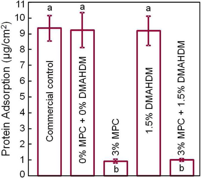
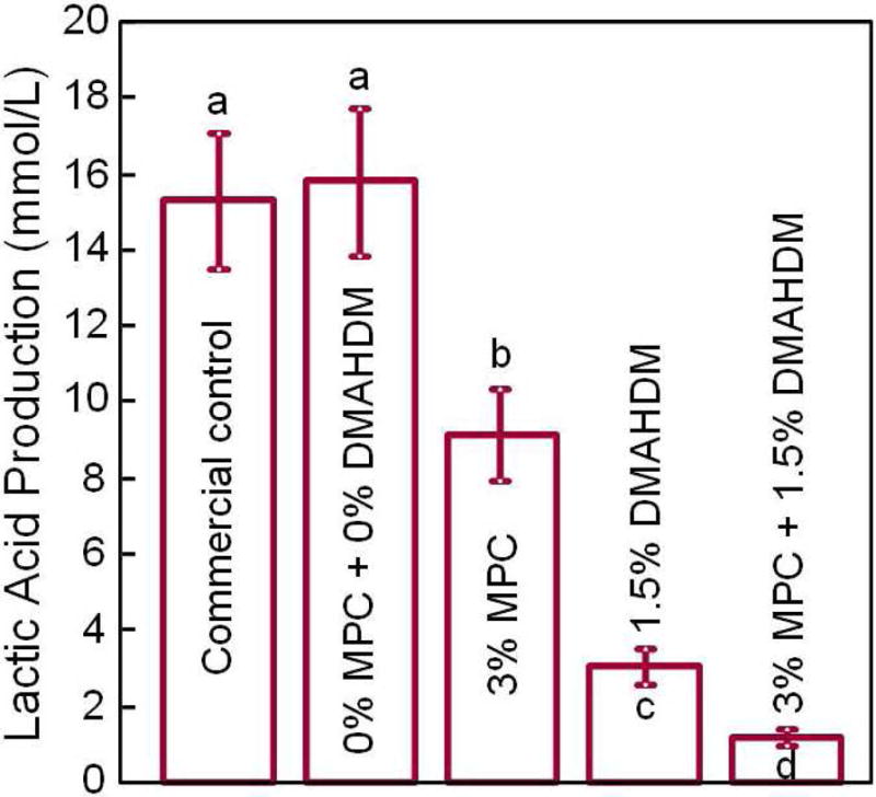
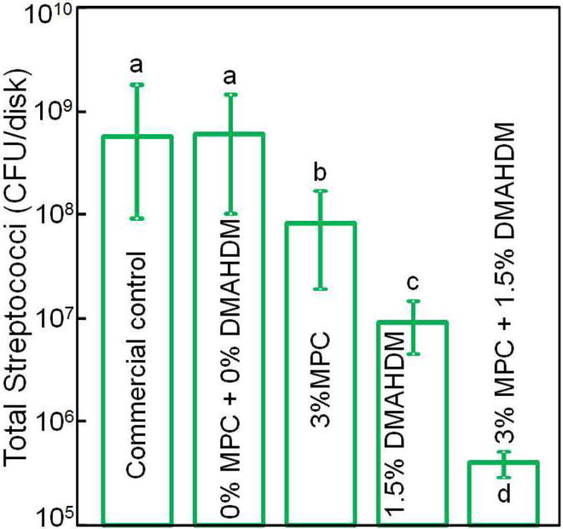
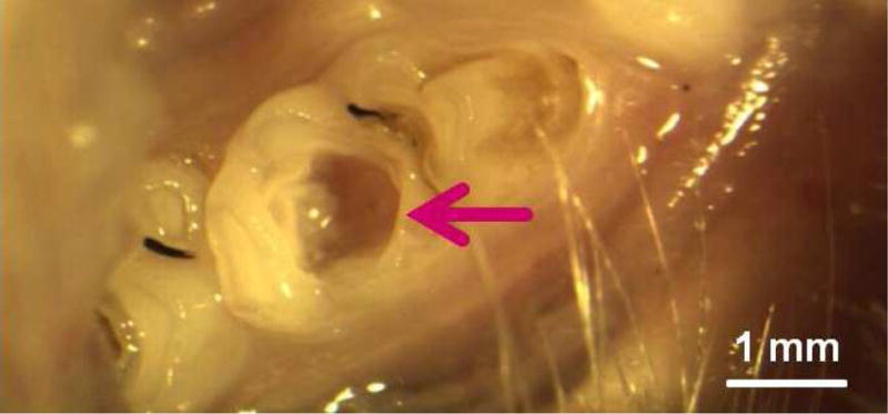
Current dental restorative materials are typically inert and replace missing tooth structures. This article reviews efforts in the development of a new generation of bioactive materials designed to not only replace the missing tooth volume but also possess therapeutic functions. Composites and bonding agents with remineralizing and antibacterial characteristics have shown promise in replacing lost minerals, inhibiting recurrent caries, neutralizing acids, repelling proteins, and suppressing biofilms and acid production. Furthermore, they have demonstrated a low cytotoxicity similar to current resins, with additional benefits to protect the dental pulp and promote tertiary dentin formation. This new class of bioactive materials shows promise in reversing lesions and inhibiting caries.

Keywords: Antibacterial monomers; Bioactive composites; Bonding agents; Calcium phosphate nanoparticles; Caries inhibition; Oral biofilms; Remineralization; Silver nanoparticles.


Conflict of interest statement

Disclosure statement

There is no conflict of interest for all authors.

Figures

Figure 1
Figure 1
Human in situ caries inhibition via NACP composite. (A) One hundred bovine enamel slabs of 5×5×2 mm were obtained. A cavity of 2 mm in diameter and 1.5 mm in depth was prepared and restored with a composite. 25 volunteers wore palatal devices each containing four slabs: two filled with NACP nanocomposite on one side, and two filled with control composite on the other side. (B) A plastic mesh with 1 mm space protected the biofilms in situ. (C) Enamel slabs were cut to sections for microradiography. (D) Enamel mineral loss around NACP nanocomposite was reduced to nearly 1/3 of that around control composite (p < 0.05). Figure 1A, 1B, 1C: From Melo MA, Weir MD, Rodrigues LK, et al. Novel calcium phosphate nanocomposite with caries-inhibition in a human in situ model. Dent Mater 2013;29(2):233; with permission. Figure 1D: Adapted from Melo MA, Weir MD, Rodrigues LK, et al. Novel calcium phosphate nanocomposite with caries-inhibition in a human in situ model. Dent Mater 2013;29(2):237; with permission.
Figure 1
Figure 1
Human in situ caries inhibition via NACP composite. (A) One hundred bovine enamel slabs of 5×5×2 mm were obtained. A cavity of 2 mm in diameter and 1.5 mm in depth was prepared and restored with a composite. 25 volunteers wore palatal devices each containing four slabs: two filled with NACP nanocomposite on one side, and two filled with control composite on the other side. (B) A plastic mesh with 1 mm space protected the biofilms in situ. (C) Enamel slabs were cut to sections for microradiography. (D) Enamel mineral loss around NACP nanocomposite was reduced to nearly 1/3 of that around control composite (p < 0.05). Figure 1A, 1B, 1C: From Melo MA, Weir MD, Rodrigues LK, et al. Novel calcium phosphate nanocomposite with caries-inhibition in a human in situ model. Dent Mater 2013;29(2):233; with permission. Figure 1D: Adapted from Melo MA, Weir MD, Rodrigues LK, et al. Novel calcium phosphate nanocomposite with caries-inhibition in a human in situ model. Dent Mater 2013;29(2):237; with permission.
Figure 2
Figure 2
Rechargeable NACP composite for long-term Ca and P ion release. (A) NACP composite was immersed in a pH 4 solution for 70 d to exhaust the ion release (lower left arrow). Then the specimens were immersed in a new pH 4 solution to confirm that the ion release was exhausted (lower middle arrow). Specimens were recharged in a recharge solution, then tested for ion re-release for 7 d (third arrow at the bottom of A). This constituted the first recharge/re-release cycle. This process was repeated for 6 cycles. (B) Three NACP nanocomposites with six cycles of recharge/re-release, showing no decrease in ion release with increasing recharge cycles (p > 0.1). Figure 2A: From Zhang L, Weir MD, Chow LC, et al. Novel rechargeable calcium phosphate dental nanocomposite. Dent Mater 2016;32(2):288; with permission. Figure 2B: From Zhang L, Weir MD, Chow LC, et al. Novel rechargeable calcium phosphate dental nanocomposite. Dent Mater 2016;32(2):289; with permission.
Figure 2
Figure 2
Rechargeable NACP composite for long-term Ca and P ion release. (A) NACP composite was immersed in a pH 4 solution for 70 d to exhaust the ion release (lower left arrow). Then the specimens were immersed in a new pH 4 solution to confirm that the ion release was exhausted (lower middle arrow). Specimens were recharged in a recharge solution, then tested for ion re-release for 7 d (third arrow at the bottom of A). This constituted the first recharge/re-release cycle. This process was repeated for 6 cycles. (B) Three NACP nanocomposites with six cycles of recharge/re-release, showing no decrease in ion release with increasing recharge cycles (p > 0.1). Figure 2A: From Zhang L, Weir MD, Chow LC, et al. Novel rechargeable calcium phosphate dental nanocomposite. Dent Mater 2016;32(2):288; with permission. Figure 2B: From Zhang L, Weir MD, Chow LC, et al. Novel rechargeable calcium phosphate dental nanocomposite. Dent Mater 2016;32(2):289; with permission.
Figure 3
Figure 3
Dental plaque microcosm biofilm model with colony-forming unit (CFU) counts of 2-day biofilms on composites: (A) total microorganisms, (B) total streptococci, and (C) mutans streptococci (mean ± sd; n = 6). In each plot, values with dissimilar letters are significantly different from each other (p < 0.05). The two control composites had the highest CFU counts. Increasing CL from 3 to 16 decreased the CFU counts significantly (p < 0.05). Note the log scale for the y-axis. Adapted from Zhang K, Cheng L, Weir MD, et al. Effects of quaternary ammonium chain length on the antibacterial and remineralizing effects of a calcium phosphate nanocomposite. Int J Oral Sci 2016; 8(1):50; with permission.
Figure 3
Figure 3
Dental plaque microcosm biofilm model with colony-forming unit (CFU) counts of 2-day biofilms on composites: (A) total microorganisms, (B) total streptococci, and (C) mutans streptococci (mean ± sd; n = 6). In each plot, values with dissimilar letters are significantly different from each other (p < 0.05). The two control composites had the highest CFU counts. Increasing CL from 3 to 16 decreased the CFU counts significantly (p < 0.05). Note the log scale for the y-axis. Adapted from Zhang K, Cheng L, Weir MD, et al. Effects of quaternary ammonium chain length on the antibacterial and remineralizing effects of a calcium phosphate nanocomposite. Int J Oral Sci 2016; 8(1):50; with permission.
Figure 4
Figure 4
Dentin bonding. (A) SEM of dentin-adhesive interface for DMADDM+NAg+NACP group (T = resin tag). (B) Higher magnification SEM of a resin tag showing NACP in tubules. (C) Higher magnification TEM indicating NAg and NACP in resin tag. (D) Dentin shear bond strengths (mean ± sd; n = 10). Dissimilar letters indicate significantly different values (p < 0.05). There was a 35% loss in bond strength for commercial group in water-aging for 6 months. There was no bond strength loss for groups containing DMADDM, NAg and NACP. From Zhang K, Cheng L, Wu EJ, et al. Effect of water-aging on dentin bond strength and anti-biofilm activity of bonding agent containing new monomer dimethylaminododecyl methacrylate. J Dent 2013;41(6):504–513; with permission.
Figure 4
Figure 4
Dentin bonding. (A) SEM of dentin-adhesive interface for DMADDM+NAg+NACP group (T = resin tag). (B) Higher magnification SEM of a resin tag showing NACP in tubules. (C) Higher magnification TEM indicating NAg and NACP in resin tag. (D) Dentin shear bond strengths (mean ± sd; n = 10). Dissimilar letters indicate significantly different values (p < 0.05). There was a 35% loss in bond strength for commercial group in water-aging for 6 months. There was no bond strength loss for groups containing DMADDM, NAg and NACP. From Zhang K, Cheng L, Wu EJ, et al. Effect of water-aging on dentin bond strength and anti-biofilm activity of bonding agent containing new monomer dimethylaminododecyl methacrylate. J Dent 2013;41(6):504–513; with permission.
Figure 5
Figure 5
Protein-repellent. (A) Bovine serum albumin adsorption onto composite surface. The composite with 3% MPC and the composite with 3% MPC + 1.5% DMAHDM both had protein adsorption about 1/10 that of commercial composite. In each plot, dissimilar letters indicate significantly different values (p < 0.05). (B) Lactic acid production by biofilms was greatly reduced via MPC and DMAHDM. (C) Biofilm total streptococci CFU on composite with 3% MPC + 1.5% DMAHDM was more than 3 orders of magnitude lower than commercial control. Adapted from Zhang N, Ma J, Melo MA, et al. Protein-repellent and antibacterial dental composite to inhibit biofilms and caries. J Dent 2015;43(2):225–234; with permission.
Figure 5
Figure 5
Protein-repellent. (A) Bovine serum albumin adsorption onto composite surface. The composite with 3% MPC and the composite with 3% MPC + 1.5% DMAHDM both had protein adsorption about 1/10 that of commercial composite. In each plot, dissimilar letters indicate significantly different values (p < 0.05). (B) Lactic acid production by biofilms was greatly reduced via MPC and DMAHDM. (C) Biofilm total streptococci CFU on composite with 3% MPC + 1.5% DMAHDM was more than 3 orders of magnitude lower than commercial control. Adapted from Zhang N, Ma J, Melo MA, et al. Protein-repellent and antibacterial dental composite to inhibit biofilms and caries. J Dent 2015;43(2):225–234; with permission.
Figure 6
Figure 6
In vivo biocompatibility of antibacterial and remineralizing composite and adhesive. (A) Rat tooth model, in which the right and left molars were used. (B) Occlusal cavity was restored with bonding agent and composite. H&E images at 30 d for (C) control, and (D) DMADDM + NACP. Star indicates inflammatory cells. Blood vessels are indicated by arrows. Control group exhibited slight inflammation. NACP group and DMADDM + NACP group showed normal pulp without inflammatory response, along with greater tertiary dentin thicknesses. (E) Tertiary dentin thickness data. Different letters indicate significantly different values (p < 0.05). Figures 6A, 6B, and 6E: Adapted from Li F, Wang P, Weir MD, et al. Evaluation of antibacterial and remineralizing nanocomposite and adhesive in rat tooth cavity model. Acta Biomater 2014;10(6):2804–2813; with permission. Figures 6C and 6D: From Li F, Wang P, Weir MD, et al. Evaluation of antibacterial and remineralizing nanocomposite and adhesive in rat tooth cavity model. Acta Biomater 2014;10(6):2810; with permission.
Figure 6
Figure 6
In vivo biocompatibility of antibacterial and remineralizing composite and adhesive. (A) Rat tooth model, in which the right and left molars were used. (B) Occlusal cavity was restored with bonding agent and composite. H&E images at 30 d for (C) control, and (D) DMADDM + NACP. Star indicates inflammatory cells. Blood vessels are indicated by arrows. Control group exhibited slight inflammation. NACP group and DMADDM + NACP group showed normal pulp without inflammatory response, along with greater tertiary dentin thicknesses. (E) Tertiary dentin thickness data. Different letters indicate significantly different values (p < 0.05). Figures 6A, 6B, and 6E: Adapted from Li F, Wang P, Weir MD, et al. Evaluation of antibacterial and remineralizing nanocomposite and adhesive in rat tooth cavity model. Acta Biomater 2014;10(6):2804–2813; with permission. Figures 6C and 6D: From Li F, Wang P, Weir MD, et al. Evaluation of antibacterial and remineralizing nanocomposite and adhesive in rat tooth cavity model. Acta Biomater 2014;10(6):2810; with permission.

References

    1. Bayne SC, Thompson JY, Swift EJ, Stamatiades P, Wilkerson M. A characterization of first-generation flowable composites. J Am Dent Assoc. 1998;129:567–577. - PubMed
    1. Watts DC, Marouf AS, Al-Hindi AM. Photo-polymerization shrinkage-stress kinetics in resin-composites: methods development. Dent Mater. 2003;19:1–11. - PubMed
    1. Drummond JL. Degradation, fatigue, and failure of resin dental composite materials. J Dent Res. 2008;87:710–719. - PMC - PubMed
    1. Lynch CD. Successful posterior composites. London: Quintessence Publishing Co; 2008.
    1. Ferracane JL. Resin composite - State of the art. Dent Mater. 2011;27:29–38. - PubMed

Publication types

MeSH terms