

The qualitative grading of muscle fat infiltration in whiplash using fat and water magnetic resonance imaging
- PMID: 28887274
- PMCID: PMC8845185
- DOI: 10.1016/j.spinee.2017.08.233
The qualitative grading of muscle fat infiltration in whiplash using fat and water magnetic resonance imaging
Abstract
Background context: The development of muscle fat infiltration (MFI) in the neck muscles is associated with poor functional recovery following whiplash injury. Custom software and time-consuming manual segmentation of magnetic resonance imaging (MRI) is required for quantitative analysis and presents as a barrier for clinical translation.
Purpose: The purpose of this work was to establish a qualitative MRI measure for MFI and evaluate its ability to differentiate between individuals with severe whiplash-associated disorder (WAD), mild or moderate WAD, and healthy controls.
Study design/setting: This is a cross-sectional study.
Patient sample: Thirty-one subjects with WAD and 31 age- and sex-matched controls were recruited from an ongoing randomized controlled trial.
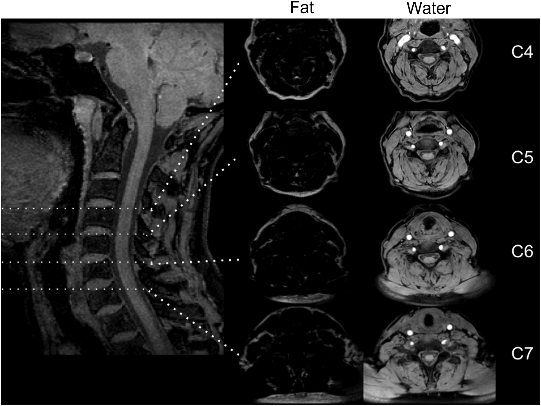
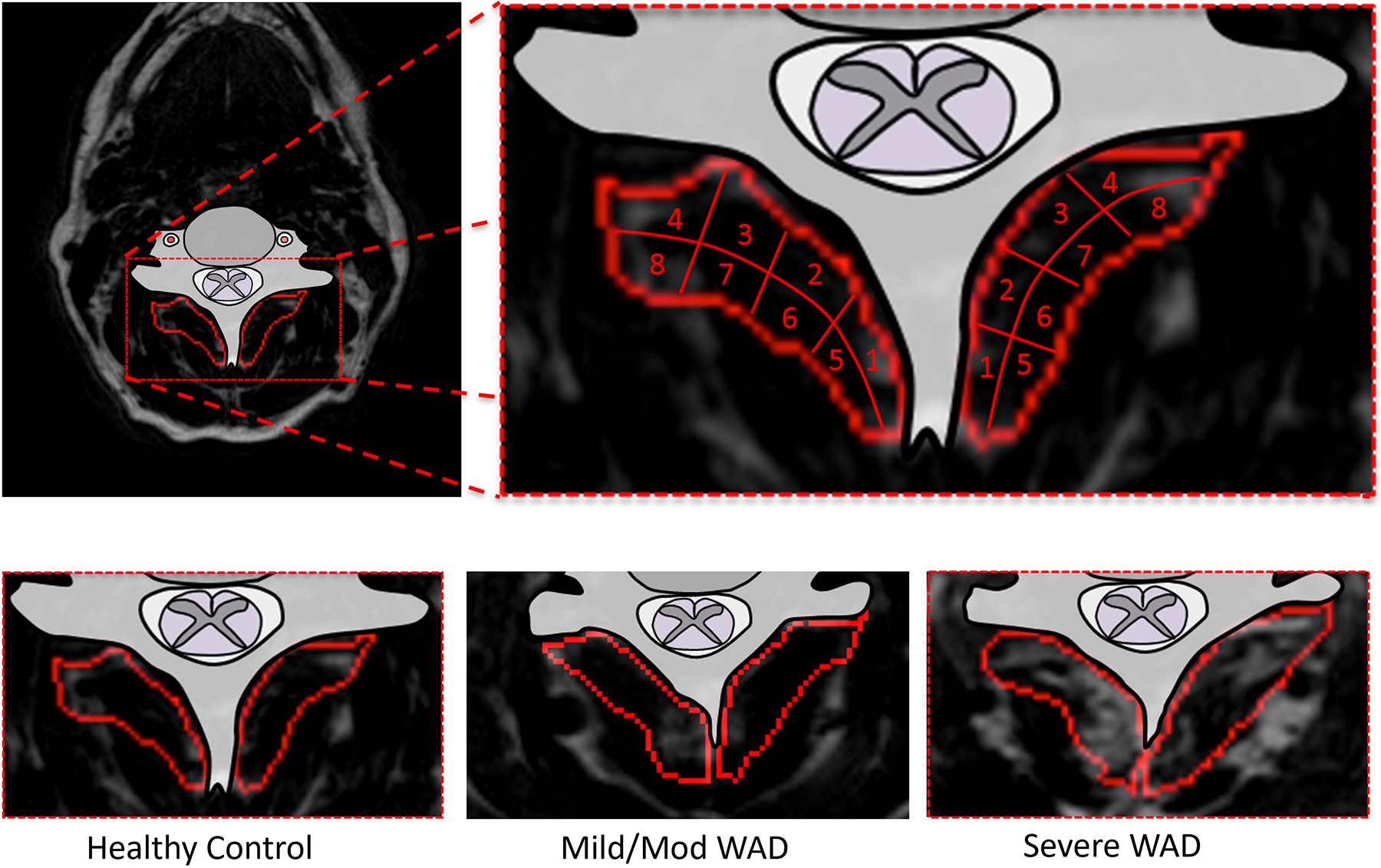
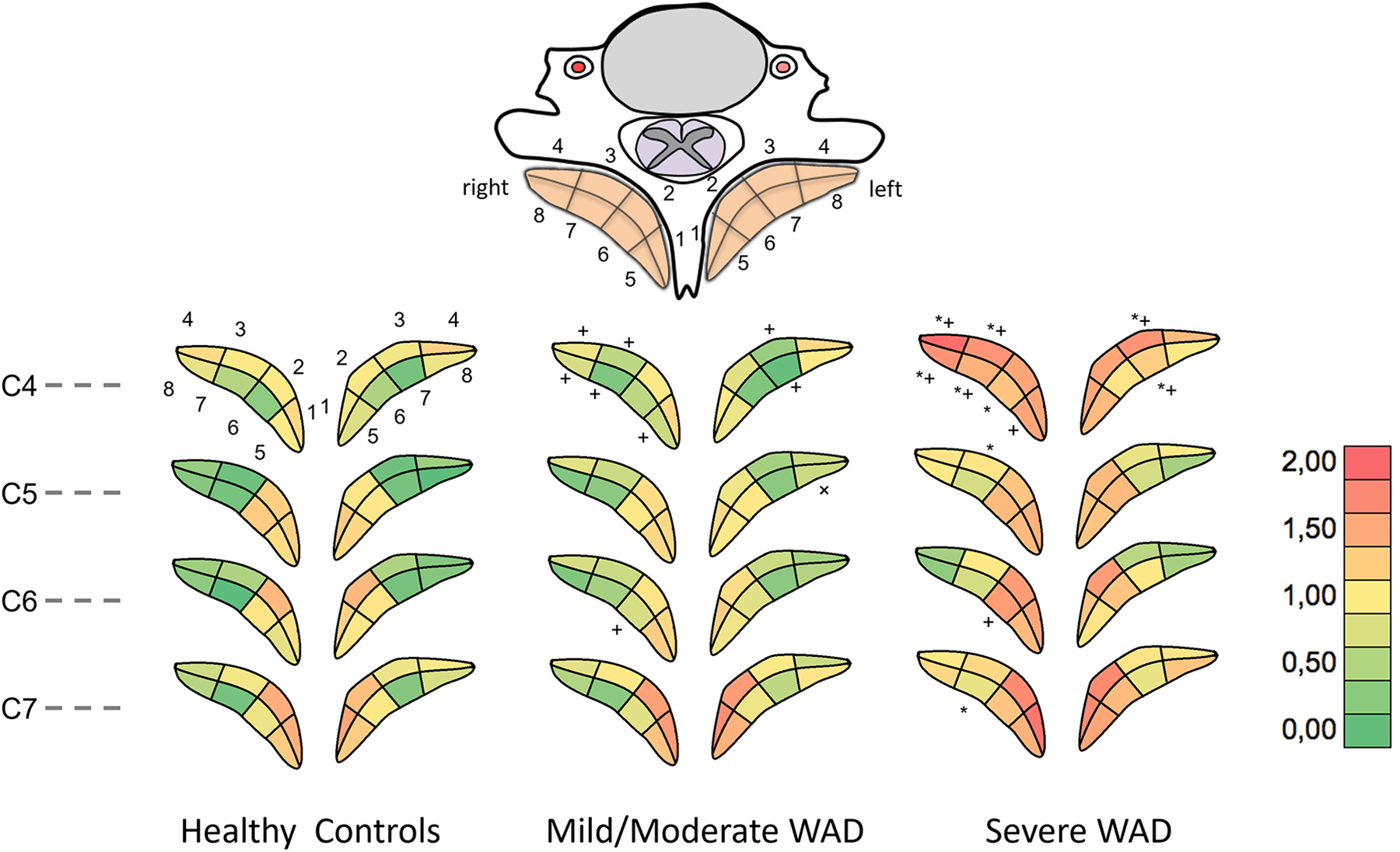
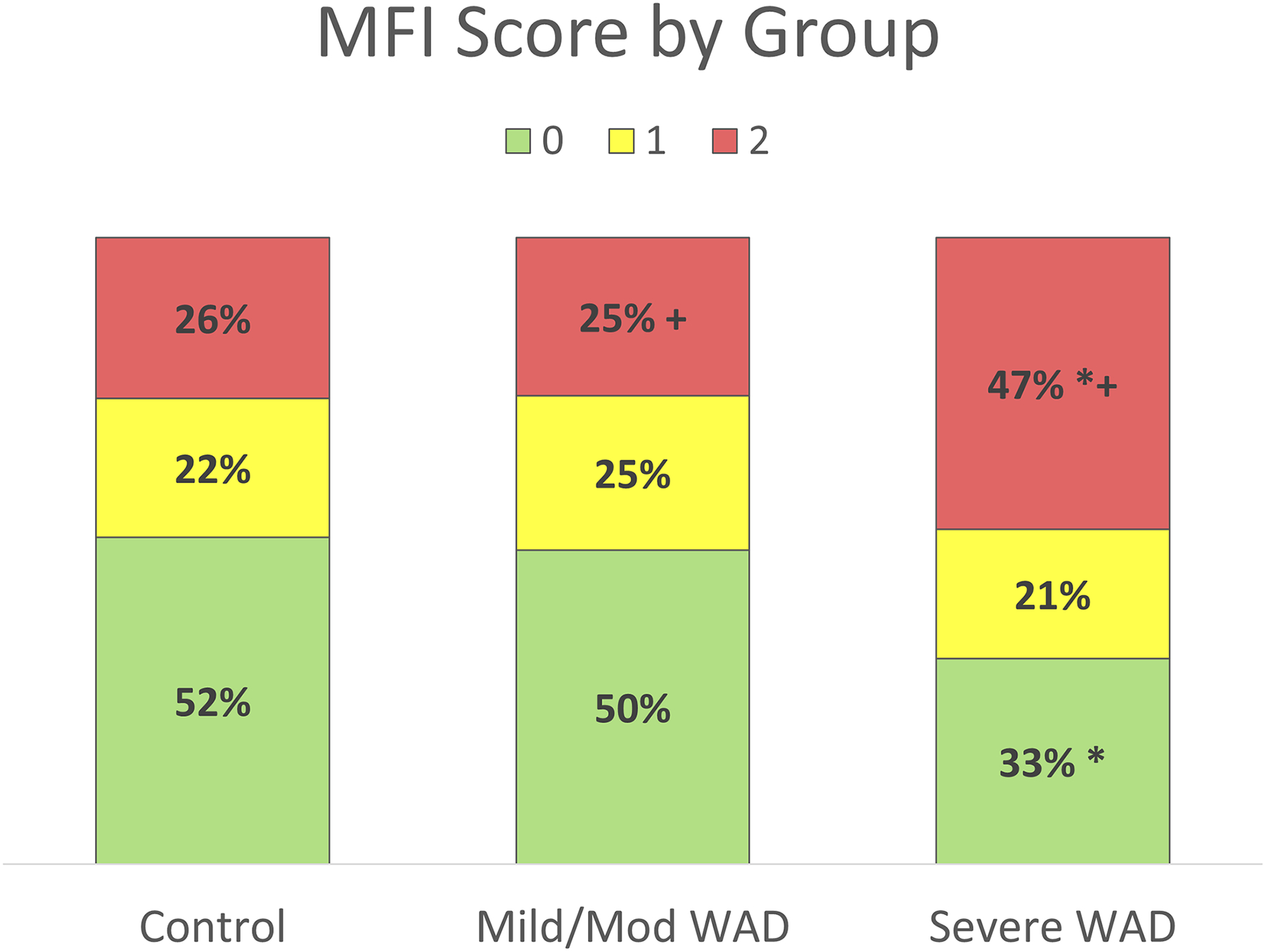
Outcome measures: The cervical multifidus was visually identified and segmented into eighths in the axial fat/water images (C4-C7). Muscle fat infiltration was assessed on a visual scale: 0 for no or marginal MFI, 1 for light MFI, and 2 for distinct MFI. The participants with WAD were divided in two groups: mild or moderate and severe based on Neck Disability Index % scores.
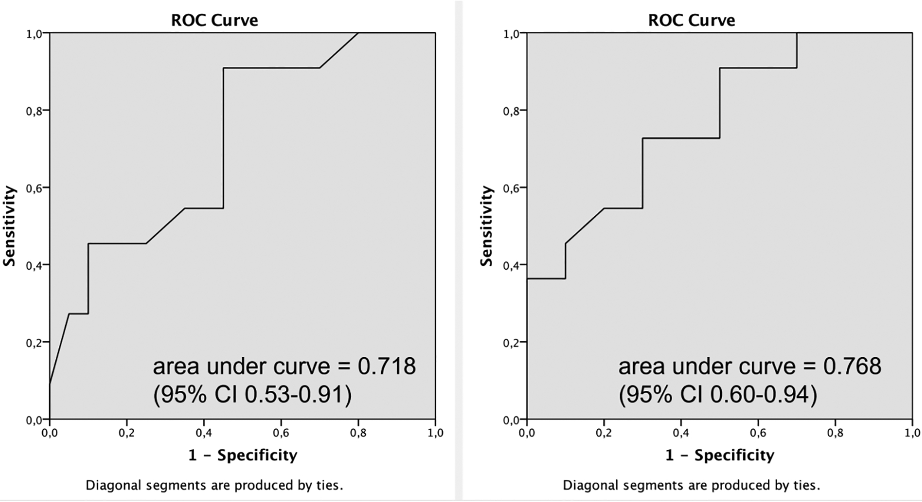
Methods: The mean regional MFI was compared between the healthy controls and each of the WAD groups using the Mann-Whitney U test. Receiver operator characteristic (ROC) analyses were carried out to evaluate the validity of the qualitative method.
Results: Twenty (65%) patients had mild or moderate disability and 11 (35%) were considered severe. Inter- and intra-rater reliability was excellent when grading was averaged by level or when frequency of grade II was considered. Statistically significant differences (p<.05) in regional MFI were particularly notable between the severe WAD group and healthy controls. The ROC curve, based on detection of distinct MFI, showed an area-under-the curve of 0.768 (95% confidence interval 0.59-0.94) for discrimination of WAD participants.
Conclusions: These preliminary results suggest a qualitative MRI measure for MFI is reliable and valid, and may prove useful toward the classification of WAD in radiology practice.
Keywords: MRI; Muscle fat; Neck; Recovery; Spine; Whiplash.
Copyright © 2017 Elsevier Inc. All rights reserved.
Figures
References
-
- Carroll LJ, Holm LW, Hogg-Johnson S, et al. Course and prognostic factors for neck pain in whiplash-associated disorders (WAD): results of the Bone and Joint Decade 2000–2010 Task Force on Neck Pain and Its Associated Disorders. Spine (Phila Pa 1976). 2008;33(4 Suppl):S83–92. - PubMed
-
- Sterling M, Hendrikz J, Kenardy J, et al. Assessment and validation of prognostic models for poor functional recovery 12 months after whiplash injury: a multicentre inception cohort study. Pain. 2012;153(8):1727–34. - PubMed
-
- Elliott J. Are there implications for morphological changes in neck muscles after whiplash injury? Spine (Phila Pa 1976). 2011;1(36(25 Suppl)):S205–10. Review. - PubMed
-
- Elliott J, Jull G, Noteboom JT, Darnell R, Galloway G, Gibbon WW. Fatty infiltration in the cervical extensor muscles in persistent whiplash-associated disorders: a magnetic resonance imaging analysis. Spine (Phila Pa 1976). 2006;31(22):E847–55. - PubMed
Publication types
MeSH terms
Grants and funding
LinkOut - more resources
Full Text Sources
Other Literature Sources
Medical
Miscellaneous
