. 2017 Sep 8;8(1):492.
doi: 10.1038/s41467-017-00536-1.

Evolution of protein-coupled RNA dynamics during hierarchical assembly of ribosomal complexes

Affiliations

Evolution of protein-coupled RNA dynamics during hierarchical assembly of ribosomal complexes

Sanjaya C Abeysirigunawardena et al. Nat Commun. .

Abstract

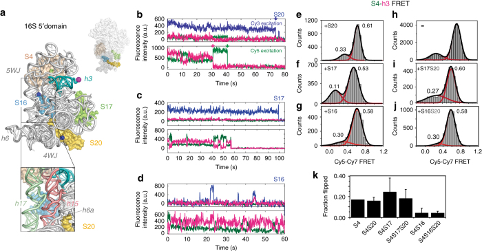
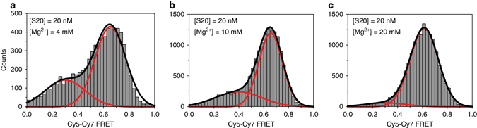
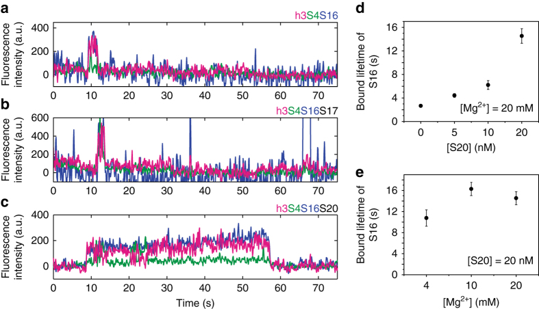
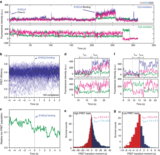
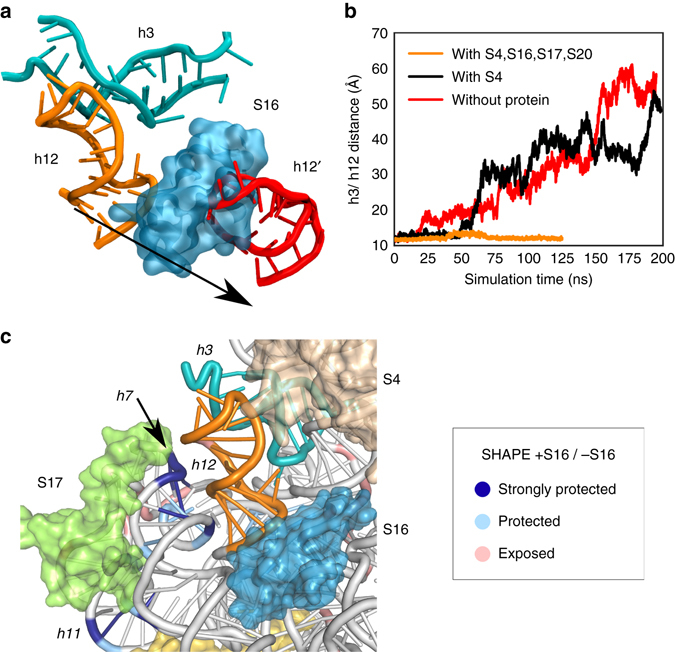
Assembly of 30S ribosomes involves the hierarchical addition of ribosomal proteins that progressively stabilize the folded 16S rRNA. Here, we use three-color single molecule FRET to show how combinations of ribosomal proteins uS4, uS17 and bS20 in the 16S 5' domain enable the recruitment of protein bS16, the next protein to join the complex. Analysis of real-time bS16 binding events shows that bS16 binds both native and non-native forms of the rRNA. The native rRNA conformation is increasingly favored after bS16 binds, explaining how bS16 drives later steps of 30S assembly. Chemical footprinting and molecular dynamics simulations show that each ribosomal protein switches the 16S conformation and dampens fluctuations at the interface between rRNA subdomains where bS16 binds. The results suggest that specific protein-induced changes in the rRNA dynamics underlie the hierarchy of 30S assembly and simplify the search for the native ribosome structure.Ribosomes assemble through the hierarchical addition of proteins to a ribosomal RNA scaffold. Here the authors use three-color single-molecule FRET to show how the dynamics of the rRNA dictate the order in which multiple proteins assemble on the 5' domain of the E. coli 16S rRNA.


Conflict of interest statement

The authors declare no competing financial interests.

Figures

Fig. 1
Fig. 1
Ribosomal proteins change the preference for rRNA conformations. a E. coli 16S 5′ domain RNA (gray ribbon, main panel) forms the 30S body (small surface; PDB accession 2I2P) and binds three primary assembly proteins (S4, S17, and S20) and secondary assembly protein S16. The RNA was fluorescently labeled with Cy7 (magenta sphere) by extension of helix 3 (h3; teal). S4 (tan surface) was labeled with Cy5 (green sphere). Proteins S16, S17, and S20 were labeled with Cy3 (blue spheres). RNA–protein complexes were excited by alternating 532 nm and 633 nm laser pulses using a custom-built multi-color single molecule FRET microscope. Inset: expansion of S16 binding site showing h15 (light red) and h17 (light green). bd Representative fluorescence traces obtained from complexes of 5′ domain RNA (h3-Cy7) and S4-Cy5 with S20-Cy3 b, S17-Cy3 c, or S16-Cy3 d. Cy3, blue; Cy5, green; Cy7, magenta. Single-step photobleaching events for each dye (colored arrows) indicate 1:1:1 stoichiometry between the components. S16-Cy3 exhibits high FRET efficiency to S4-Cy5 and h3-Cy7 upon specific binding to the complex (at 43 s in d). ej. Histograms of FRET between S4-Cy5 and h3-Cy7 in the presence of the additional proteins in eg, i and j were obtained from 110, 50, 30, 20, and 37 individual complexes, respectively. The Cy3 intensity was used to verify the presence of S20-Cy3, S16-Cy3 and S17-Cy3 eg; the presence of unlabeled S20 i, j was inferred from the frequency of S20-Cy3 binding in b. Data in h are from Ref. and represent Cy3-Cy5 FRET. k Population of the flipped conformation (low FRET) from the histograms in ej. Error bars represent the s.d. between three data sets of each sample
Fig. 2
Fig. 2
S4-S16 complexes sample the non-native conformation in physiological Mg2+. Populations of native (high FRET) and flipped (low FRET) complexes containing 5′ domain h3-Cy7, S4-Cy5, and S16-Cy3 in 20 nM S20, at different Mg2+ concentrations. Histograms for Cy5-Cy7 FRET are based on a 68, b 97, and c 105 trajectories from three-color smFRET experiments as in Fig. 1
Fig. 3
Fig. 3
Lifetime of S16 binding depends on the composition of preassembled RNPs. ac Examples of three-color fluorescence traces showing S16 binding in the presence of a S4, b S4 + S17 (20 nM), and c S4 + S20 (20 nM). These reveal the short lifetime of bound S16 when S20 is missing from the primary assembly complex. Fluorescence intensities are from S16-Cy3 (blue), S4-Cy5 (green), or h3-Cy7 (magenta) upon excitation of Cy3. The intensity jumps when S16-Cy3 localizes at immobilized preassembled 5′ domain RNPs. Anti-correlated fluctuations of Cy7 and Cy5 intensities indicate that 16S h3 remains dynamic after S16 binding. d Average lifetime of bound S16 vs. S20 concentration, in 20 mM MgCl2. Each measurement is based on 75–154 molecules. Error bars indicate the s.e.m. from triplicate measurements. e Average lifetime of bound S16 vs. Mg2+ concentration with 20 nM S20, as in d. Each measurement is based on 68–105 molecules. The lifetime of S16 binding strongly depends on the presence of S20 but not on the Mg2+-dependent stability of the RNA structure
Fig. 4
Fig. 4
S16 indirectly induces the folding of the RNA. a Real-time binding of S16 to RNA–S4–S20 complexes. S16-Cy3 was flowed into slide chambers containing immobilized RNA h3-Cy7·S4-Cy5·S20 complexes. Three-color fluorescence traces with alternating Cy3 and Cy5 excitation demonstrate successful S16 binding following several trials, as indicated by the increase in Cy3 signal (top). 16S h3 fluctuates before and after S16 binding (bottom). b Time-dependent population map of S4-h3 FRET was obtained from 194 traces that are post-synchronized at the moment of S16 binding (dashed line). c Low FRET population (E < 0.35) from the map in b gradually decreases before and after S16 binding, demonstrating the preference of S16 for binding the complex in the high FRET state and its additional stabilization of the high-FRET complex. dg Lifetimes of the high and low FRET states of h3 before and after the moment of S16 binding were compared. d The lifetimes of the high FRET state were calculated from the interval between S16 binding and the low-to-high FRET transition prior to it (τ pre) or the high-to-low FRET transition following it (τ post). e The average lifetime of the high FRET state slightly increased upon S16 binding (83 traces). f, g Analysis as in d, e showing that the lifetime of the low FRET state decreased significantly after S16 binding (28 traces)
Fig. 5
Fig. 5
Molecular dynamics simulation of 5’ domain. a Snapshots from two molecular dynamics trajectories of h3 (cyan) and h12 (red: without protein; orange: with S4, S16, S17, S20). b Center of mass distance between residues at the interface of h3 and h12. When S16 is bound to the 5′ domain, h12 is pressed against h3 and the two helices remain packed together. Without S16, however, h12 becomes more pliable and dissociates from h3, causing the helix conformation to deviate from the crystallographic structure (Supplementary Fig. 5). c Differences in SHAPE modification caused by protein S16. SHAPE chemical modification of the ribose 2′OH increases with backbone flexibility and accessibility. Colors show the relative 5′ domain RNA modification ρ in the presence of S4, S16, S17 and S20 (+S16) compared to S4, S17, and S20 (–S16): dark blue, strongly protected (log(ρ +S16/ ρ –S16) = –1 ~ –1.5); light blue, moderately protected (−0.5 ~ −1), pink, moderately exposed (0.5 ~ 1). See Supplementary Fig. 6 for further data
Fig. 6
Fig. 6
Protein-dependent switch in RNA dynamics promotes the hierarchical assembly of multi-protein complexes. Heterogeneously fluctuating encounter complexes between protein S4 (tan) and the 16S 5′ domain RNA (gray) transition to a slow equilibrium between a non-native low FRET conformation, in which 16S h3 flips away from S4, and a native high FRET conformation in which h3 docks against S4 (pink arrow). Binding of protein S20 does not alter the h3 equilibrium, but increases the probability of stable S16 binding by switching the conformation of h15 and neighboring helices (Supplementary Fig. 7). In the absence of S20, S16 dissociates from the RNA within a few seconds. Although h3 continues to fluctuate between low and high FRET conformations in S4–S20–S16 complexes, S16 binding progressively stabilizes the docked h3 complex that is competent to bind protein S12 during late steps of 30S assembly

References

    1. Karbstein K. Quality control mechanisms during ribosome maturation. Trends. Cell Biol. 2013;23:242–250. doi: 10.1016/j.tcb.2013.01.004. - DOI - PMC - PubMed
    1. Shajani Z, Sykes MT, Williamson JR. Assembly of bacterial ribosomes. Annu. Rev. Biochem. 2011;80:501–526. doi: 10.1146/annurev-biochem-062608-160432. - DOI - PubMed
    1. Stern S, Weiser B, Noller HF. Model for the three-dimensional folding of 16 S ribosomal RNA. J. Mol. Biol. 1988;204:447–481. doi: 10.1016/0022-2836(88)90588-8. - DOI - PubMed
    1. Ha T, et al. Ligand-induced conformational changes observed in single RNA molecules. Proc. Natl Acad. Sci. USA. 1999;96:9077–9082. doi: 10.1073/pnas.96.16.9077. - DOI - PMC - PubMed
    1. Batey RT, Williamson JR. Effects of polyvalent cations on the folding of an rRNA three-way junction and binding of ribosomal protein S15. RNA. 1998;4:984–997. doi: 10.1017/S1355838298980426. - DOI - PMC - PubMed

Publication types