Review
. 2017 Dec;74(23):4293-4303.
doi: 10.1007/s00018-017-2648-0. Epub 2017 Sep 8.

Periostin in inflammation and allergy

Affiliations
Review

Periostin in inflammation and allergy

Kenji Izuhara et al. Cell Mol Life Sci. 2017 Dec.

Abstract

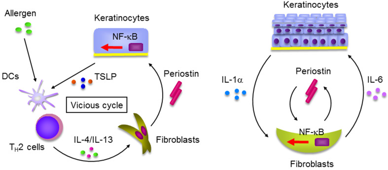
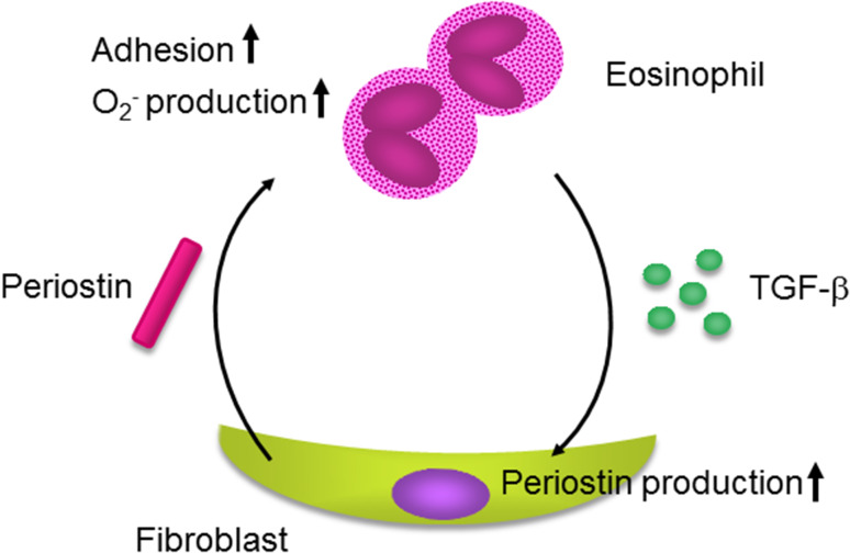
We found for the first time that IL-4 and IL-13, signature type 2 cytokines, are able to induce periostin expression. We and others have subsequently shown that periostin is highly expressed in chronic inflammatory diseases-asthma, atopic dermatitis, eosinophilc chronic sinusitis/chronic rhinosinusitis with nasal polyp, and allergic conjunctivitis-and that periostin plays important roles in the pathogenesis of these diseases. The epithelial/mesenchymal interaction via periostin is important for the onset of allergic inflammation, in which periostin derived from fibroblasts acts on epithelial cells or fibroblasts, activating their NF-κB. Moreover, the immune cell/non-immune cell interaction via periostin may be also involved. Now the significance of periostin has been expanded into other inflammatory or fibrotic diseases such as scleroderma and pulmonary fibrosis. The cross-talk of periostin with TGF-β or pro-inflammatory cytokines is important for the underlying mechanism of these diseases. Because of its pathogenic importance and broad expression, diagnostics or therapeutic drugs can be potentially developed to target periostin as a means of treating these diseases.

Keywords: Allergic conjunctivitis; Allergy; Asthma; Atopic dermatitis; Biomarker; Cross-talk; Epithelial/mesenchymal interaction; IL-13; IL-4; Matricellular protein; Periostin; Pulmonary fibrosis; Scleroderma; TGF-β.


Figures

Fig. 1
Fig. 1
High expression of periostin in inflamed sites of allergic diseases. Expression of periostin in bronchial tissue from an asthma patient (a) [15], in skin tissue from an AD patient (b) [16], in a nasal polyp from an eosinophilc chronic sinusitis/CRSwNP patient (c) [17], and in conjunctival tissue from an AKC patient (d) [19]. It is of note that periostin is highly expressed in the subepithelial regions of each tissue
Fig. 2
Fig. 2
Epithelial/mesenchymal interaction via periostin in the pathogenesis of skin allergic diseases (modified from [16, 29]). a IL-4/IL-13 produced by TH2 cells activated by exposure to allergens induces periostin production in fibroblasts. Periostin acts on keratinocytes activating NF-κB followed by production of pro-inflammatory cytokines including TSLP, which acts on dendritic cells (DCs), accelerating type 2 inflammation. Thus, IL-4/IL-13, periostin, and TSLP generate a vicious cycle in the pathogenesis of skin allergic diseases. b IL-1α and periostin produced by keratinocytes and fibroblasts, respectively, cooperate to act on fibroblasts activating NF-κB. Activated fibroblasts produce IL-6 accelerating proliferation of keratinocytes
Fig. 3
Fig. 3
Immune cell/non-immune cell interaction via periostin in the pathogenesis of allergic diseases. Periostin augments adhesion, superoxide anion (O2 ) generation, and TGF-β production in eosinophils. Reciprocally, activated eosinophils may induce periostin protein in fibroblasts
Fig. 4
Fig. 4
Advantages and disadvantages of periostin as a biomarker. Several advantages and one disadvantage of periostin as a biomarker are depicted
Fig. 5
Fig. 5
Algorithm for the treatment of asthma (modified from [10]). A first-line of anti-asthma drugs is inhaled corticosteroids. If they are ineffective, measurement of serum periostin is recommended. If the level is high, anti-IL-4/IL-13 antagonists should be added
Fig. 6
Fig. 6
Cross-talk of periostin and TGF-β in the pathogenesis of scleroderma. Periostin cooperates with TGF-β activating the phosphatidylinositol-3 kinase (PI3-K)/Akt pathway in fibroblasts followed by induction of collagen 1α1 and α-SMA, which would accelerate scleroderma
Fig. 7
Fig. 7
Cross-talk of periostin and TNFα/IL-1α in the pathogenesis of pulmonary fibrosis. Periostin cooperates with TNFα or IL-1α derived from epithelial cells or inflammatory cells activating NF-κB in fibroblasts followed by induction of various chemokines/pro-inflammatory cytokines such as Ccl2/MCP1, Ccl4/MCP1, Ccl7/MCP3, Cxcl1/KC, Cxcl2/MIP-1α, and IL-1β. These chemokines/pro-inflammatory cytokines recruit neutrophils and macrophages, accelerating pulmonary fibrosis

References

    1. Conway SJ, Izuhara K, Kudo Y, Litvin J, Markwald R, Ouyang G, Arron JR, Holweg CT, Kudo A. The role of periostin in tissue remodeling across health and disease. Cell Mol Life Sci. 2014;71:1279–1288. doi: 10.1007/s00018-013-1494-y. - DOI - PMC - PubMed
    1. Ontsuka K, Kotobuki Y, Shiraishi H, Serada S, Ohta S, Tanemura A, Yang L, Fujimoto M, Arima K, Suzuki S, Murota H, Toda S, Kudo A, Conway SJ, Narisawa Y, Katayama I, Izuhara K, Naka T. Periostin, a matricellular protein, accelerates cutaneous wound repair by activating dermal fibroblasts. Exp Dermatol. 2012;21:331–336. doi: 10.1111/j.1600-0625.2012.01454.x. - DOI - PubMed
    1. Nishiyama T, Kii I, Kashima TG, Kikuchi Y, Ohazama A, Shimazaki M, Fukayama M, Kudo A. Delayed re-epithelialization in periostin-deficient mice during cutaneous wound healing. PLoS One. 2011;6:e18410. doi: 10.1371/journal.pone.0018410. - DOI - PMC - PubMed
    1. Elliott CG, Wang J, Guo X, Xu SW, Eastwood M, Guan J, Leask A, Conway SJ, Hamilton DW. Periostin modulates myofibroblast differentiation during full-thickness cutaneous wound repair. J Cell Sci. 2012;125:121–132. doi: 10.1242/jcs.087841. - DOI - PMC - PubMed
    1. Yuyama N, Davies DE, Akaiwa M, Matsui K, Hamasaki Y, Suminami Y, Yoshida NL, Maeda M, Pandit A, Lordan JL, Kamogawa Y, Arima K, Nagumo F, Sugimachi M, Berger A, Richards I, Roberds SL, Yamashita T, Kishi F, Kato H, Arai K, Ohshima K, Tadano J, Hamasaki N, Miyatake S, Sugita Y, Holgate ST, Izuhara K. Analysis of novel disease-related genes in bronchial asthma. Cytokine. 2002;19:287–296. doi: 10.1006/cyto.2002.1972. - DOI - PubMed

MeSH terms