Review
. 2017 Sep;32(9):1264-1310.
doi: 10.1002/mds.27115.

Past, present, and future of Parkinson's disease: A special essay on the 200th Anniversary of the Shaking Palsy

Affiliations
Review

Past, present, and future of Parkinson's disease: A special essay on the 200th Anniversary of the Shaking Palsy

J A Obeso et al. Mov Disord. 2017 Sep.

Abstract

This article reviews and summarizes 200 years of Parkinson's disease. It comprises a relevant history of Dr. James Parkinson's himself and what he described accurately and what he missed from today's perspective. Parkinson's disease today is understood as a multietiological condition with uncertain etiopathogenesis. Many advances have occurred regarding pathophysiology and symptomatic treatments, but critically important issues are still pending resolution. Among the latter, the need to modify disease progression is undoubtedly a priority. In sum, this multiple-author article, prepared to commemorate the bicentenary of the shaking palsy, provides a historical state-of-the-art account of what has been achieved, the current situation, and how to progress toward resolving Parkinson's disease. © 2017 International Parkinson and Movement Disorder Society.

Keywords: 200 years anniversary; Parkinson's disease; Shaking Palsy.


Conflict of interest statement

Relevant conflicts of interests/financial disclosures: Nothing to report.

Figures

FIG. 1
FIG. 1
Current picture of the house where James Parkinson lived and worked in East London and the commemorative plaque.
FIG. 2
FIG. 2
The main pathologies in patients with clinical Parkinson’s disease and the pathological progression. (A) Transverse hemisection of the midbrain of a control at left and a patient with clinical Parkinson’s disease (PD) at right showing the marked reduction in the black pigment within the substantia nigra region. (BC) Haematoxylin and eosin stained section of the ventrolateral region identified by the box in A showing at higher magnification the pigmented neurons of the substantia nigra in a control without PD (B) and a person with clinical PD (C). (DE) Intracytoplasmic Lewy bodies in remaining pigmented neuron of the substantia nigra of a patient with PD showing the eosinophilic core and paler halo in haematoxylin and eosin stain (D) and the dark aggregation of α-synuclein using immunoperoxidase with cresyl violet counterstaining (E). (F) Cartoon representation (based on data from Toledo et al) of the two major patterns of Lewy-body pathology in patients with (below) and without (above) Alzheimer’s disease (AD) pathology. In those with clinical PD and little AD pathology, Lewy bodies begin in the olfactory bulb and medulla oblongata then infiltrate higher brain stem regions, then limbic brain regions, and finally the neocortex. In those with AD pathology, this pattern is different. Lewy bodies concentrate in limbic regions of the brain prior to infiltrating to other regions.
FIG. 3
FIG. 3
(A) Spin echo T1-weighted 3T images sensitive to neuromelanin showing a reduction of the area of hyperintensity of the substantia nigra (arrow) in the PD patient as compared with the healthy control (HC). (B) T2*-weighted 7T images showing the normal dorsal nigral hyperintensity (DNH) in the substantia nigra of the HC (arrow) that is not visible in the PD patient. (C) Quantitative susceptibility map of the SN in a control subject showing the substantia nigra as an area of high signal intensity indicating high susceptibility as result of iron deposition (arrow). (D) Fractional anisotropy map of the Substantia Nigra (SN) in a control subject. The arrow indicates the substantia nigra.
FIG. 4
FIG. 4
Tracers for presynaptic dopaminergic function. The vesicular monoamine transporter 2 (VMAT2) is responsible for packaging monoamine transmitters into synaptic vesicles. 6-18F-fluoro-L-dopa is a radioactive analog of levodopa that is decarboxylated into 6-18F-fluoro-L-dopamine, which is subsequently stored in synaptic vesicles but then undergoes slow egress and enzymatic degradation. Once dopamine is release from the synapse, it is taken up by the dopamine transporter (DaT), which can be labeled using a variety of 11C and 18F (for PET) and 131I or 99mTc (for SPECT) tracers. For each tracer, the left panel shows a healthy control subject, whereas the right shows a patient with mild Parkinson’s disease. In the latter, there is asymmetric reduction of tracer uptake, maximally affecting the posterior striatum. From Chandran & Stoessl, in Jankovic & Tolosa, Parkinson’s Disease and Movement Disorders, Wolters Kluwer, 2015.
FIG. 5
FIG. 5
Glucose metabolism in parkinsonian disorders. PD is associated with increased metabolism in the basal ganglia, thalamus, pons, and cerebellum, with concomitant reductions of metabolism in premotor and parietal cortex (the so-called PD related pattern or PDRP, right panel), whereas multiple system atrophy (MSA) is associated with reduced metabolism in basal ganglia and cerebellum, progressive supranuclear palsy (PSP) with reduced metabolism in medial frontal cortex and thalamus, and corticobasal degeneration (CBD) with asymmetrically reduced metabolism in cortex and basal ganglia. Taken from (left) Eckert T, et al. FDG PET in the differential diagnosis of parkinsonian disorders. Neuroimage 2005;26:912–921 and (right) Asanuma K, et al. Network modulation in the treatment of Parkinson’s disease. Brain 2006;129:2667–2678.
FIG. 6
FIG. 6
Classic scheme of cortico-basal ganglia connectivity highlighting the main motor, associative-cognitive, and emotional-limbic domains. M1, primary motor cortex; SMA, supplementary motor area; PMC, pre-motor cortex; SNc, substantia nigra pars compacta; GPe, globus pallidus pars externa; STN, subthalamic nucleus; GPi, globus pallidus pars interna; SNr, substantia nigra pars reticulate; VL, ventralis lateralis; VA, ventralis anterior; MD, medio-dorsal.
FIG. 7
FIG. 7
Bermuda triangle of disease mechanisms implicated in monogenic (and idiopathic) Parkinson’s disease, highlighting the role of confirmed genes for monogenic PD and parkinsonism, as well as for the GBA gene in the context of protein degradation, mitochondrial function, and synaptic and endosomal vesicle and protein recycling.
FIG. 8
FIG. 8
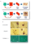
(A) Schematic illustration of the similarity between the PrP and alpha-synuclein proteins in terms of their potential to misfold to form betarich sheets, and polymerize to form oligomers/rod, and amyloid plaques/Lewy Bodies (adapted from ref. ). (B) Comparison of Lewy pathology in grafted embryonic dopamine neurons (graft) and in the host substantia nigra (host). Note the similarity in staining for alpha-synuclein, ubiquin, and thioflavin-S (indicative of beta sheet formation). These observations raise the possibility that misfolded alpha-synuclein has spread from affected neurons in the PD brain to unaffected implanted dopamine neurons in a prion-like manner (adapted from ref. ).

Similar articles

Cited by

References

    1. Goldman JG, Goetz CG. History of Parkinson’s disease. Handb Clin Neurol. 2007;83:107–128. - PubMed
    1. Parkinson J, Hunter J. Hunterian Reminiscences; Being the Substance of a Course of Lectures on the Principles and Practice of Surgery, Delivered by J. Hunter, in the Year 1785. London: Sherwood, Gilbert, and Piper; 1833.
    1. Parkinson J. The Villager’s Friend and Physician. London: C Whittingham; 1804.
    1. Parkinson J. Dangerous Sports: A Tale. London: HD Symonds; 1807.
    1. Parkinson J. Some account of the effects of lightning. Mem M Soc Lon. 1789;2:490–500.