. 2017 Dec;174(23):4295-4307.
doi: 10.1111/bph.14027. Epub 2017 Oct 19.

Magnesium therapy improves outcome in Streptococcus pneumoniae meningitis by altering pneumolysin pore formation

Affiliations

Magnesium therapy improves outcome in Streptococcus pneumoniae meningitis by altering pneumolysin pore formation

Sabrina Hupp et al. Br J Pharmacol. 2017 Dec.

Abstract

Background and purpose: Streptococcus pneumoniae is the most common cause of bacterial meningitis in adults and is characterized by high lethality and substantial cognitive disabilities in survivors. Here, we have studied the capacity of an established therapeutic agent, magnesium, to improve survival in pneumococcal meningitis by modulating the neurological effects of the major pneumococcal pathogenic factor, pneumolysin.

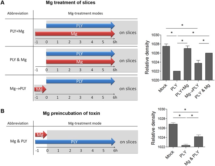
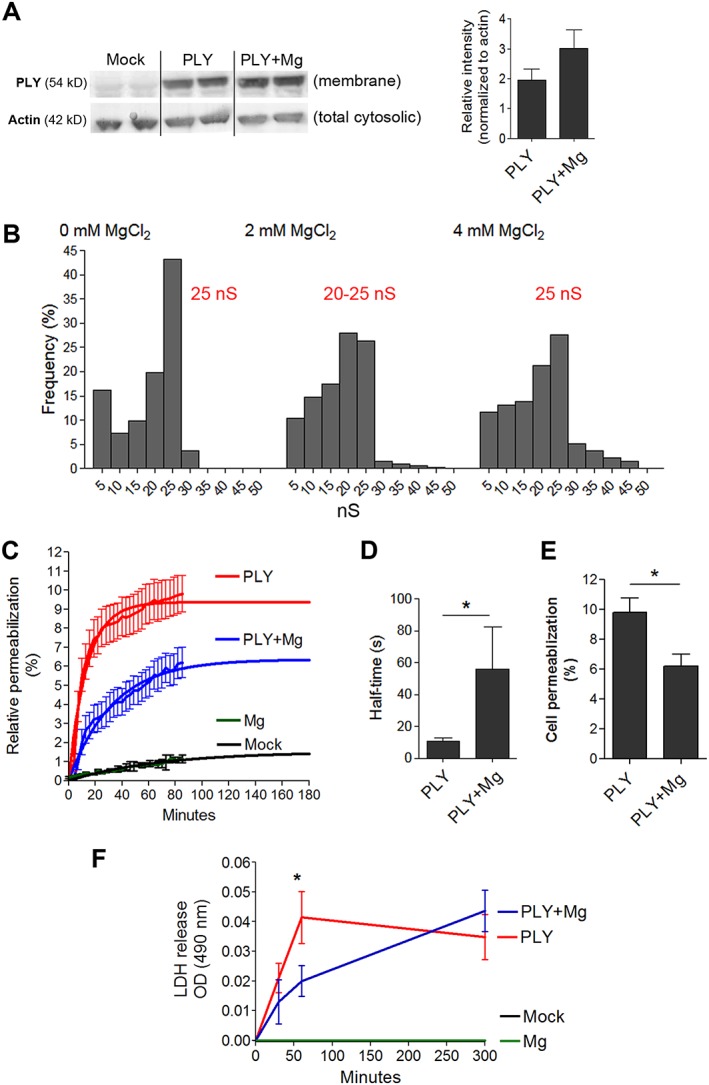
Experimental approach: We used mixed primary glial and acute brain slice cultures, pneumolysin injection in infant rats, a mouse meningitis model and complementary approaches such as Western blot, a black lipid bilayer conductance assay and live imaging of primary glial cells.

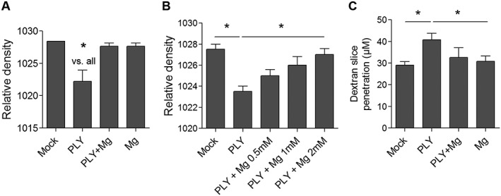
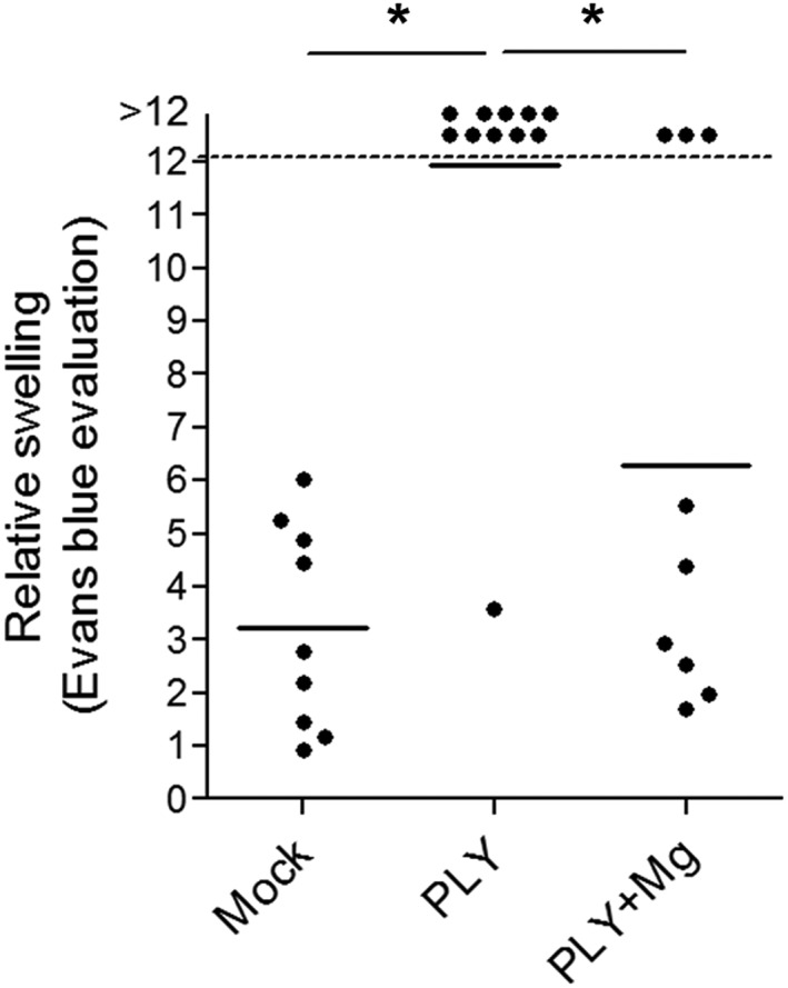
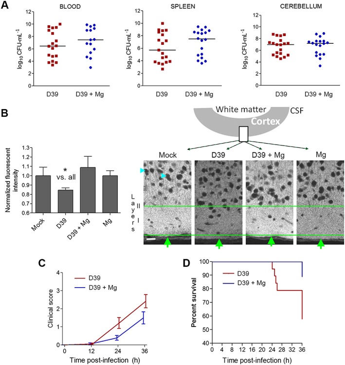
Key results: Treatment with therapeutic concentrations of magnesium chloride (500 mg·kg-1 in animals and 2 mM in cultures) prevented pneumolysin-induced brain swelling and tissue remodelling both in brain slices and in animal models. In contrast to other divalent ions, which diminish the membrane binding of pneumolysin in non-therapeutic concentrations, magnesium delayed toxin-driven pore formation without affecting its membrane binding or the conductance profile of its pores. Finally, magnesium prolonged the survival and improved clinical condition of mice with pneumococcal meningitis, in the absence of antibiotic treatment.

Conclusions and implications: Magnesium is a well-established and safe therapeutic agent that has demonstrated capacity for attenuating pneumolysin-triggered pathogenic effects on the brain. The improved animal survival and clinical condition in the meningitis model identifies magnesium as a promising candidate for adjunctive treatment of pneumococcal meningitis, together with antibiotic therapy.


Figures

Figure 1
Figure 1
Ex vivo inhibition of PLY‐induced swelling by MgCl2. (A) Relative density (lower density indicates higher water content and swelling) of brain slices after 6 h incubation without treatment (mock), with exposure to 4 HU·mL−1 PLY, treatment with 2 mM Mg only (Mg) or combined exposure to PLY and Mg (n = 6 independent experiments). (B) Concentration‐dependent inhibition by Mg2+ of rat brain swelling after 6 h co‐exposure of 4 HU·mL−1 PLY (n = 5 independent experiments). (C) Effect of 2 mM Mg to 4 HU·mL−1 PLY on penetration of dextran‐TRITC in rat brain slices (n = 5 independent experiments). *P<0.05, significantly different as indicated.
Figure 2
Figure 2
Attenuation of brain swelling by different schedules for magnesium application. (A) Schematic diagram of the different modes of magnesium (Mg) application (left). Inhibition of PLY‐induced oedema by 4 HU·mL−1 for 6 h by simultaneous incubation with 2 mM Mg2+ with 1 h pre‐incubation of the slices with Mg2+ (PLY + Mg) and without slice pre‐incubation (PLY and Mg). Pre‐incubation of the slices with Mg2+ for 1 h and removal before adding PLY (Mg→PLY) did not alter toxin‐triggered oedema (n = 5 independent experiments). (B) Partial inhibition of rat brain slice swelling by pre‐incubation of PLY with 2 mM Mg2+ for 1 h, followed by treatment of slices in normal medium without additional Mg2+ with 4 HU·mL−1 PLY for 6 h (n = 5 independent experiments). *P<0.05, significantly different as indicated.
Figure 3
Figure 3
Mechanisms of modulation of PLY properties by magnesium. (A) The presence of Mg2+ does not inhibit toxin binding to mouse glial cells (Western blot) 15 min after challenge with 4 HU·mL−1 (4 independent experiments). (B) The conductance profile of PLY in a black lipid bilayer demonstrates an unchanged conductance pattern that is independent of the Mg concentration. The number of measured events is as follows: mock – 130 events, 2 mM Mg – 330 events, 4 mM – 127 events with peak conductance for mock at 25 nS, for 2 mM Mg at 20–25 nS and for 4 mM Mg at 25 nS. (C) Live‐cell imaging analysis of mouse glial cell membrane permeabilization (as judged by PI staining) by 4 HU·mL−1 PLY reveals delayed permeabilization and diminished number of permeabilized cells during treatment with 2 mM Mg. The curves are extrapolated beyond 120 min using non‐linear regression curves fitted with one‐phase exponential association (n = 5 independent experiments). (D) Increased half‐time of PLY permeabilization during treatment with 2 mM Mg (n = 5 independent experiments). (E) Diminished permeabilization at the plateau of the regression curve in the presence of 2 mM Mg treatment (n = 5 independent experiments). (F) Diminished LDH release by Mg at 60 min after PLY challenge, followed by equalized release at 6 h (n = 5 independent experiments). *P<0.05, significantly different as indicated; in (F), *P<0.05, significant effect of Mg.
Figure 4
Figure 4
Effect of Mg on PLY brain oedema model in infant rats. The amount of Evans Blue (expressed as mm2 after filter paper absorption) displaced out of the intracranial space (as a marker of elevated intracranial pressure) at 6 h after treatment with PLY (see Methods) in rats with or without treatment with MgCl2 (500 mg·kg−1 i.p) at the beginning of the experiment, reveals ameliorated intracranial pressure increase after treatment with Mg2+. The intracranial pressure of the PLY‐treated animals is significantly higher than that in the mock and PLY + Mg groups. Data shown are individual values with mean indicated by the horizontal line. *P<0.05, significantly different as indicated.
Figure 5
Figure 5
Effect of magnesium treatment in mice with experimental S. pneumoniae D39 meningitis. (A) S. pneumoniae D39 concentrations (as CFUs) in blood, cerebellum and spleen homogenates after 36 h demonstrated comparable growth in infected mice treated i.p. with 0.45% NaCl (mock) or treated i.p. with MgCl2 (Mg). (B) Relative fluorescent intensity measurement of the PSD95 immunofluorescence in layers 1–3 of the neocortex at the level of the postcentral gyrus (mock (n = 4 animals): NaCl‐injected and NaCl‐treated group; D39 (n = 19 animals): Spn D39‐infected and NaCl‐treated group; D39 + Mg (n = 18 animals): Spn D39‐infected and MgCl2‐treated group; Mg (n = 3 animals): NaCl‐injected and MgCl2‐treated group) and corresponding fluorescent images (cyan arrows indicate staining‐negative nuclear regions; green arrows – cortical surface; green lines limit the region of interest, including layer I and partially layer II; schematic diagram above indicates the position of the imaged fragment). Scale bar: 20 μm. (C) Clinical score (0 = no apparent behavioural abnormality; 1, moderate lethargy; 2 = severe lethargy; 3 = unable to walk; 4 = dead) of the animals. Mock and Mg controls demonstrate score of 0 (not included in the graph, overlap with axis). For statistical analysis, the area under the curve is calculated and compared (see Methods). There was a significant effect of treatment with Mg2+. (D) Survival curves of MgCl2 (Mg, n = 18) or NaCl‐treated (mock, n = 19) infected animals. All mock and Mg controls demonstrate 100% survival at 36 h (not included in the graph, overlap of multiple lines). There was a significantly increased survival after treatment with Mg2+.

References

    1. Alexander SPH, Peters JA, Kelly E, Marrion N, Benson HE, Faccenda E et al (2015a). The Concise Guide to PHARMACOLOGY 2015/16: Ligand‐gated ion channels. Br J Pharmacol 172: 5870–5903. - PMC - PubMed
    1. Alexander SPH, Fabbro D, Kelly E, Marrion N, Peters JA, Benson HE et al (2015b). The Concise Guide to PHARMACOLOGY 2015/16: Enzymes. Br J Pharmacol 172: 6024–6109. - PMC - PubMed
    1. Alouf JE (2000). Cholesterol‐binding cytolytic protein toxins. Int J Med Microbiol 290: 351–356. - PubMed
    1. Amagasa M, Ogawa A, Yoshimoto T (1990). Effects of calcium and calcium antagonists against deprivation of glucose and oxygen in guinea pig hippocampal slices. Brain Res 526: 1–7. - PubMed
    1. Awad MM, Ellemor DM, Boyd RL, Emmins JJ, Rood JI (2001). Synergistic effects of alpha‐toxin and perfringolysin O in Clostridium perfringens‐mediated gas gangrene. Infect Immun 69: 7904–7910. - PMC - PubMed

MeSH terms

LinkOut - more resources