. 2017 Nov:59:107-120.
doi: 10.1016/j.neurobiolaging.2017.07.006. Epub 2017 Aug 1.

Aged chimpanzees exhibit pathologic hallmarks of Alzheimer's disease

Affiliations

Aged chimpanzees exhibit pathologic hallmarks of Alzheimer's disease

Melissa K Edler et al. Neurobiol Aging. 2017 Nov.

Abstract

Alzheimer's disease (AD) is a uniquely human brain disorder characterized by the accumulation of amyloid-beta protein (Aβ) into extracellular plaques, neurofibrillary tangles (NFT) made from intracellular, abnormally phosphorylated tau, and selective neuronal loss. We analyzed a large group of aged chimpanzees (n = 20, age 37-62 years) for evidence of Aβ and tau lesions in brain regions affected by AD in humans. Aβ was observed in plaques and blood vessels, and tau lesions were found in the form of pretangles, NFT, and tau-immunoreactive neuritic clusters. Aβ deposition was higher in vessels than in plaques and correlated with increases in tau lesions, suggesting that amyloid build-up in the brain's microvasculature precedes plaque formation in chimpanzees. Age was correlated to greater volumes of Aβ plaques and vessels. Tangle pathology was observed in individuals that exhibited plaques and moderate or severe cerebral amyloid angiopathy, a condition in which amyloid accumulates in the brain's vasculature. Amyloid and tau pathology in aged chimpanzees suggests these AD lesions are not specific to the human brain.

Keywords: Alzheimer's disease; Amyloid-beta protein; Chimpanzee; Neurofibrillary tangle; Primate; Tau.


Figures

Figure 1.
Figure 1.
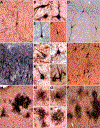
APP/Aβ and Aβ42-ir plaques and vessels in aged chimpanzees (subject 12: A-B,D-F,H,M-O; subject 9: C; subject 20: G,J,L; subject 19: I,K): (A-B,E) APP/Aβ-ir plaques, (C-D,F) Aβ42-ir plaques, (G) APP/Aβ-ir cortical artery and arterioles, (H) APP/Aβ-ir leptomeningeal arteries, (I) Aβ42-ir cortical artery (J) Aβ42-ir cortical arterioles, (K) APP/Aβ-ir cortical arterioles, (L) Aβ42-ir cortical arterioles, and (M-O) APP/Aβ-ir plaques (black arrows) near immunopositive vessels (white arrows). Scale bars = 250 µm (A, D, G, J-L, M-O) or 25 µm (B-C, E-F, H-I).
Figure 2.
Figure 2.
Thioflavin S staining in the temporal cortex (A-B, I-J, L), CA1 subfield of the hippocampus (C-F, H, K), and prefrontal cortex (G) of aged chimpanzees (subject 19: A,D-F,H-J; subject 20: B,L; subject 12: C,K; subject 13: G): (A) diffuse plaque, (B) dense-core plaque, (C) plaque near positive vessel (white arrow), (D) positive arteriole, (E-G) NFT, (H) NFT (yellow arrow) near vessel, (I-J) cortical arteries, and (K-L) cortical arterioles. Scale bars = 20 µm (A-B, E-H) or 200 µm (C-D, I-L).
Figure 3.
Figure 3.
Co-occurrence of Aβ and tau reactivity in hippocampus (CA1, pyramidal layer) of aged chimpanzees (subject 19: A-E; subject 12: F): (A-F) AT8-ir pretangles (red arrows, NFT (yellow arrows), and NC (blue arrows) (DAB with nickel enhancement, black) associated with Aβ42-ir (NovaRED) vessels (white arrows) and plaques (black arrows). Scale bars = 25 µm (A-B) or 250 µm (C-F).
Figure 4.
Figure 4.
AT8-ir lesions in the prefrontal cortex (C, H, J-K, L-N, P-R) and hippocampus (A-B, D-G, I, O) of aged chimpanzees (subject 11: A,F; subject 19: B-E,G,I,M-R; subject 13: H,J-L): (A-F) pretangles, (G-J) NFT, and (M-R) AT8-ir NC. Scale bars = 250 µm (A, D, G, J, M, P) or 25 µm (B-C, E-F, H-I, K-L, N-O, QR).
Figure 5.
Figure 5.
Plaque and vessel volumes in 20 chimpanzees aged 37–62 years old (mean age = 46 years): (A) Average total plaque and vessel volume (%) (rs = 0.70, p ≤ 0.01, small circles represent outliers), (B) correlation of total APP/Aβ plaque volume vs total Aβ42 plaque volume across brain regions (R2 = 0.54, p < 0.01), and (C) total plaque volume vs total vessel volume by type (APP/Aβ: R2 = 0.66, Aβ42: R2 = 0.87, p’s < 0.01).
Figure 6.
Figure 6.
Significant correlations of APP/Aβ (%) and tau (mm3) lesions (p’s ≤ 0.05): (A) HC pretangle density vs NC and HC APP/Aβ vessel volume (NC: R2 = 0.22, HC: R2 = 0.18), (B) HC pretangle density vs HC APP/Aβ plaque volume (R2 = 0.19), (C) HC NFT density vs NC APP/Aβ vessel volume (R2 = 0.23), (D) HC AT8-ir tau NC density vs NC and HC APP/Aβ vessel volume (NC: R2 = 0.20, HC: R2 = 0.20), and (E) NC AT8-ir tau NC density and HC APP/Aβ plaque volume (R2 = 0.27).
Figure 7.
Figure 7.
Severity of CAA was associated with higher Aβ plaque volume (%) and increasing levels of tau lesions (per mm3) in aged chimpanzees: (A) total APP/Aβ plaque volume vs CAA stage (p ≤ 0.01), (B) total pretangle density vs CAA stage (p ≤ 0.01), (C) total NFT density vs CAA stage (p = 0.47), and (D) total AT8-ir tau NC density vs CAA stage (p = 0.02).

References

    1. Akram A, Christoffel D, Rocher AB, Bouras C, Kovari E, Perl DP, Morrison JH, Herrmann FR, Haroutunian V, Giannakopoulos P, Hof PR, 2008. Stereologic estimates of total spinophilinimmunoreactive spine number in area 9 and the CA1 field: relationship with the progression of Alzheimer’s disease. Neurobiol. Aging 29, 1296–1307. doi:10.1016/j.neurobiolaging.2007.03.007 - DOI - PMC - PubMed
    1. Anderson US, Stoinski TS, Bloomsmith MA, Marr MJ, Smith AD, Maple TL, 2005. Relative numerousness judgment and summation in young and old Western lowland gorillas. J. Comp. Psychol 119, 285–295. doi:10.1037/0735-7036.119.3.285 - DOI - PubMed
    1. Arriagada PV, Marzloff K, Hyman BT, 1992. Distribution of Alzheimer-type pathologic changes in nondemented elderly individuals matches the pattern in Alzheimer’s disease. Neurology 42, 1681–1688. - PubMed
    1. Attems J, Quass M, Jellinger KA, Lintner F, 2007. Topographical distribution of cerebral amyloid angiopathy and its effect on cognitive decline are influenced by Alzheimer disease pathology. J. Neurol. Sci 257, 49–55. doi:10.1016/j.jns.2007.01.013 - DOI - PubMed
    1. Bachevalier J, Landis LS, Walker LC, Brickson M, Mishkin M, Price DL, Cork LC, 1991. Aged monkeys exhibit behavioral deficits indicative of widespread cerebral dysfunction. Neurobiol. Aging 12, 99–111. - PubMed

Publication types