. 2017 Sep 9;6(9):e006465.
doi: 10.1161/JAHA.117.006465.

The Impact of Heterozygous KCNK3 Mutations Associated With Pulmonary Arterial Hypertension on Channel Function and Pharmacological Recovery

Affiliations

The Impact of Heterozygous KCNK3 Mutations Associated With Pulmonary Arterial Hypertension on Channel Function and Pharmacological Recovery

Michael S Bohnen et al. J Am Heart Assoc. .

Abstract

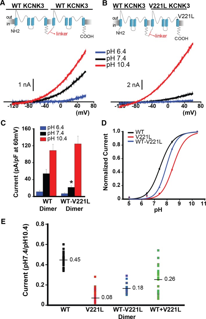
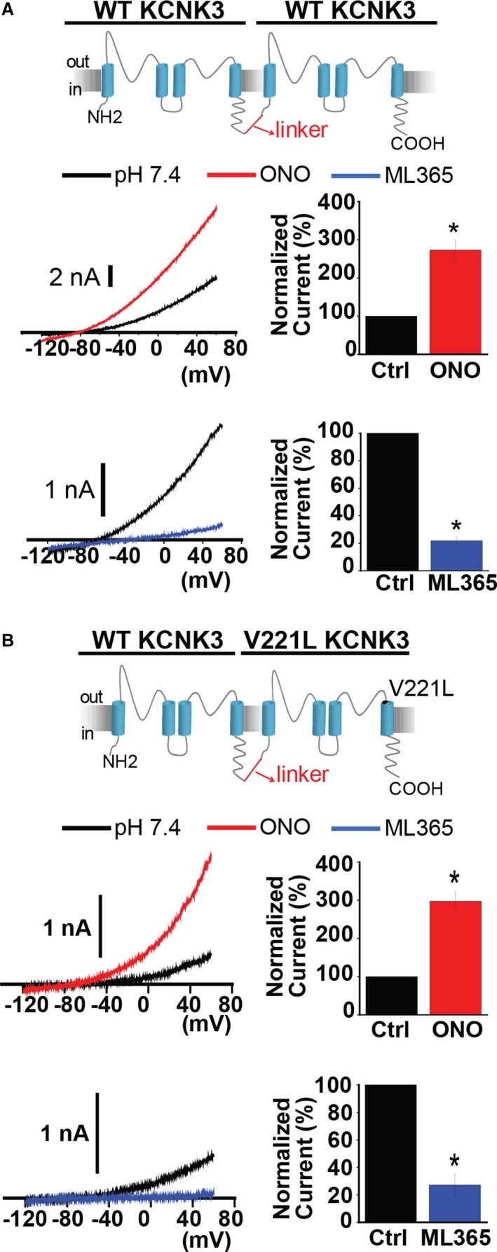
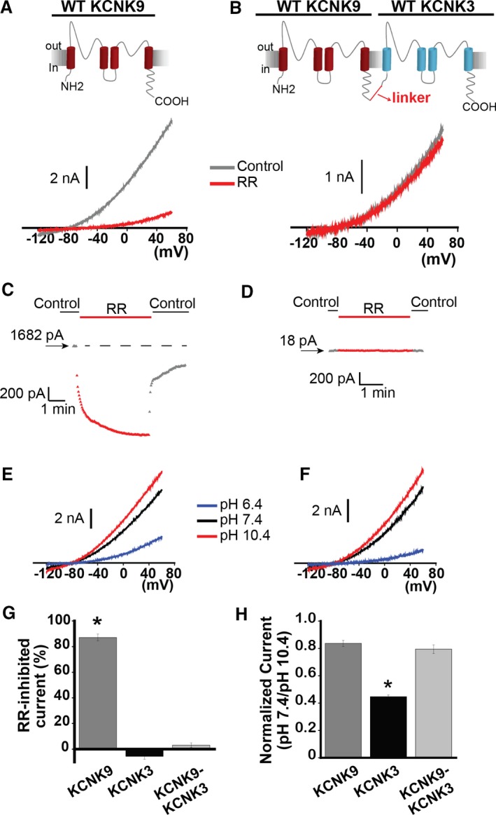
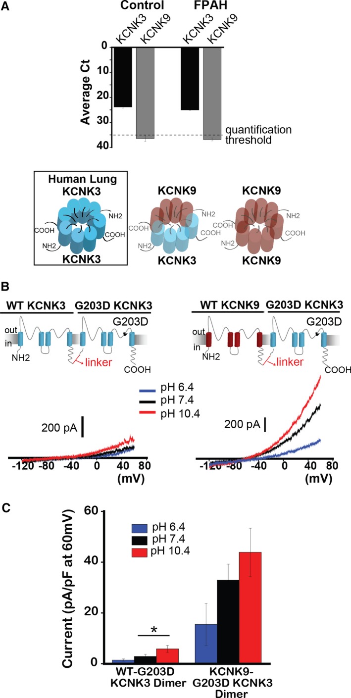
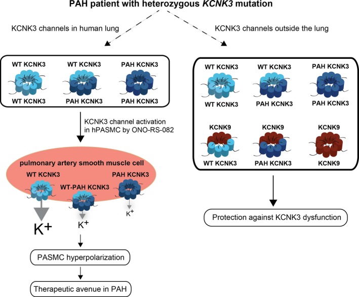
Background: Heterozygous loss of function mutations in the KCNK3 gene cause hereditary pulmonary arterial hypertension (PAH). KCNK3 encodes an acid-sensitive potassium channel, which contributes to the resting potential of human pulmonary artery smooth muscle cells. KCNK3 is widely expressed in the body, and dimerizes with other KCNK3 subunits, or the closely related, acid-sensitive KCNK9 channel.

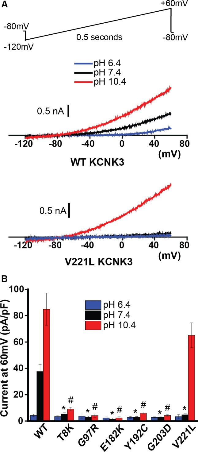
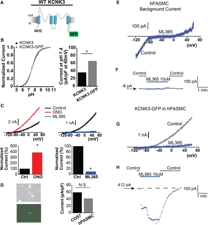
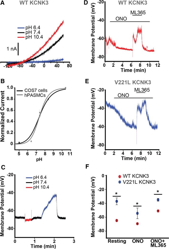
Methods and results: We engineered homomeric and heterodimeric mutant and nonmutant KCNK3 channels associated with PAH. Using whole-cell patch-clamp electrophysiology in human pulmonary artery smooth muscle and COS7 cell lines, we determined that homomeric and heterodimeric mutant channels in heterozygous KCNK3 conditions lead to mutation-specific severity of channel dysfunction. Both wildtype and mutant KCNK3 channels were activated by ONO-RS-082 (10 μmol/L), causing cell hyperpolarization. We observed robust gene expression of KCNK3 in healthy and familial PAH patient lungs, but no quantifiable expression of KCNK9, and demonstrated in functional studies that KCNK9 minimizes the impact of select KCNK3 mutations when the 2 channel subunits co-assemble.

Conclusions: Heterozygous KCNK3 mutations in PAH lead to variable loss of channel function via distinct mechanisms. Homomeric and heterodimeric mutant KCNK3 channels represent novel therapeutic substrates in PAH. Pharmacological and pH-dependent activation of wildtype and mutant KCNK3 channels in pulmonary artery smooth muscle cells leads to membrane hyperpolarization. Co-assembly of KCNK3 with KCNK9 subunits may provide protection against KCNK3 loss of function in tissues where both KCNK9 and KCNK3 are expressed, contributing to the lung-specific phenotype observed clinically in patients with PAH because of KCNK3 mutations.

Keywords: ion channel; pathophysiology; pharmacology; potassium channels; pulmonary hypertension.


Figures

Figure 1
Figure 1
Pulmonary arterial hypertension (PAH)‐associated mutant KCNK3 channels demonstrate mutation‐specific severity of loss of function, across a broad pH range in COS7 cells. A, Typical voltage clamp recordings of wildtype (WT, top) and V221L (bottom) KCNK3. Sample current traces at pH 6.4 (blue), 7.4 (black), and 10.4 (red) are shown. A voltage ramp (top) was applied, −120 mV to +60 mV over 0.5 s, every 3 s, from a holding potential of −80 mV for all voltage clamp recordings in this study. B, Summary of current density (pA/pF at 60 mV) of cells expressing WT or one of the PAH‐associated mutant KCNK3 channels (n=3–9 cells at pH 6.4; n=6–33 cells at pH 7.4; n=6–32 cells at pH 10.4). Bars show mean±SEM. *P<0.05 at pH 7.4; # P<0.05 at pH 10.4, for the comparison of WT and each KCNK3 mutant channel by 1‐way ANOVA (P<0.05) and post hoc Tukey test.
Figure 2
Figure 2
KCNK3 expression platform in human pulmonary artery smooth muscle cells (hPASMCs). A, KCNK3, tagged with GFP at the C‐terminus, was engineered (KCNK3‐GFP). B, KCNK3‐GFP (gray curve) vs KCNK3 (black curve) expression in COS7 cells across a broad pH range, with current normalized to max current at pH 10.4 (n=9–25 cells per pH data point for KCNK3; n=9–12 cells for KCNK3‐GFP; fitted by the Hill equation. C, KCNK3‐GFP is activated by ONORS‐082 (ONO) 10 μmol/L (red trace, left) and inhibited by ML365 10 μmol/L (blue trace, right), under voltage‐clamp shown in COS7 cells. Predrug (control, black traces) and drug conditions are at pH 7.4. Bar graphs (bottom) show percent change in current at −50 mV after ONO (n=8 cells) or ML365 (n=12 cells) application, compared with control. D, Cultured hPASMCs (left, top) expressing KCNK3‐GFP fluoresce green (left, bottom). Bar graph (right) shows KCNK3‐GFP current activity at pH 7.4 (pA/pF at 60 mV) in hPASMCs (gray, n=20 cells) vs COS7 cells (black, n=26 cells). E, Background hPASMC current at pH 7.4 (control, gray trace), and after application of ML365 10 μmol/L (blue trace). F, Sample ML365 time course of action in control and drug conditions, measured at −50 mV, from a starting current amplitude of −6 pA indicated by the arrow. G, Current from hPASMC expressing KCNK3‐GFP is shown at pH 7.4 (control, gray trace), and in ML365 10 μmol/L (blue trace). H, Sample ML365 time course of action in control and drug conditions, measured at −50 mV, from a starting current amplitude of 412 pA indicated by the arrow. Horizontal dashed line is drawn at the starting level of current in control solution. Bar graphs show mean±SEM. *P<0.05 by the paired (B) and unpaired (C) Student t test. N.S. indicates no significant difference.
Figure 3
Figure 3
Robust response of KCNK3 channels to pH changes and pharmacological modulators in human pulmonary artery smooth muscle cells (hPASMCs). A, Voltage clamp recording of KCNK3‐GFP expressed in hPASMCs at pH 6.4 (blue), pH 7.4 (black), and pH 10.4 (red). B, KCNK3‐GFP expression in hPASMCs (gray curve) vs COS7 cells (black curve) across a broad pH range, with current normalized to max current at pH 10.4 (n=9–12 cells per pH data point in COS7; n=2–6 cells per pH data point in hPASMCs; fitted by the Hill equation). C, Current clamp recording of wildtype (WT) KCNK3‐GFP, with changes in membrane potential (mV) measured at pH 6.4 (blue), pH 7.4 (black), and pH 10.4 (red). D, Current clamp recording of WT KCNK3‐GFP (red trace), showing changes in membrane potential (mV) upon application of ONORS‐082 (ONO), ONO+ML365, or pH 8.4. E, Current clamp recording of V221L KCNK3‐GFP (blue trace), showing changes in membrane potential (mV) upon application of ONO, or ONO+ML365. F, Current clamp summary of WT (red) and V221L (blue) KCNK3‐GFP for resting potential, ONO, and ONO+ML365 conditions (n=3–11 cells per condition). Drugs applied at a concentration of 10 μmol/L in all experiments. Data plots represent means±SEM. *P<0.05 by the unpaired Student t test.
Figure 4
Figure 4
Tandem‐linked KCNK3 heterodimeric channels are functional reporters of KCNK3 heterozygosity. A and B, KCNK3 dimers were engineered by interconnecting 2 KCNK3 subunits with a glycine‐rich linker. The wildtype (WT) KCNK3 homodimer (A) and the WT−V221L KCNK3 heterodimer (B) are depicted, with sample voltage clamp recordings for each condition. Current traces at pH 6.4 (blue), 7.4 (black), and 10.4 (red) are shown. C, Summary of current densities (pA/pF at 60 mV) at pH 6.4, 7.4, and 10.4 (n=4–16 cells per pH bar). D, KCNK3 current activity is depicted for WT (black curve), V221L (red curve), and WT−V221L heterodimer (blue curve), at extracellular pH 5.0 through 10.4, with current normalized to max current at pH 10.4 (n=5–33 cells per pH value plotted; fitted by the Hill equation). E, Scatterplot of current at pH 7.4 normalized to current at pH 10.4, for WT, V221L, WT−V221L heterodimer, and WT+V221L co‐expression. Each dot represents an independent cell recording. Mean current in each condition is displayed at the horizontal line (n=16–33 cells per condition, measured at 60 mV). Bar graphs and pH curve values show means±SEM. *P<0.05 for the comparison of WT vs WT−V221L KCNK3 dimer conditions in panel C by the unpaired Student t test; (E) P<0.05 for the comparison of all KCNK3 conditions, calculated by 1‐way ANOVA (P<0.05) and post hoc Tukey test.
Figure 5
Figure 5
Wildtype (WT) and mutant KCNK3 dimers respond to pharmacological modulation. A, WT KCNK3 dimer (top) is activated by ONORS‐082 (ONO) 10 μmol/L (red trace) and inhibited by ML365 10 μmol/L (blue trace), in current recordings from voltage clamp experiments. Control (predrug, pH 7.4) traces are shown in black. Bar graphs show fold change in current at −50 mV for the WT KCNK3 dimer, after ONO (red, n=4 cells) or ML365 (blue, n=7 cells) application. B, WT−V221L KCNK3 heterodimer (top) is activated by ONO 10 μmol/L (red trace), and inhibited by ML365 10 μmol/L (blue trace), and heterodimer channel activity was confirmed by channel activation at extracellular pH 10.4 (gray dotted traces). Control (predrug, pH 7.4) traces are shown in black. Bar graphs show fold change in current at −50 mV for the WT−V221L heterodimer, after ONO (red, n=5 cells) or ML365 (blue, n=3 cells) application. Bar graphs display mean±SEM. *P<0.05 by the paired Student t test.
Figure 6
Figure 6
KCNK9 forms functional heterodimers with KCNK3. A, The effect of ruthenium red (RR) 10 μmol/L (red trace) on KCNK9 channels. Control trace (predrug, pH 7.4) shown in gray. B, The effect of RR 10 μmol/L (red trace) on KCNK9‐KCNK3 heterodimeric channels. Control trace (predrug, pH 7.4) shown in gray. C, Sample RR time course of action on KCNK9 in control and drug conditions, measured at −50 mV, from a starting current amplitude of 1682 pA indicated by the arrow. Horizontal dashed line is drawn at the starting level of current in control solution. D, Sample RR time course of action on KCNK9‐KCNK3 heterodimers in control and drug conditions, measured at −50 mV, from a starting current amplitude of 18 pA indicated by the arrow. E, Voltage clamp recording of KCNK9. F, Voltage clamp recording of KCNK9‐KCNK3. E and F, sample current traces at pH 6.4 (blue), 7.4 (black), and 10.4 (red) are shown. G, Summary of RR's effect on KCNK9, KCNK3, and KCNK9‐KCNK3, measured by percent‐inhibited current at −50 mV (n=5–8 cells per condition). H, Summary of mean current at pH 7.4 normalized to current at pH 10.4, measured at 60 mV, for KCNK9, KCNK3, and KCNK9‐KCNK3 (n=10–25 cells per condition). Bar graphs show mean±SEM. *P<0.05 by the paired Student t test for the comparison of control vs RR (G), and * indicates significance by 1‐way ANOVA (P<0.05) and post hoc Tukey test for the comparison of KCNK9, KCNK3, and KCNK9‐KCNK3 (H).
Figure 7
Figure 7
KCNK9 protects against KCNK3 dysfunction. A, Quantitative real‐time PCR analysis of human lung samples from healthy (Control) and familial PAH (FPAH) patient lungs. Expression of KCNK3 (black bars), and KCNK9 (gray bars) are compared, based on mean cycle threshold (Ct) values observed for each gene; Ct>35 indicates no quantifiable gene expression (n=5 patient lungs for each lane). Of the possible KCNK3+KCNK9 channel combinations, only KCNK3 homomeric channels (boxed, left) are predicted to form in human lungs. B, Voltage clamp recordings of the WT‐G203D KCNK3 heterodimer (left), and the KCNK9‐G203D KCNK3 heterodimer (right), showing current traces at pH 6.4 (blue), 7.4 (black), and 10.4 (red). C, Summary of current densities (pA/pF at 60 mV) at pH 6.4, 7.4, and 10.4 (n=4–14 cells per pH bar). Bar graphs show mean±SEM. *P<0.05 by the unpaired Student t test. PAH indicates pulmonary arterial hypertension; PCR, polymerase chain reaction; WT, wildtype.
Figure 8
Figure 8
Schematic of the proposed impact of heterozygous potassium channel subfamily K member 3 (KCNK3) mutation in pulmonary arterial hypertension (PAH). Wildtype KCNK3 (light blue) and mutant (“PAH”) KCNK3 (dark blue) homomeric, and heterodimeric channels are expressed in human lung. Additional interactions of KCNK3 with KCNK9 (brown) channel subunits occur outside of the lung, protecting against KCNK3 loss of function. However, in human pulmonary artery smooth muscle cells (hPASMCs), only KCNK3 (and not KCNK9) is expressed, and the greater proportion of mutant KCNK3 channels in hPASMCs promotes membrane depolarization. ONORS‐082, a KCNK3 activator, recovers function of some mutant and wildtype KCNK3 channels leading to PASMC hyperpolarization, which may represent a therapeutic avenue in PAH. PAH indicates pulmonary arterial hypertension; PASMC, pulmonary artery smooth muscle cell; WT, wildtype.

References

    1. Galie N, Humbert M, Vachiery JL, Gibbs S, Lang I, Torbicki A, Simonneau G, Peacock A, Vonk Noordegraaf A, Beghetti M, Ghofrani A, Gomez Sanchez MA, Hansmann G, Klepetko W, Lancellotti P, Matucci M, McDonagh T, Pierard LA, Trindade PT, Zompatori M, Hoeper M, Aboyans V, Vaz Carneiro A, Achenbach S, Agewall S, Allanore Y, Asteggiano R, Paolo Badano L, Albert Barbera J, Bouvaist H, Bueno H, Byrne RA, Carerj S, Castro G, Erol C, Falk V, Funck‐Brentano C, Gorenflo M, Granton J, Iung B, Kiely DG, Kirchhof P, Kjellstrom B, Landmesser U, Lekakis J, Lionis C, Lip GY, Orfanos SE, Park MH, Piepoli MF, Ponikowski P, Revel MP, Rigau D, Rosenkranz S, Voller H, Luis Zamorano J. 2015 ESC/ERS Guidelines for the diagnosis and treatment of pulmonary hypertension: the Joint Task Force for the Diagnosis and Treatment of Pulmonary Hypertension of the European Society of Cardiology (ESC) and the European Respiratory Society (ERS): endorsed by: association for European Paediatric and Congenital Cardiology (AEPC), International Society for Heart and Lung Transplantation (ISHLT). Eur Heart J. 2016;37:67–119. - PubMed
    1. Ma L, Chung WK. The role of genetics in pulmonary arterial hypertension. J Pathol. 2017;241:273–280. - PMC - PubMed
    1. Ma L, Roman‐Campos D, Austin ED, Eyries M, Sampson KS, Soubrier F, Germain M, Tregouet DA, Borczuk A, Rosenzweig EB, Girerd B, Montani D, Humbert M, Loyd JE, Kass RS, Chung WK. A novel channelopathy in pulmonary arterial hypertension. N Engl J Med. 2013;369:351–361. - PMC - PubMed
    1. Duprat F, Lesage F, Fink M, Reyes R, Heurteaux C, Lazdunski M. TASK, a human background K+ channel to sense external pH variations near physiological pH. EMBO J. 1997;16:5464–5471. - PMC - PubMed
    1. Olschewski A, Li Y, Tang B, Hanze J, Eul B, Bohle RM, Wilhelm J, Morty RE, Brau ME, Weir EK, Kwapiszewska G, Klepetko W, Seeger W, Olschewski H. Impact of TASK‐1 in human pulmonary artery smooth muscle cells. Circ Res. 2006;98:1072–1080. - PubMed

MeSH terms

Substances

LinkOut - more resources