Review
. 2018 Apr;176(4):804-841.
doi: 10.1002/ajmg.a.38418. Epub 2017 Sep 10.

Treating pediatric neuromuscular disorders: The future is now

Affiliations
Review

Treating pediatric neuromuscular disorders: The future is now

James J Dowling et al. Am J Med Genet A. 2018 Apr.

Abstract

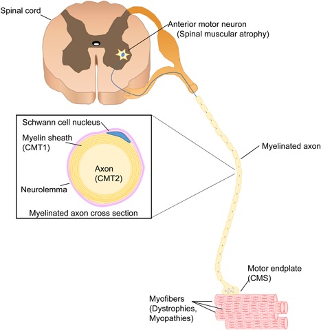
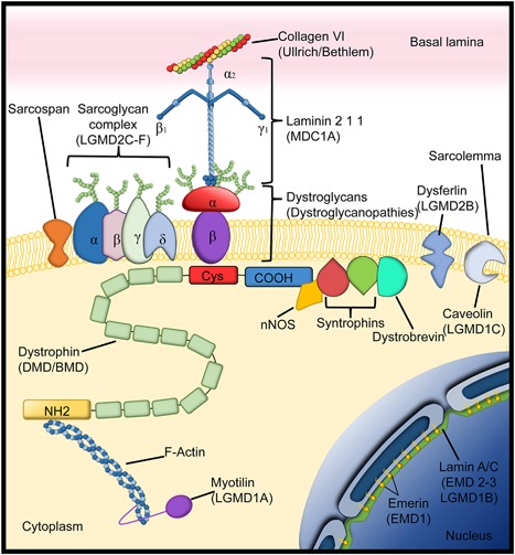
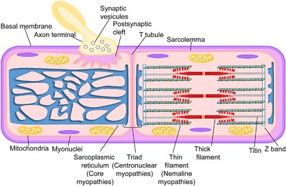
Pediatric neuromuscular diseases encompass all disorders with onset in childhood and where the primary area of pathology is in the peripheral nervous system. These conditions are largely genetic in etiology, and only those with a genetic underpinning will be presented in this review. This includes disorders of the anterior horn cell (e.g., spinal muscular atrophy), peripheral nerve (e.g., Charcot-Marie-Tooth disease), the neuromuscular junction (e.g., congenital myasthenic syndrome), and the muscle (myopathies and muscular dystrophies). Historically, pediatric neuromuscular disorders have uniformly been considered to be without treatment possibilities and to have dire prognoses. This perception has gradually changed, starting in part with the discovery and widespread application of corticosteroids for Duchenne muscular dystrophy. At present, several exciting therapeutic avenues are under investigation for a range of conditions, offering the potential for significant improvements in patient morbidities and mortality and, in some cases, curative intervention. In this review, we will present the current state of treatment for the most common pediatric neuromuscular conditions, and detail the treatment strategies with the greatest potential for helping with these devastating diseases.

Keywords: Charcot-Marie-Tooth disease; congenital myopathies; muscular dystrophies; neuromuscular disorders.


Figures

Figure 1
Figure 1
The motor unit. The motor unit is composed by the anterior horn cell (motor neuron) and the skeletal muscle fibers that are innervated by it. All myofibers in one motor unit are of the same type (I, IIA, IIB). The main genetic paediatric disorders of each part of the motor unit are within brackets (CMS, congenital myasthenicsyndromes; CMT, Charcot–Marie–Tooth type 1 or type 2).
Figure 2
Figure 2
The neuromuscular junction. Components of the neuromuscular junction. In the presynaptic terminal, acetylcholine is synthesized by the enzyme choline acetyltransferase (ChAT) from the compounds choline and acetyl‐CoA (1). When an action potential arrives at the endplate it activates voltage gated Ca2+ channels allowing Ca2+ ions flow into the axon terminal (2) and the release of the acetylcholine into the synaptic cleft (3). Acetylcholine binds to the alpha subunit of the acetylcholine receptor (AchR) to create a Na+ current into the myofiber (4), which then generates an action potential through the activation of the voltage‐gate Na+ channels (5), These leads to activation of the dihydropyridine receptor (DHPR, another Voltage gate Ca2+ channel) and then activation of the ryanodine type 1 receptor (RyR1) that releases Ca2+ from the sarcoplasmic reticulum (SR) into the cytoplasm (6). The acetylcholine will be broken down by the enzyme acetylcholinesterase (7) and choline is then transported into the axon terminal by a high affinity transporter (8). On the postsynaptic membrane AchRs are clustered by a complex of proteins (Rapsyn; docking protein 7, (Dok7); Muscle‐specific kinase (MuSK); Agrin; LDL receptor related protein (Lrp4)). Nerve derived Agrin binds to an LRP4 MuSK complex and induces the Rapsyn mediated clustering of AChR. Twelve catalytic subunits of AchE are attached to Collagen Q (ColQ) to the postsynaptic membrane via binding to MuSK. The congenial myasthenic syndromes (CMS) can be classified by the localization of the protein affected in the neuromuscular junction (presynaptic, synaptic, postsynaptic). The main drugs use as treatment for the CMS are within bracket under the protein/channel where they are acting. Pyridostigmine inhibits the AchE. Fluoxetine and quinidine blocks the AchR. 3–4 diaminopyridine (3–4 DAP) acts in the Voltage gate K+ channel by blocking the repolarization of the terminal axon. It is not know the exact mechanism of action of salbutamol, but it is thought that produce activation of second messenger signaling that partially compensates the instability of the Agrin‐MuSK‐LRP4‐Rapsyn‐DOK7 (Beeson, 2016; Ravenscroft, Laing, & Bönnemann, 2015).
Figure 3
Figure 3
Dystrophin associated glycoprotein complex. Dystrophin associated glycoprotein complex and related proteins that help the anchoring of the sarcolemma to the basal lamina. Within brackets under the different proteins are the different diseases that result from deficiency of the respective proteins. (Limb girdle muscle dystrophies (LGDMD); Duchenne muscular dystrophy DMD; Becker muscular dystrophy (BMD); Congenital muscular dystrophy type 1A (MDC1A); Emery–Dreifuss muscular dystrophy (EMD)) (Adapted from Diseases of Muscle and the Neuromuscular Junction Part 1).
Figure 4
Figure 4
The myofiber. Muscle fiber and it different components. Within brackets are the congenital myopathies associated with defects in the different muscle substructures. (Adapted from Ravenscroft et al., 2015).

References

    1. Abicht A., Muller J. S., Lochmuller H. (1993). Congenital Myasthenic syndromes. In R. A. Pagon, M. P. Adam, H. H. Ardinger, S. E.Wallace, A. Amemiya, L. J. H. Bean, T. D. Bird, C. T. Fong, H. C. Mefford, R. J. H. Smith, & K. Stephens (Eds). Seattle WA: GeneReviews(R).
    1. Accornero, F. , Kanisicak, O. , Tjondrokoesoemo, A. , Attia, A. C. , McNally, E. M. , & Molkentin, J. D. (2014). Myofiber‐specific inhibition of TGFbeta signaling protects skeletal muscle from injury and dystrophic disease in mice. Human Molecular Genetics, 23(25), 6903–6915. - PMC - PubMed
    1. Allen, D. G. , Whitehead, N. P. , & Froehner, S. C. (2016). Absence of dystrophin disrupts skeletal muscle signaling: Roles of Ca2+, reactive oxygen species, and nitric oxide in the development of muscular dystrophy. Physiological Reviews, 96(1), 253–305. - PMC - PubMed
    1. Allen, H. D. , Flanigan, K. M. , Thrush, P. T. , Dvorchik, I. , Yin, H. , Canter, C. , … Mendell, J. R. (2013). A randomized, double‐blind trial of lisinopril and losartan for the treatment of cardiomyopathy in duchenne muscular dystrophy. PLoS Currents, 5, pii: ecurrents.md.2cc69a1dae4be7dfe2bcb420024ea865 https://doi.org/10.1371/currents.md.2cc69a1dae4be7dfe2bcb420024ea865 - DOI - PMC - PubMed
    1. Amthor, H. , & Hoogaars, W. M. (2012). Interference with myostatin/ActRIIB signaling as a therapeutic strategy for Duchenne muscular dystrophy. Current Gene Therapy, 12(3), 245–259. - PubMed