. 2017 Sep 21;171(1):217-228.e13.
doi: 10.1016/j.cell.2017.08.006. Epub 2017 Sep 7.

Sensory Neurons Co-opt Classical Immune Signaling Pathways to Mediate Chronic Itch

Affiliations

Sensory Neurons Co-opt Classical Immune Signaling Pathways to Mediate Chronic Itch

Landon K Oetjen et al. Cell. .

Abstract

Mammals have evolved neurophysiologic reflexes, such as coughing and scratching, to expel invading pathogens and noxious environmental stimuli. It is well established that these responses are also associated with chronic inflammatory diseases, including asthma and atopic dermatitis. However, the mechanisms by which inflammatory pathways promote sensations such as itch remain poorly understood. Here, we show that type 2 cytokines directly activate sensory neurons in both mice and humans. Further, we demonstrate that chronic itch is dependent on neuronal IL-4Rα and JAK1 signaling. We also observe that patients with recalcitrant chronic itch that failed other immunosuppressive therapies markedly improve when treated with JAK inhibitors. Thus, signaling mechanisms previously ascribed to the immune system may represent novel therapeutic targets within the nervous system. Collectively, this study reveals an evolutionarily conserved paradigm in which the sensory nervous system employs classical immune signaling pathways to influence mammalian behavior.

Keywords: IL-13; IL-4; IL-4Rα; JAK1; atopic dermatitis; itch; pruriceptor; pruritus; type 2 cytokines.


Conflict of interest statement

The authors have no conflicts of interest to declare.

Figures
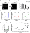
Figure 1
Figure 1. Type 2 cytokines activate mouse and human sensory neurons
(A) Representative gel of RT-PCR product of whole mouse dorsal root ganglia (DRG), n = 4 wild type (WT) mice. (B) Representative gel of RT-PCR product of whole human DRG, N = 3 donors. (C) Comparison of Il4ra, Il13ra1, and Il31ra expression by RT-qPCR in whole mouse DRG and trigeminal ganglia (TG), n = 4 WT mice. (D–F) Representative calcium imaging trace of mouse DRG neurons in response to (D) recombinant murine (rm)IL-4 (300 nM), (E) rmIL-13 (300 nM), and (F) rmIL-31 (300 nM) and potassium chloride (KCl, 100 mM). (G) rmIL-4-, rmIL-13-, rmIL-31-, and histamine-responsive DRG neurons as a percentage of total KCl-responsive neurons, n > 500 neurons from 2 WT mice. (H) Representative calcium imaging trace of human DRG neurons in response to recombinant human IL-4 (300 nM), capsaicin (500 nM), and KCl (50 mM), n > 40 neurons from a male donor. Data are represented as mean ± SEM. Black bars in calcium imaging traces indicate timing of challenges. See also Figure S1.
Figure 2
Figure 2. Type 2 cytokines activate itch-sensory pathways
(A) Expression of selected genes in mouse DRG neuron populations clustered into functional subsets based on single-cell RNA-seq data. Numbers indicate fraction of positive cells within individual clusters by thresholding method. Full data set and methods available in Usoskin et al., 2015. (B) Representative size distribution of rmIL-4-, rmIL-13-, and histamine-responsive mouse DRG neurons classified as small-, medium-, and large-diameter neurons (<18 μm, 18–25 μm, and >25 μm), n > 500 neurons from a WT mouse. (C–D) Representative Venn diagrams of overlapping responses of mouse DRG neurons to stimulation with rmIL-4 with subsequent challenge with (C) rmIL-31 (300 nM) and histamine (50 μM) or with (D) rmIL-13 (4.0 μg/mL) and capsaicin (300 nM), n > 300 neurons from a WT mouse. (E–F) Responses to (E) rmIL-4 (4 μg/mL) and (F) rmIL-13 (4 μg/mL) of mouse DRG neurons from Trpa1−/− mice, Trpv1−/− mice, and in calcium-free conditions as a percentage of total KCl-responsive neurons compared to WT control mice, n > 300 neurons per group. (G) Total scratching bouts elicited in response to intradermal (i.d.) cheek injection of vehicle control (0.1% BSA in PBS, 10 μL), rmIL-4, rmIL-13, or rmIL-31 (all 2.5 μg/10 μL), n ≥ 5 mice per group. (H) Representative calcium imaging trace of mouse DRG neurons responding to low levels of histamine (5 μM) after vehicle control or rmIL-4 (4 μg/mL) challenge, n > 200 neurons from 2 WT mice. (I) Cheek-directed scratching bouts elicited in response to i.d. cheek injection of a low dose of histamine (4 mM, 10 μL) with or without rmIL-4 (2.5 μg), n ≥ 7 mice per group. Data are represented as mean ± SEM. Black bars in calcium imaging traces indicate timing of challenges. See also Figure S2.
Figure 3
Figure 3. Neuronal IL-4Rα expression is necessary for chronic itch
(A) Quantification of Il4ra, Il2rg, and Il13ra1 expression by RT-qPCR in whole sensory trigeminal ganglia (TG) of IL-4RαΔneuron mice, n ≥ 4 mice per group. (B–E) Representative responses of DRG neurons from an IL-4RαΔneuron mouse to (B) rmIL-4 (1 μg/mL), (C) rmIL-13 (4 μg/mL), (D) rmIL-31 (3 μM), and (E) histamine (50 μM) as a percentage of total KCl-responsive neurons compared to neurons from a littermate control mouse, n > 300 neurons per group. (F) Experimental schematic indicating daily topical treatment with MC903 to the ears of IL-4RαΔneuron and littermate control mice. (G) Scratching behavior of IL-4RαΔneuron mice compared to littermate control mice over the course of MC903 treatment, n ≥ 7 mice per group. (H) Ear thickness measurements, (I) representative H&E histopathology, and (J) histology score of MC903-treated IL-4RαΔneuron mice compared to littermate control mice, n ≥ 4 mice per group. Scale bars indicate 100 μm. Data are represented as mean ± SEM. See also Figures S3–S5.
Figure 4
Figure 4. Disruption of neuronal JAK1 reduces chronic itch
(A) Representative responses to rmIL-4 (1 μg/mL) of DRG neurons from a JAK1Δneuron mouse compared to a littermate control mouse as a percentage of total KCl-responsive neurons, n ≥ 200 neurons per group. (B) Experimental schematic indicating MC903 treatment of JAK1Δneuron and littermate control mice. (C) Scratching behavior, (D) ear thickness measurement, (E) representative H&E histopathology, and (F) histology score of JAK1Δneuron mice compared to littermate control mice on Day 7, n ≥ 8 mice per group. (G) Representative responses to rmIL-4 (1 μg/mL) of DRG neurons from a WT mouse after incubation with ruxolitinib (Rux, 1 μg/mL) compared to vehicle control as a percentage of total KCl-responsive neurons, n ≥ 200 neurons per group. (H) Experimental schematic indicating twice daily intraperitoneal (i.p.) injection of vehicle control or Rux (100 μg) during MC903 treatment to the ears of WT mice. (I) Scratching behavior, (J) ear thickness measurement, (K) representative H&E histopathology, and (L) histology score of vehicle control and Rux-treated mice on Day 7, n ≥ 10 mice per group. Scale bars indicate 100 μm. Data are represented as mean ± SEM. See also Figure S6.
Figure 5
Figure 5. CIP is a distinct chronic itch disorder that exhibits severe itch despite minimal skin inflammation
(A–B) Representative clinical pictures of (A) atopic dermatitis (AD) and (B) chronic idiopathic pruritus (CIP). (C–D) Representative H&E histopathology of pruritic sites from (C) AD and (D) CIP. (E) Histology score of AD and CIP patient biopsies, N ≥ 4 biopsies per group. (F) Numerical Rating Scale (NRS) itch scores of AD and CIP patients, N ≥ 22 patients per group. (G) Clustering of AD, CIP, and control skin samples by row Z-scores of the regularized logarithm of gene expression values of the top 100 differentially expressed genes from RNA-seq of AD versus control skin, N = 4 donors per condition. Scale bars indicate 100 μm. Data are represented as mean ± SEM. See also Tables S1–S3.
Figure 6
Figure 6. Patients with refractory CIP improve when treated with JAK inhibition
(A) NRS itch scores for a cohort of CIP patients (N = 5) given the JAK inhibitor tofacitinib. Individual patients are shown by unique colors. Significance was calculated using a paired t-test. (B) Daily NRS itch scores of three CIP patients treated with tofacitinib including one patient treated with cyclosporine immediately preceding tofacitinib treatment (black). See also Table S4.

Comment in

References

    1. Agarwal N, Offermanns S, Kuner R. Conditional gene deletion in primary nociceptive neurons of trigeminal ganglia and dorsal root ganglia. Genesis. 2004;38:122–129. - PubMed
    1. Andersen HH, Elberling J, Sølvsten H, Yosipovitch G, Arendt-Nielsen L. Non-histaminergic and mechanical itch sensitization in atopic dermatitis. Pain. 2017 doi: 10.1097/j.pain.0000000000000980. - DOI - PubMed
    1. Artis D, Spits H. The biology of innate lymphoid cells. Nature. 2015;517:293–301. - PubMed
    1. Bautista DM, Wilson SR, Hoon MA. Why we scratch an itch: the molecules, cells and circuits of itch. Nat Neurosci. 2014;17:175–182. - PMC - PubMed
    1. Beck LA, Thaçi D, Hamilton JD, Graham NM, Bieber T, Rocklin R, Ming JE, Ren H, Kao R, Simpson E, et al. Dupilumab treatment in adults with moderate-to-severe atopic dermatitis. N Engl J Med. 2014;371:130–139. - PubMed