Review
. 2017 Dec;27(12):895-905.
doi: 10.1016/j.tcb.2017.08.002. Epub 2017 Sep 7.

Rethinking HSF1 in Stress, Development, and Organismal Health

Affiliations
Review

Rethinking HSF1 in Stress, Development, and Organismal Health

Jian Li et al. Trends Cell Biol. 2017 Dec.

Abstract

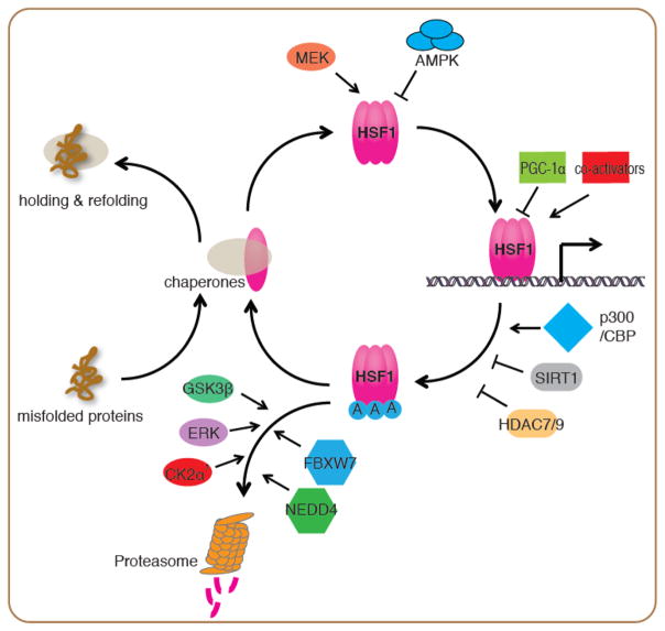
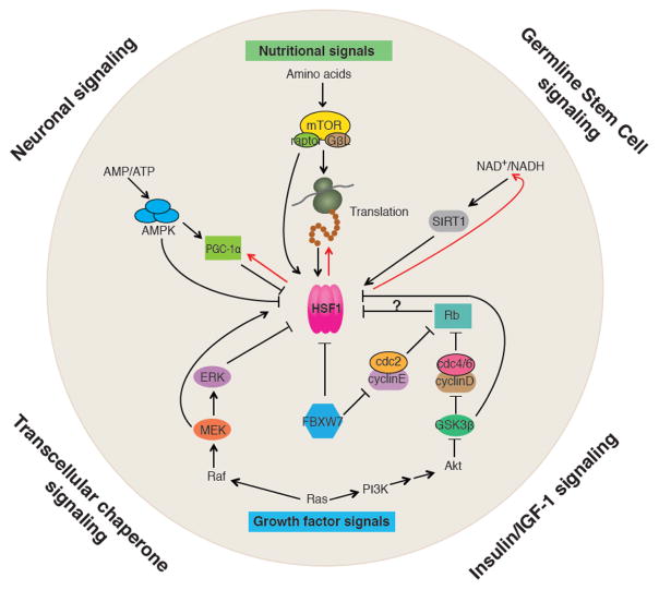
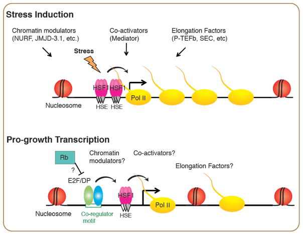
The heat shock response (HSR) was originally discovered as a transcriptional response to elevated temperature shock and led to the identification of heat shock proteins and heat shock factor 1 (HSF1). Since then HSF1 has been shown to be important for combating other forms of environmental perturbations as well as genetic variations that cause proteotoxic stress. The HSR has long been thought to be an absolute response to conditions of cell stress and the primary mechanism by which HSF1 promotes organismal health by preventing protein aggregation and subsequent proteome imbalance. Accumulating evidence now shows that HSF1, the central player in the HSR, is regulated according to specific cellular requirements through cell-autonomous and non-autonomous signals, and directs transcriptional programs distinct from the HSR during development and in carcinogenesis. We discuss here these 'non-canonical' roles of HSF1, its regulation in diverse conditions of development, reproduction, metabolism, and aging, and posit that HSF1 serves to integrate diverse biological and pathological responses.

Keywords: HSF1; cell proliferation; heat shock response (HSR); metabolism; organismal health; proteostasis.


Figures

Figure 1
Figure 1
Regulation of the HSF1 activation and attenuation cycle in the HSR Upon stress, misfolded proteins dissociate chaperones from HSF1 and allow HSF1 to form DNA-binding competent trimers. MEK promotes HSF1 nuclear translocation and transcriptional activity through phosphorylation at Ser326. Conversely, AMPK inhibits HSF1 nuclear translocation through phosphorylation at Ser121. HSF1 transcriptional activity is regulated by co-activators such as the mediator complex, and repressors including PGC-1α at its target promoters. Attenuation of the HSR is controlled by acetylation of HSF1 at its DNA binding domain by p300/CBP. The histone deacetylases SIRT1, HDAC7 and HDAC9 prevent this acetylation and stabilize HSF1 DNA binding. The E3 ligases FBXW7 and NEDD4 target HSF1 for degradation through the ubiquitin proteasome system, with FBXW7 mediated degradation being promoted by phosphorylation of HSF1 by GSK3β, ERK and CK2 α′ kinases.
Figure 2
Figure 2
Intercellular and intracellular signaling pathways converging on HSF1 At the organismal level, HSF1 is subject to cell non-autonomous regulation by neuronal signaling (serotonin), insulin/IGF-1 signaling, germline stem cell (GSC) signaling and transcellular chaperone signaling. Within the cell, HSF1’s activity is regulated by signal pathways that sense nutrients and control cell proliferation. The three major metabolic sensors, AMPK, mTORC1 and SIRT1 control HSF1’s activity directly through post-translational modifications. AMPK also inhibits HSF1 indirectly through PGC-1α. mTORC1 activates translation, and the nascent polypeptides and newly synthesized proteins titrate chaperones from HSF1, leading to de-repression of HSF1. HSF1 also influence metabolism by activating expression of PGC-1α, promoting protein synthesis through co-translational folding, and maintaining cellular levels of NAD+ via the NAD+ salvage pathway (indicated by the red arrows). HSF1 is also regulated by key components of the RAS/MAPK and RAS/PI3K pathways including MEK, ERK and GSK3β through phosphorylation. The pocket protein Rb, an important cell cycle regulator, may also influence HSF1 activity considering it is a repressor of E2F/DP that serves as a co-activator of HSF1 in C. elegans larval development.
Figure 3
Figure 3
Models of HSF1 in stress-induced and pro-growth transcription Binding of HSF1 induced by stress is through cooperative binding of HSF1 at clusters of canonical HSEs, which accessibility is controlled by chromatin modulators such as the chromatin remodeler NURF [65] and the histone demethylase JMJD-3.1. HSF1 cooperates with Mediator, and transcription elongation factors such as the P-TEFb kinase and the Super Elongation Complex to robustly induce the HSR. Binding of HSF1 in the pro-growth transcriptional program, however, relies on co-regulators such as the active E2F/DP heterodimer that binds to the same promoters with HSF1 in C. elegans larval development. How the co-activators, chromatin modulators and elongation factors in the HSR contribute to the pro-growth transcriptional program of HSF1 are of interests for future studies.

References

    1. Labbadia J, Morimoto RI. The biology of proteostasis in aging and disease. Annu Rev Biochem. 2015;84:435–64. - PMC - PubMed
    1. Akerfelt M, et al. Heat shock factors: integrators of cell stress, development and lifespan. Nat Rev Mol Cell Biol. 2010;11(8):545–55. - PMC - PubMed
    1. Ben-Zvi A, et al. Collapse of proteostasis represents an early molecular event in Caenorhabditis elegans aging. Proc Natl Acad Sci U S A. 2009;106(35):14914–9. - PMC - PubMed
    1. Cohen E, et al. Opposing activities protect against age-onset proteotoxicity. Science. 2006;313(5793):1604–10. - PubMed
    1. Brandman O, et al. A ribosome-bound quality control complex triggers degradation of nascent peptides and signals translation stress. Cell. 2012;151(5):1042–54. - PMC - PubMed

Publication types

LinkOut - more resources