. 2017 Sep 21;67(6):1068-1079.e4.
doi: 10.1016/j.molcel.2017.08.008. Epub 2017 Sep 7.

CRISPR-Mediated Base Editing Enables Efficient Disruption of Eukaryotic Genes through Induction of STOP Codons

Affiliations

CRISPR-Mediated Base Editing Enables Efficient Disruption of Eukaryotic Genes through Induction of STOP Codons

Pierre Billon et al. Mol Cell. .

Abstract

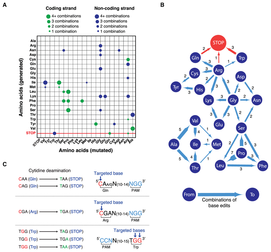
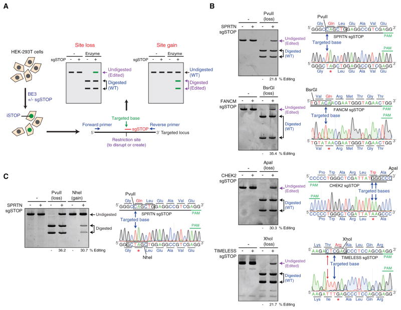
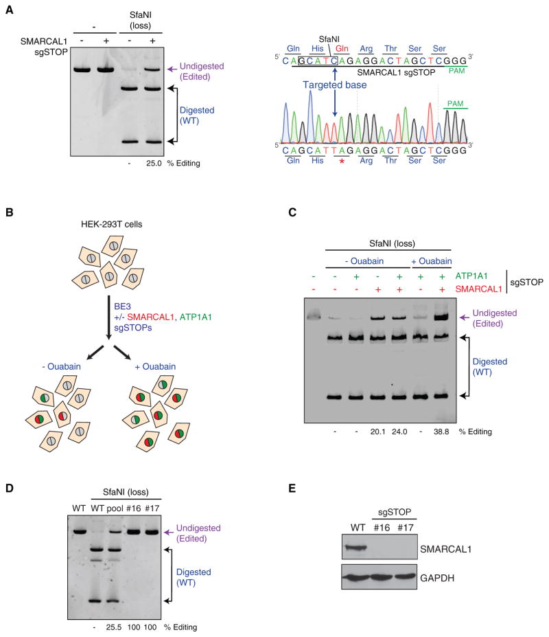
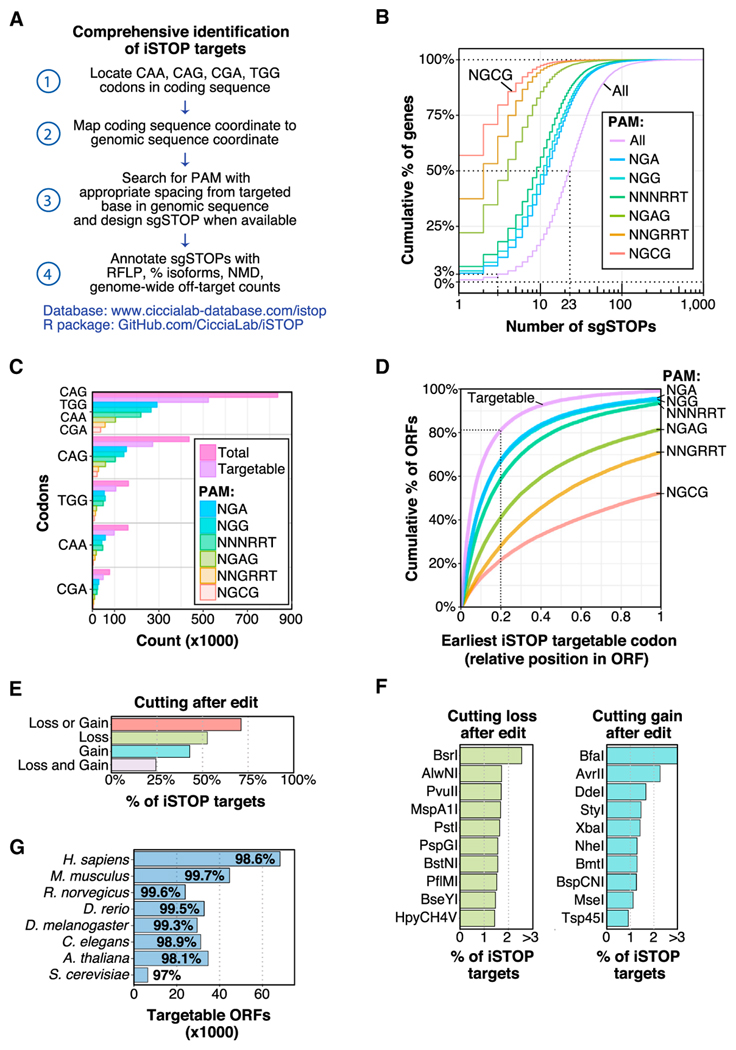
Standard CRISPR-mediated gene disruption strategies rely on Cas9-induced DNA double-strand breaks (DSBs). Here, we show that CRISPR-dependent base editing efficiently inactivates genes by precisely converting four codons (CAA, CAG, CGA, and TGG) into STOP codons without DSB formation. To facilitate gene inactivation by induction of STOP codons (iSTOP), we provide access to a database of over 3.4 million single guide RNAs (sgRNAs) for iSTOP (sgSTOPs) targeting 97%-99% of genes in eight eukaryotic species, and we describe a restriction fragment length polymorphism (RFLP) assay that allows the rapid detection of iSTOP-mediated editing in cell populations and clones. To simplify the selection of sgSTOPs, our resource includes annotations for off-target propensity, percentage of isoforms targeted, prediction of nonsense-mediated decay, and restriction enzymes for RFLP analysis. Additionally, our database includes sgSTOPs that could be employed to precisely model over 32,000 cancer-associated nonsense mutations. Altogether, this work provides a comprehensive resource for DSB-free gene disruption by iSTOP.

Keywords: CRISPR-mediated base editing; RFLP assay; STOP codons; cancer; genome-wide sgRNA analysis; iSTOP; nonsense mutations.


Figures

Figure 1
Figure 1. Generation of STOP Codons Using CRISPR-Mediated Base Editing
(A) Representation of the repertoire of mutations generated by cytidine deaminase-dependent CRISPR base editors. Mutated amino acids (x axis) and generated amino acids (y axis) either from coding (green) or non-coding (blue) strands are represented. The size of the circle indicates the number of combinations that generate each modification. Ala, alanine; Arg, arginine; Asn, asparagine; Asp, aspartic acid; Cys, cysteine; Gln, glutamine; Glu, glutamic acid; Gly, glycine; His, histidine; Ile, isoleucine; Met, methionine; Leu, leucine; Lys, lysine; Phe, phenylalanine; Pro, proline; Ser, serine; Thr, threonine; Trp, tryptophan; Tyr, tyrosine; Val, valine; STOP, STOP codon. See also Figures S1C and S1D and Table S1. (B) Representation of amino acid substitutions generated by cytidine deaminase-dependent CRISPR base editors. Dark blue circles indicate amino acids, blue lines show the direction of amino acid substitutions induced by CRISPR-dependent base editing and the number of possible combinations to obtain the indicated substitutions. See also Figures S1C and S1D and Table S1. (C) Representation of the cytidine deamination reactions induced by CRISPR-dependent base editors to generate STOP codons. The CRISPR base editor BE3 converts CAA, CAG, CGA, and TGG codons into STOP codons when the targeted base(s) (red) is at the correct distance (13–17 bps) from a protospacer adjacent motif (PAM, blue). See also Figures S1A and S1B and Table S1.
Figure 2
Figure 2. Restriction Fragment Length Polymorphism Assay to Detect iSTOP-Edited Cells
(A) Schematic representation of the protocol utilized to disrupt genes by iSTOP. HEK293T cells are transfected with BE3 with or without sgSTOP, thus resulting in the generation of cells edited by iSTOP (green). The targeted locus is then amplified by PCR and digested with a restriction enzyme that recognizes a restriction site containing the base targeted by iSTOP or a restriction site generated by iSTOP-mediated base editing. Base editing by iSTOP results in PCR products refractory to restriction digestion (Site loss) or induces the formation of new restriction sites (Site gain). (B) Conversion of CAG, CAA, TGG, and CGA codons into STOP codons by iSTOP in human cells. PCR products amplified from four different genomic loci (SPRTN, FANCM, CHEK2, and TIMELESS) edited by iSTOP were subjected to restriction digest with enzymes (PvuII, BsrGI, ApaI, and XhoI) that recognize sites containing the targeted bases. Products of the restriction digest reactions were run on polyacrylamide gels (left) and editing efficiency was determined by the percentage of undigested PCR amplicons (purple). Sequencing profiles of undigested PCR products (right) are compared to the wild-type genomic sequence, including the targeted base (blue arrow) and PAM (green). Editing of a cytosine that generates a missense mutation in the TIMELESS locus is indicated by red arrows. The sequencing profiles are representative of four to eight sequences for each targeted locus. Each base is colored according to the sequencing peaks (A in green, G in black, T in red, and C in blue). PAM, protospacer adjacent motif. See also Figures S2A and S2B. (C) Detection of iSTOP-induced events by both loss and gain of restriction sites. The SPRTN locus was PCR-amplified and left undigested or digested with either PvuII or NheI (left). Sequencing profiles of PCR amplicons from sgSTOP-treated cells, including the targeted base (blue arrow) and PAM (green), are shown on the right. Editing efficiency was determined by the percentage of PCR amplicons refractory to PvuII digestion, as in (B), or by the percentage of NheI-digested PCR amplicons.
Figure 3
Figure 3. Generation of Knockout Human Cell Lines Using iSTOP
(A) Editing of the SMARCAL1 locus by iSTOP. The SMARCAL1 locus was PCR-amplified after transfection of BE3 with or without SMARCAL1 sgSTOP. The SMARCAL1 amplicon was then digested with SfaNI, which recognizes a restriction site containing the base targeted by iSTOP and the products of the restriction digest reaction were run on a polyacrylamide gel (left). Editing efficiency at the SMARCAL1 locus was estimated by the percentage of SMARCAL1 PCR amplicons refractory to SfaNI digestion (purple), as indicated in Figure 2B. One sequencing profile representative of four sequences of undigested SMARCAL1 PCR products, including the targeted base, is indicated on the right inside. (B) Schematic representation of the ATP1A1 co-selection strategy. HEK293T cells were transfected with BE3 with or without sgSTOPs targeting SMARCAL1 and/or ATP1A1. Cell populations were subsequently left untreated or treated with 1 μM ouabain, resulting in the enrichment of cells that had undergone genome editing at the ATP1A1 and SMARCAL1 loci. (C) Polyacrylamide gel showing the SfaNI digestion products of SMARCAL1 PCR amplicons from HEK293T transfected with BE3 with or without sgSTOPs targeting SMARCAL1 and/or ATP1A1 and subjected to ouabain treatment, as represented in (B). Percentage of editing at the SMARCAL1 locus with or without ouabain treatment was assessed as indicated in (A). (D) RFLP analysis of HEK293T clones edited by iSTOP in the SMARCAL1 locus. Clones (#16 and #17) were isolated from cells transfected with BE3 and sgSTOPs targeting SMARCAL1 and ATP1A1 and subjected to ouabain selection, as described in (B). Amplicons of the SMARCAL1 locus were digested by SfaNI and analyzed on polyacrylamide gel, as in (A). Restriction digest products of SMARCAL1 amplicons from wild-type (WT) cells, iSTOP-edited cellular pool and clones #16 and #17 are indicated. See also Figures S3E–S3G. (E) Western blot analysis of SMARCAL1 protein levels in whole cellular extracts obtained from WT cells and clones #16 and #17, as shown in (D). GAPDH levels are used as loading controls.
Figure 4
Figure 4. Comprehensive Detection of iSTOP Targets in Eukaryotic Genomes
(A) Workflow utilized to identify iSTOP targetable sites in all ORFs with CDS coordinates available from the UCSC genome browser (https://genome.ucsc.edu/). Targetable sites were identified by first locating all CAA, CAG, CGA, and TGG codons in each coding sequences, then mapping the coordinates of the targeted base(s) in each codon to a genomic coordinate (steps 1–2). In total, 150 bases of genomic sequence flanking the targeted site were used to search for an appropriately spaced PAM (13–17 nucleotides from a targeted base) for all validated BE3 variants and for unique cutting of restriction enzymes (steps 3–4). Targeted isoforms and NMD predictions were determined as described in the STAR Methods. (B) Cumulative distribution of the number of sgSTOPs designed per gene. The number of sgSTOPs for an average ORF (50%) in the human genome is indicated by a dotted line. Distributions for distinct PAM specificities (NGA, NGG, NNNRRT, NGAG, NNGRRT, and NGCG) are also shown. (C) Number of CAG, TGG, CAA, and CGA codons in the GRCh38 human reference genome targetable by iSTOP using BE3 variants with distinct PAM specificities (NGA, NGG, NNNRRT, NGAG, NNGRRT, and NGCG). (D) Relative position of the earliest iSTOP codon targetable in human ORFs (cumulative percentage) by BE3 variants with distinct PAM specificities (NGA, NGG, NNNRRT, NGAG, NNGRRT, and NGCG). The purple line takes into account all iSTOP targetable codons. (E) Percentage of human iSTOP sites verifiable by RFLP analysis using restriction enzymes that cut only once within a genomic region of ±50 bps flanking the targeted site. Bars indicate percentage of sites that can be verified by restriction enzyme cutting loss and/or gain. (F) Top ten restriction enzymes that can be utilized to validate iSTOP targets in the human genome by loss or gain of cutting within a genomic region of ±150 bps flanking the targeted site. The complete enzyme list is available in Table S3. (G) Number and percentage of iSTOP targetable ORFs in eight different eukaryotic species. See also Figures S4C–S4E.
Figure 5
Figure 5. Modeling of Cancer-Associated Nonsense Mutations by iSTOP
(A) Percentage of unique nonsense coordinates in cancer types, as observed in COSMIC. The percentage of base substitutions in CAA, CAG, CGA, or TGG codons that result in nonsense mutations in each cancer type is indicated. The total number of iSTOP targetable sites in each cancer type is annotated in white text. (B) Genes with frequently observed nonsense mutations at CAA, CAG, CGA, and TGG codons (iSTOPers) and their prevalence in different cancer types. The size and opacity of each circle represents the percentage of possible CAA, CAG, CGA, and TGG codons in the gene that were observed mutated to nonsense in each cancer type. See also Figures S5A and S5B. (C) Percentage of CAA, CAG, CGA, and TGG codons observed mutated to nonsense in the genes shown in (B) across all cancers. The total number of iSTOP targetable sites in each gene is annotated in white text. See also Figures S5A and S5B. (D) Maps of three iSTOPers (ATM, SETD2, and EZH2) indicating locations of (1) nonsense base substitutions in cancer (red tick marks), (2) CAA, CAG, CGA, and TGG codons (black tick marks), (3) iSTOP targetable codons (blue tick marks), and (4) iSTOP targetable codons that are verifiable via RFLP (green tick marks). The largest isoform for each gene is shown with exon numbers indicated below the gene. The size of the maps is not proportional to the length of the ORFs.

References

    1. Agudelo D, Duringer A, Bozoyan L, Huard CC, Carter S, Loehr J, Synodinou D, Drouin M, Salsman J, Dellaire G, et al. Markerfree coselection for CRISPR-driven genome editing in human cells. Nat Methods. 2017;14:615–620. - PubMed
    1. Aguirre AJ, Meyers RM, Weir BA, Vazquez F, Zhang CZ, Ben-David U, Cook A, Ha G, Harrington WF, Doshi MB, et al. Genomic copy number dictates a gene-independent cell response to CRISPR/Cas9 targeting. Cancer Discov. 2016;6:914–929. - PMC - PubMed
    1. Andrews SJ, Rothnagel JA. Emerging evidence for functional peptides encoded by short open reading frames. Nat Rev Genet. 2014;15:193–204. - PubMed
    1. Bansbach CE, Bétous R, Lovejoy CA, Glick GG, Cortez D. The annealing helicase SMARCAL1 maintains genome integrity at stalled replication forks. Genes Dev. 2009;23:2405–2414. - PMC - PubMed
    1. Barrangou R, Doudna JA. Applications of CRISPR technologies in research and beyond. Nat Biotechnol. 2016;34:933–941. - PubMed

MeSH terms