

Biofilm formation and determination of minimum biofilm eradication concentration of antibiotics in Mycoplasma hyopneumoniae
- PMID: 28890520
- PMCID: PMC5658566
- DOI: 10.1292/jvms.17-0279
Biofilm formation and determination of minimum biofilm eradication concentration of antibiotics in Mycoplasma hyopneumoniae
Abstract
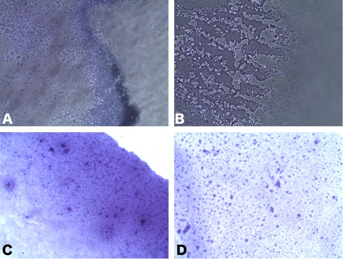
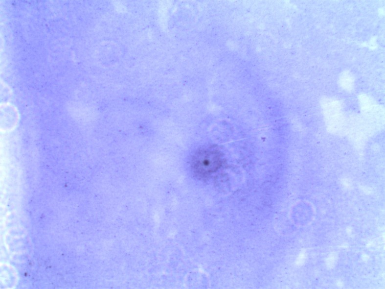
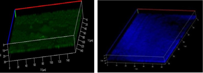
The study was aimed to investigate biofilm forming ability of Mycoplasma hyopneumoniae and to determine the minimum biofilm eradication concentrations of antibiotics. Biofilm forming ability of six strains of M. hyopneumoniae was examined using crystal violet staining on coverslips. The results demonstrated an apparent line of biofilm growth in 3 of the strains isolated from swine with confirmed cases of enzootic pneumonia. BacLight bacterial viability assay revealed that the majority of the cells were viable after 336 hr of incubation. Moreover, M. hyopneumoniae persists in the biofilm after being exposed to 10 fold higher concentration of antibiotics than the minimum inhibitory concentrations in planktonic cells. To the best of our knowledge, this is the first report of biofilm formation in M. hyopneumoniae. However, comprehensive studies on the mechanisms of biofilm formation are needed to combat swine enzootic pneumonia caused by resistant M. hyopneumoniae.
Keywords: Mycoplasma hyopneumoniae; biofilm; eradication; inhibition.
Figures
References
-
- Balish M., Krause D. C.2002. Molecular Biology and Pathogenicity of Mycoplasmas. (Herrmann, R, Razin, S. eds.), Kluwer Academic/Plenum, New York.
-
- Damte D., Suh J. W., Lee S. J., Yohannes S. B., Hossain M. A., Park S. C.2013. Putative drug and vaccine target protein identification using comparative genomic analysis of KEGG annotated metabolic pathways of Mycoplasma hyopneumoniae. Genomics 102: 47–56. doi: 10.1016/j.ygeno.2013.04.011 - DOI - PubMed
MeSH terms
Substances
LinkOut - more resources
Full Text Sources
Other Literature Sources
Medical