. 2017 Nov;95(10):933-942.
doi: 10.1038/icb.2017.74. Epub 2017 Sep 11.

A key role for IL-7R in the generation of microenvironments required for thymic dendritic cells

Affiliations

A key role for IL-7R in the generation of microenvironments required for thymic dendritic cells

Amanda J Moore et al. Immunol Cell Biol. 2017 Nov.

Abstract

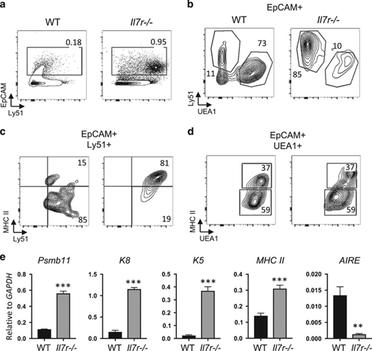
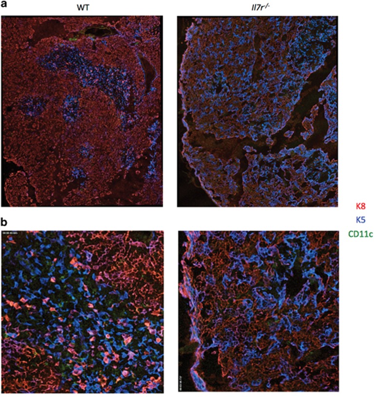
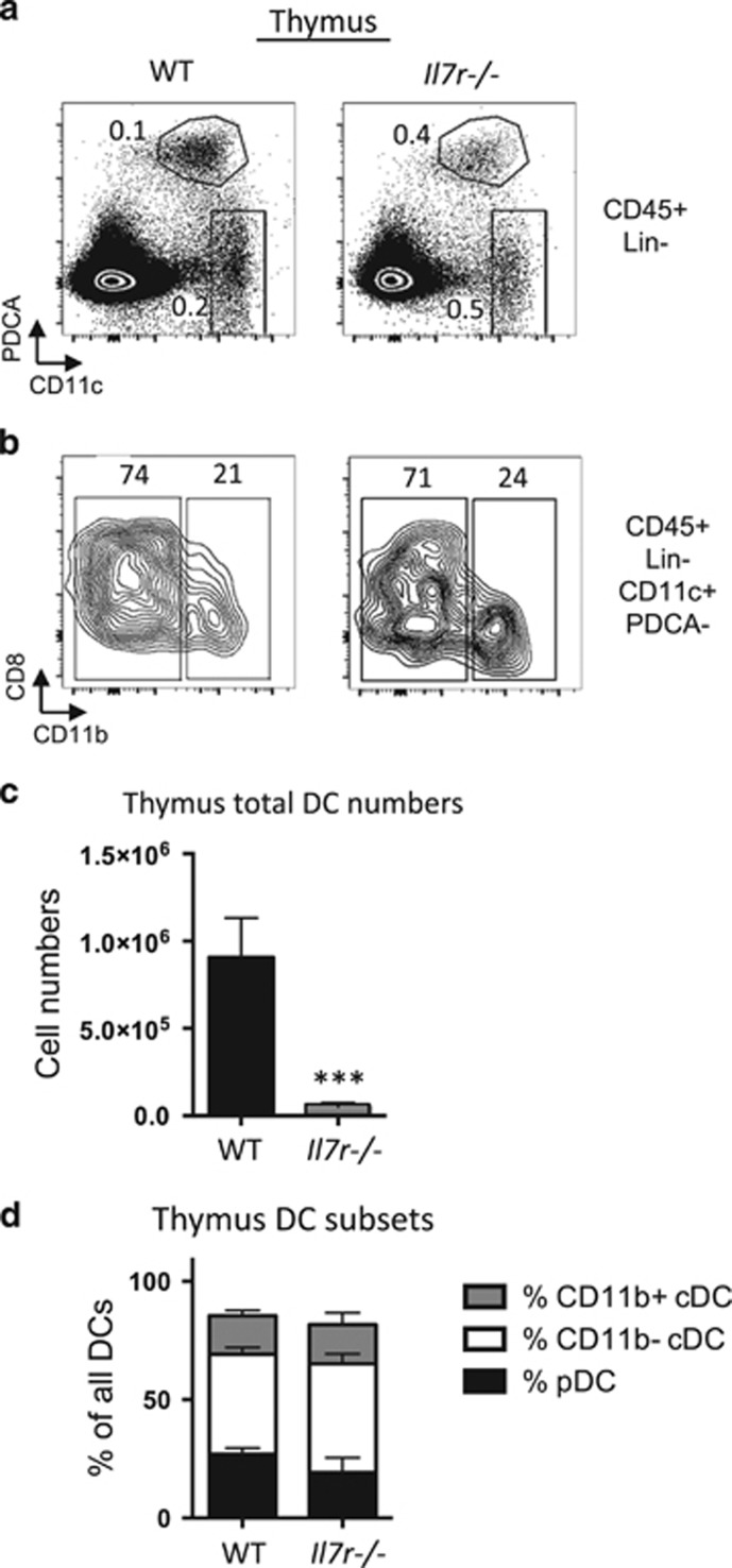
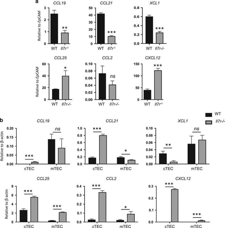
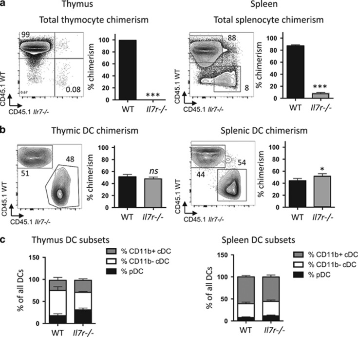
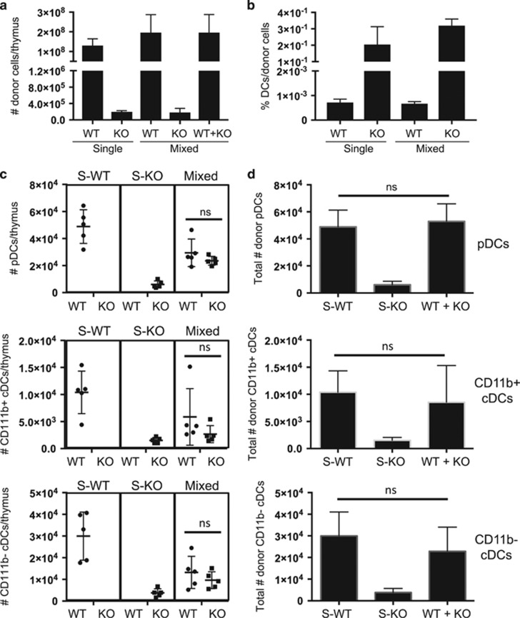
Interleukin-7 receptor (IL-7R) signaling is critical for multiple stages of T-cell development, but a role in the establishment of the mature thymic architecture needed for T-cell development and thymocyte selection has not been established. Crosstalk signals between developing thymocytes and thymic epithelial cell (TEC) precursors are critical for their differentiation into cortical TECs (cTECs) and medullary TECs (mTECs). In addition, mTEC-derived factors have been implicated in the recruitment of thymic dendritic cells (DCs) and intrathymic DC development. We therefore examined corticomedullary structure and DC populations in the thymus of Il7r-/- mice. Analysis of TEC phenotype and spatial organization revealed a striking shift in the mTEC to cTEC ratio, accompanied by disorganized corticomedullary structure. Several of the thymic subsets known to have DC potential were nearly absent, accompanied by reductions in DC cell numbers. We also examined chemokine expression in the Il7r-/- thymus, and found a significant decrease in mTEC-derived CCR7 ligand expression, and high levels of cTEC-derived chemokines, including CCL25 and CXCL12. Although splenic DCs were similarly affected, bone marrow (BM) precursors capable of giving rise to DCs were unperturbed. Finally, BM chimeras showed that there was no intrinsic need for IL-7R signaling in the development or recruitment of thymic DCs, but that the provision of wild-type progenitors enhanced reconstitution of thymic DCs from Il7r-/- progenitors. Our results are therefore supportive of a model in which Il7r-dependent cells are required to set up the microenvironments that allow accumulation of thymic DCs.


Conflict of interest statement

The authors declare no conflict of interest.

Figures

Figure 1
Figure 1
Defects in thymic epithelial cell subset ratios in the Il7r−/− thymus. (a) Thymus single-cell suspensions were made by collagenase digestion and analyzed by flow cytometry. EpCAM staining was used to gate on the TECs. (b) EpCAM-gated cells were stained with Ly51 to identify cTECs, and UEA-1 to identify mTECs. (c, d) cTECs (c) or mTECs (d) were further gated to analyze the expression of MHC II as a marker of maturity. Numbers in quadrants indicate percentages. (e) Whole WT and Il7r−/− thymuses were homogenized, RNA was extracted and first-strand cDNA was generated to be used as template for quantitative reverse transcription-PCR. The mRNA expression of the cTEC markers Psbm11 (β5t) and cytokeratin-8 (K8), the mTEC marker cytokeratin-5 (K5), MHC II and the mature mTEC marker AIRE were measured. All values shown are relative to GAPDH levels. Graphs depict means±s.e.m., n=3. Statistical significance was calculated using a t-test; ***P<0.005. Data are representative of at least two separate experiments.
Figure 2
Figure 2
The Il7r−/− thymus has disorganized corticomedullary structure and a paucity of DCs. Thymuses were dissected from WT and Il7r−/− mice and snap-frozen in optimal cutting temperature (OCT) freezing media. Ten-micrometer sections were cut and stained with anti-cytokeratin-8 (K8)/anti-rat conjugated to Cy3 (cTECs, red), anti-cytokeratin-5 (K5)/anti-rabbit conjugated to Cy5 (mTECs, blue) and anti-CD11c directly conjugated to fluoroscein isothiocyanate (DCs, green). Tiled images were gathered using an Axiovert microscope, shown at either × 10 (a) or × 20 (b).
Figure 3
Figure 3
Marked reduction of thymic DC numbers in Il7r−/− mice. WT and Il7r−/− mice were analyzed at 4.5–5.5 weeks of age. (a) Gates used to analyze pDCs and cDCs. Cells were first gated on the lineage (Lin)-negative, DAPI, CD45+ populations, where Lin=Dx5, NK1.1, F4/80, CD3ε, TCRγδ, TCRβ, CD19 and Ter119. Within this gate, two populations were analyzed: pDCs (CD11cint, PDCA-1+); and cDCs (CD11chigh, PDCA-1). (b) To further differentiate between cDC subsets, cells within the cDC gate were analyzed for expression of CD11b and CD8α. CD8α staining was consistently heterogeneous within the CD11b population. Numbers within the quadrants represent percentages. (c) Total numbers of DCs per thymus, as calculated from manual counting from single-cell suspensions multiplied by the percentages of CD45+LinCD11c+ cells as determined by FlowJo. (d) Ratios of the three DC subsets out of all DCs were calculated by taking the numbers of thymic pDCs, CD11b+ cDCs and CD11b cDCs per thymus, and dividing by the total number of DCs. n=4. ***P<0.005. Data are representative of three separate experiments.
Figure 4
Figure 4
Perturbation in chemokine expression in the Il7r−/− thymus. (a) Whole homogenized thymus was used as a template for quantitative reverse transcription-PCR (qRT-PCR) to measure the amount of chemokine mRNA produced in the adult WT versus Il7r−/− thymus. To account for the fivefold greater number of TECs in the Il7r−/− thymus versus the WT thymus, results were normalized to EpCAM mRNA levels. (b) TECs were sorted based on expression of EpCAM, and Ly51 (cTECs) or UEA-1 (mTECs). cDNA was generated from sorted TEC subsets and used as template for qRT-PCR. n=3. Values were normalized to β-actin mRNA levels. Data are representative of at least two separate experiments. Graphs depict means±s.e.m. Statistical significance was calculated using a t-test; ***P<0.005, **P<0.01, *P>0.05, ns=nonsignificant.
Figure 5
Figure 5
Contribution of Il7r−/− BM-derived cells to thymic and splenic DCs in mixed radiation chimeras. Lethally irradiated CD45.1+ × CD45.2+ F1 hybrid mice were reconstituted with a 1:1 mix of CD45.1+WT:CD45.2+ Il7r−/− BM. Tissues were analyzed 3 weeks later. (a, b) Chimerism in whole thymus (a) or in DCs (b) were calculated as the percentage contribution from Il7r−/− versus WT BM-derived cells as assessed by CD45.1 (WT) or CD45.2 (Il7r−/−) expression, in thymus (left) or spleen (right). The graph depicts the mean±s.e.m. Numbers in the quadrants indicates percentages. (c) Ratios of pDCs, CD11b cDCs and CD11b+ cDC subsets among all DCs in the thymus (left) or spleen (right), calculated by dividing the number in each subset per organ by the total number of DCs. Statistical significance was calculated using a t-test. ***P<0.005, *P<0.05, ns, nonsignificant. n=4. Data are representative of two separate experiments.
Figure 6
Figure 6
Increase of Il7r−/− donor-derived thymic DC cell numbers by the provision of WT donor cells. Lethally irradiated CD45.1+ × CD45.2+ F1 hybrid mice were reconstituted with whole BM from CD45.1+ WT mice (S-WT), CD45.2+ Il7r−/− (S-KO) mice or a 1:1 mix of each (mixed). The thymus was analyzed by manual counting and flow cytometry 3 weeks later. (a) Numbers of CD45+ donor cells of the indicated genotypes per thymus in the single and mixed chimeras. (b) Percentages of total DCs within the total donor populations of the indicated genotypes (WT or KO) in the single and mixed chimeras. (c) Numbers of pDCs, CD11b+ cDCs and CD11b cDCs per thymus of the indicated genotypes (indicated below the graph) in the single and mixed chimeras (indicated above the graph). (d) Comparison of the sum of WT and KO cell numbers in the mixed chimeras to the WT or KO cell numbers in the single chimeras. The graphs depict the mean±s.e.m. Statistical significance was calculated using a t-test. ns, nonsignificant, n=5. Data are representative of two separate experiments.

References

    1. Anderson G, Takahama Y. Thymic epithelial cells: working class heroes for T cell development and repertoire selection. Trends Immunol 2012; 33: 256–263. - PubMed
    1. Koble C, Kyewski B. The thymic medulla: a unique microenvironment for intercellular self-antigen transfer. J Exp Med 2009; 206: 1505–1513. - PMC - PubMed
    1. Ouabed A, Hubert FX, Chabannes D, Gautreau L, Heslan M, Josien R. Differential control of T regulatory cell proliferation and suppressive activity by mature plasmacytoid versus conventional spleen dendritic cells. J Immunol 2008; 180: 5862–5870. - PubMed
    1. Donskoy E, Goldschneider I. Two developmentally distinct populations of dendritic cells inhabit the adult mouse thymus: demonstration by differential importation of hematogenous precursors under steady state conditions. J Immunol 2003; 170: 3514–3521. - PubMed
    1. Luche H, Ardouin L, Teo P, See P, Henri S, Merad M et al. The earliest intrathymic precursors of CD8α(+) thymic dendritic cells correspond to myeloid-type double-negative 1c cells. Eur J Immunol 2011; 41: 2165–2175. - PMC - PubMed

Publication types

LinkOut - more resources