. 2017 Aug 25:8:1017.
doi: 10.3389/fimmu.2017.01017. eCollection 2017.

Outer Membrane Vesicles Prime and Activate Macrophage Inflammasomes and Cytokine Secretion In Vitro and In Vivo

Affiliations

Outer Membrane Vesicles Prime and Activate Macrophage Inflammasomes and Cytokine Secretion In Vitro and In Vivo

Jessica D Cecil et al. Front Immunol. .

Abstract

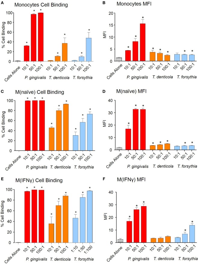
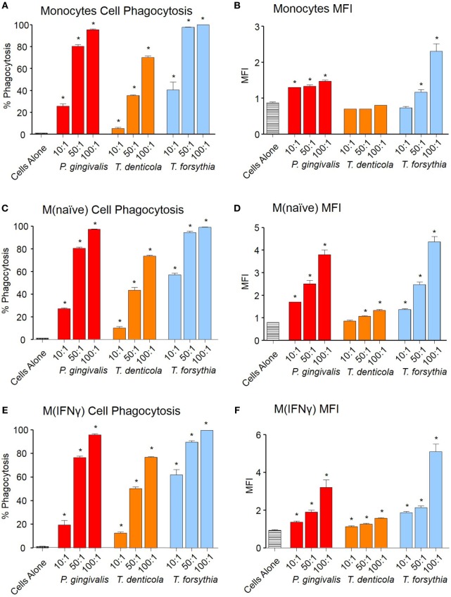
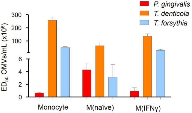
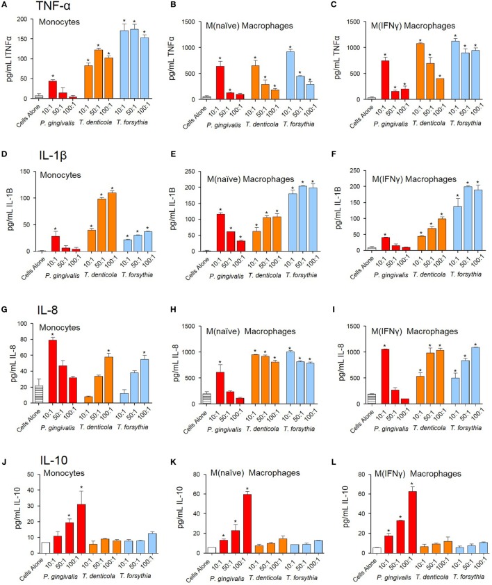
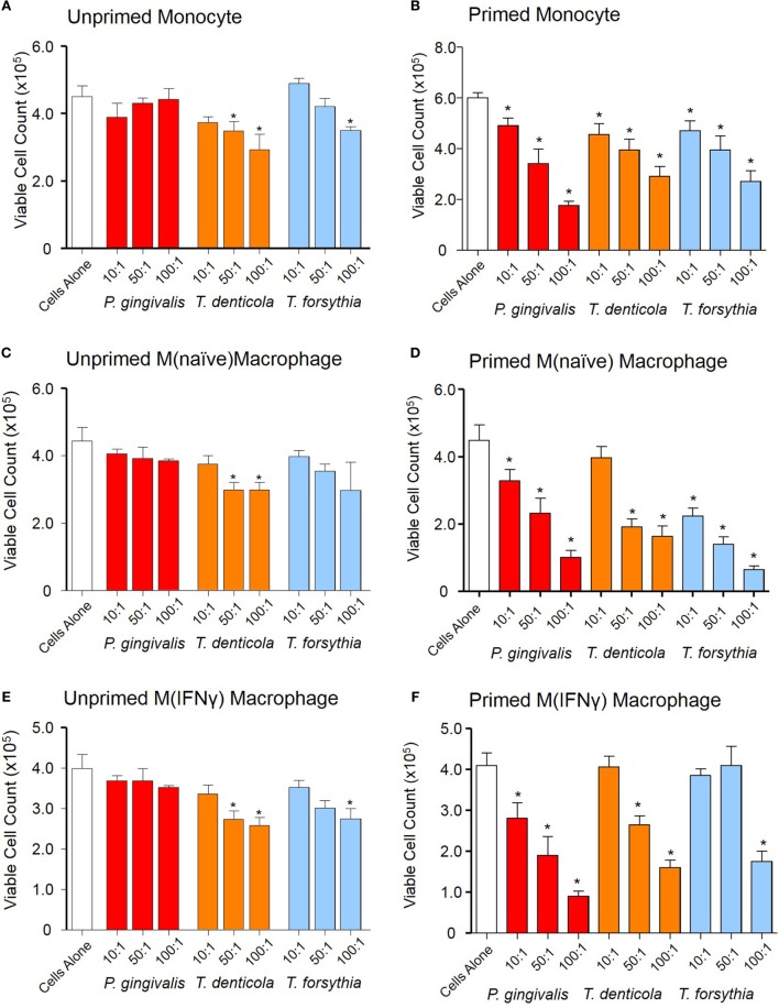
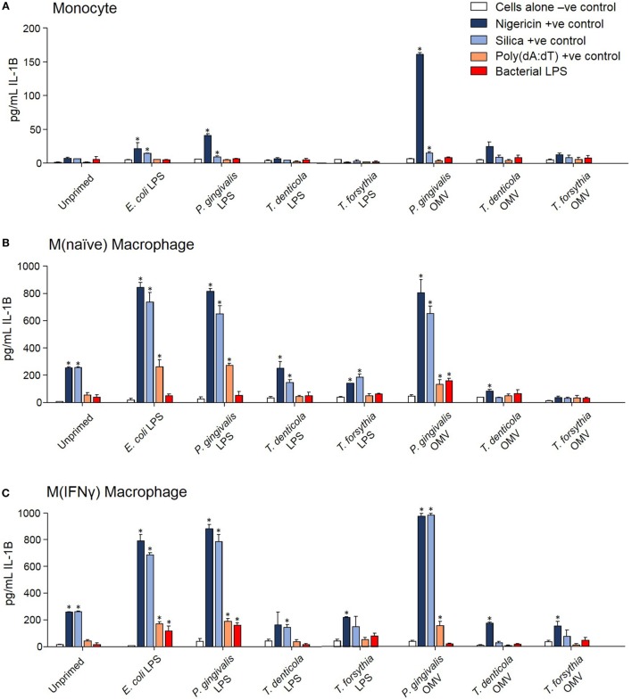
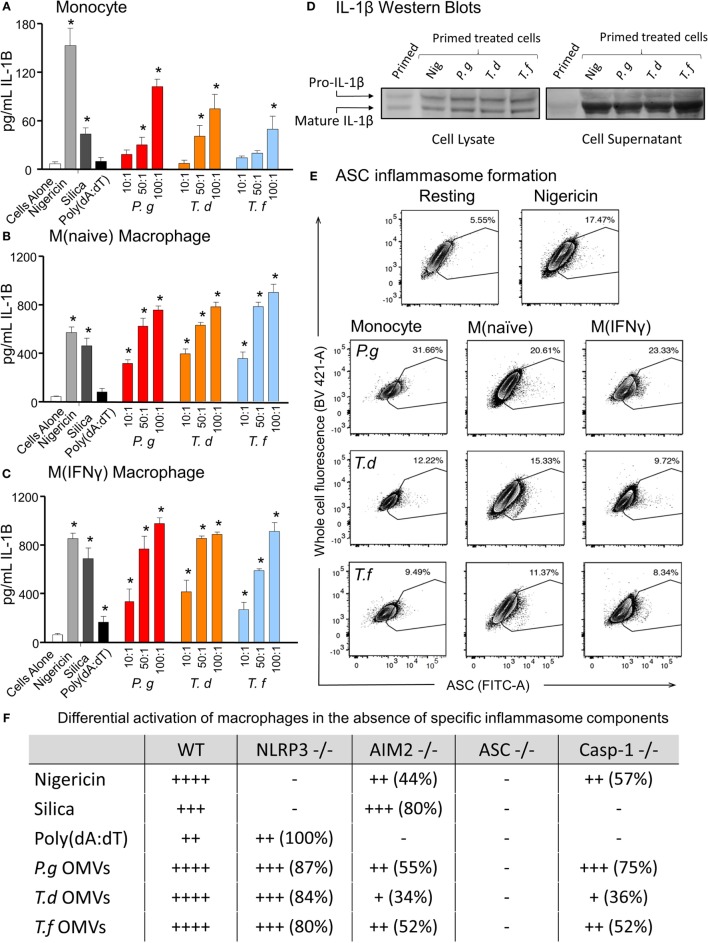
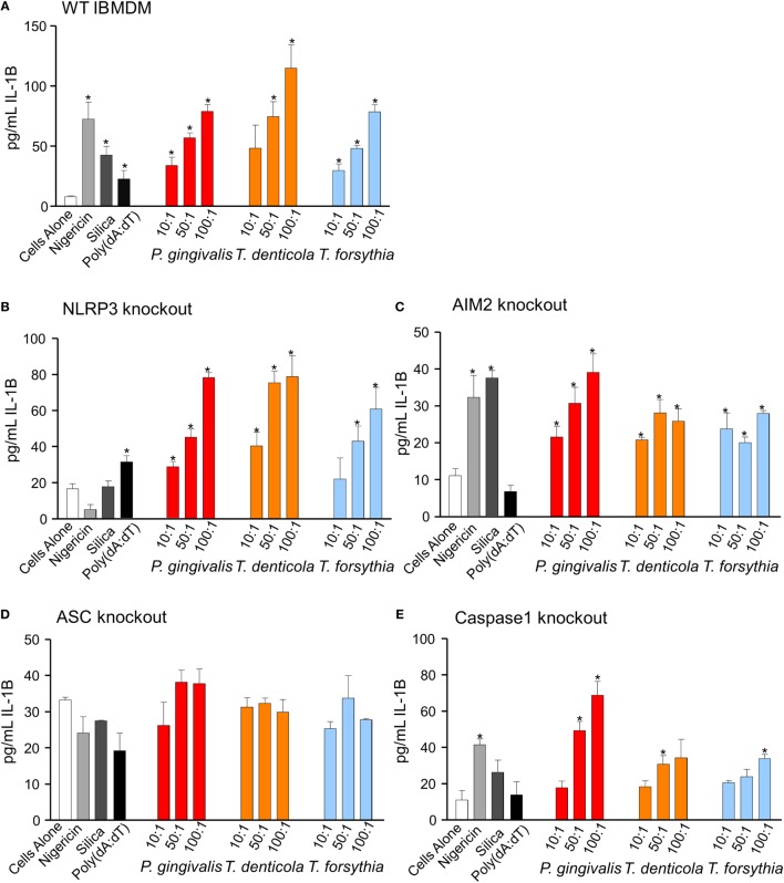
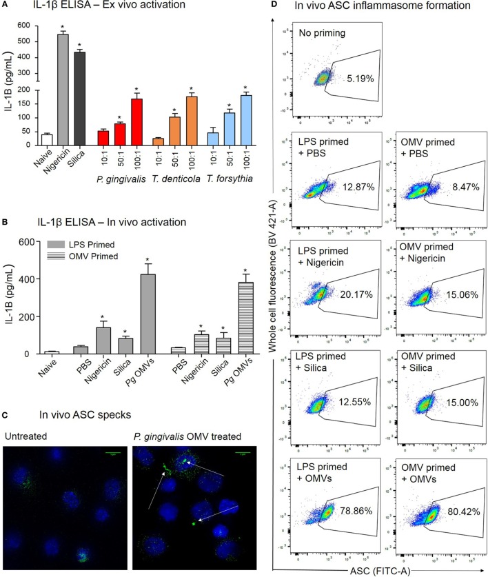
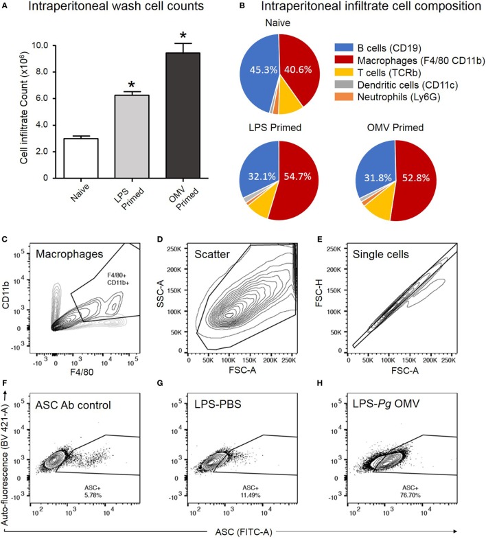
Outer membrane vesicles (OMVs) are proteoliposomes blebbed from the surface of Gram-negative bacteria. Chronic periodontitis is associated with an increase in subgingival plaque of Gram-negative bacteria, Porphyromonas gingivalis, Treponema denticola, and Tannerella forsythia. In this study, we investigated the immune-modulatory effects of P. gingivalis, T. denticola, and T. forsythia OMVs on monocytes and differentiated macrophages. All of the bacterial OMVs were phagocytosed by monocytes, M(naïve) and M(IFNγ) macrophages in a dose-dependent manner. They also induced NF-κB activation and increased TNFα, IL-8, and IL-1β cytokine secretion. P. gingivalis OMVs were also found to induce anti-inflammatory IL-10 secretion. Although unprimed monocytes and macrophages were resistant to OMV-induced cell death, lipopolysaccharide or OMV priming resulted in a significantly reduced cell viability. P. gingivalis, T. denticola, and T. forsythia OMVs all activated inflammasome complexes, as monitored by IL-1β secretion and ASC speck formation. ASC was critical for OMV-induced inflammasome formation, while AIM2-/- and Caspase-1-/- cells had significantly reduced inflammasome formation and NLRP3-/- cells exhibited a slight reduction. OMVs were also found to provide both priming and activation of the inflammasome complex. High-resolution microscopy and flow cytometry showed that P. gingivalis OMVs primed and activated macrophage inflammasomes in vivo with 80% of macrophages exhibiting inflammasome complex formation. In conclusion, periodontal pathogen OMVs were found to have significant immunomodulatory effects upon monocytes and macrophages and should therefore influence pro-inflammatory host responses associated with disease.

Keywords: Porphyromonas gingivalis; Tannerella forsythia; Treponema denticola; inflammasomes; macrophages; outer membrane vesicles; periodontitis.


Figures

Figure 1
Figure 1
Periodontal pathogen outer membrane vesicles (OMVs) bind THP-1 cell subsets. Cell binding assays were performed with THP-1 cell suspensions of monocytes and monolayers of M(naïve) and M(IFNγ) macrophages incubated with PKH26-labeled Porphyromonas gingivalis, Treponema denticola, and Tannerella forsythia OMVs at three OMV to cell ratios (10:1, 50:1, and 100:1). Cells were incubated for 60 min, washed, and OMV binding determined by flow cytometry. Results are expressed as the percentage of THP-1 cells with at least one bound OMV (A,C,E) or the mean fluorescence intensity (MFI) of each sample (B,D,F), which indicates the quantity of OMVs bound per cell. Data are represented as mean ± SEM of three replicates. *represents a result significantly higher (p < 0.05) than the fluorescence of untreated cells.
Figure 2
Figure 2
Periodontal pathogen outer membrane vesicles (OMVs) are phagocytosed by THP-1 cell subsets. Cell phagocytosis assays were performed with THP-1 cell suspensions (monocytes) and monolayers [M(naïve) and M(IFNγ)] incubated with pHrodo-labeled Porphyromonas gingivalis, Treponema denticola, and Tannerella forsythia OMVs at three OMV to cell ratios (10:1, 50:1, and 100:1). Cells were incubated for 60 min, washed, and OMV phagocytosis determined by flow cytometry. Results are expressed as the percentage of THP-1 cells with at least one bound OMV (A,C,E) or the mean fluorescence intensity (MFI) of each sample (B,D,F), which indicates the quantity of OMVs bound per cell. Data are represented as mean ± SEM of three replicates. *represents a result significantly higher (p < 0.05) than the fluorescence of untreated cells.
Figure 3
Figure 3
Periodontal pathogen outer membrane vesicles (OMVs) differentially activate NF-κB in THP-1-differentiated cells. THP-Blue cells [monocytes, M(naïve) and M(IFNγ) cell subsets] were incubated with either Porphyromonas gingivalis, Treponema denticola, or Tannerella forsythia OMVs (in fivefold dilutions) or positive control ligands Pam3CSK4 and lipopolysaccharide (LPS)-EB. Alkaline phosphatase secretion was determined with Quanti-Blue after 20 h incubation at 620 nm on a spectrophotometer. Data are represented as mean ± SEM of three biological replicates and results are presented as the number of OMVs required to achieve ED50. All Pam3CSK4 and LPS-EB controls gave positive results and were not included in the final data.
Figure 4
Figure 4
Periodontal pathogen outer membrane vesicles (OMVs) induce cytokine release from THP-1 cell subsets. THP-1 cytokine responses to Porphyromonas gingivalis, Treponema denticola, and Tannerella forsythia were determined following a 60 min incubation of OMVs (10:1, 50:1, and 100:1 OMV to cell ratios) on a THP-1 suspension (monocytes) or cell monolayer [M(naïve) and M(IFNγ)], followed by a 20-h incubation at 37°C. Results are expressed as picograms per milliliter of cytokine/chemokine TNFα (A–C), IL-1β (D–F), IL-8 (G–I), and IL-10 (J–L) in the resulting supernatant. Data are represented as mean ± SEM of three replicates. *represents a significant (p < 0.05) increase from the cytokine secretion of unstimulated cells.
Figure 5
Figure 5
Periodontal outer membrane vesicle (OMV) cytotoxicity differs between unprimed and primed THP-1 cells. Cytotoxicity assays were performed on THP-1 monocytes, M(naïve), and M(IFNγ) macrophages left unprimed (A,C,E) or primed for 3 h with a low dose of Porphyromonas gingivalis OMVs (B,D,F). Cells were treated with periodontal OMVs in increasing OMV to cell ratios (10:1, 50:1, and 100:1) in cell suspension for 4 h. Cell viability was determined by trypan blue exclusion and deteriorating cell counts determined by hemocytometer and a Z1 Coulter Particle Counter. Results are displayed as the number of remaining viable cells after 4 h. Data are represented as mean ± SEM of three replicates. *represents a significant (p < 0.05) decrease from the count of untreated cells.
Figure 6
Figure 6
Porphyromonas gingivalis outer membrane vesicles (OMVs) and lipopolysaccharide (LPS) are priming agents for macrophage inflammasome activation. THP-1 monocytes (A), M(naïve) (B), and M(IFNγ) (C) macrophages were primed for 3 h with Escherichia coli, P. gingivalis, Treponema denticola, and Tannerella forsythia LPS and OMVs or left unprimed. Cells were then stimulated with positive controls nigericin, silica, and Poly(dA:dT) or E. coli, P. gingivalis, T. denticola, and T. forsythia LPS as required to match the priming material. Cellular supernatants were collected and IL-1β secretion detected by ELISA. Data are represented as mean ± SEM of three replicates. *represents a significant (p < 0.05) increase from the IL-1β secretion of untreated cells.
Figure 7
Figure 7
Periodontal outer membrane vesicles (OMVs) prime and activate inflammasome formation in vitro. THP-1 monocytes (A), M(naïve) (B), and M(IFNγ) (C) macrophages were primed with Porphyromonas gingivalis OMVs for 3 h then stimulated with either positive controls nigericin, silica, and Poly(dA:dT) or P. gingivalis, Treponema denticola, and Tannerella forsythia OMVs. Data are represented as mean ± SEM of three replicates. *represents a significant (p < 0.05) increase from the IL-1β secretion of untreated cells. (D) IL-1β western blots were performed on P. gingivalis OMV-primed (3 h) M(naïve) cell supernatant and cell lysate following treatment with nigericin, P. gingivalis, T. denticola, and T. forsythia OMVs. (E) ASC speck formation was detected by flow cytometry for THP-1 monocytes, M(naïve), and M(IFNγ) macrophages stimulated with P. gingivalis, T. denticola, and T. forsythia OMVs. OMV-induced increases in ASC fluorescence intensity were captured by the percentage of ASC+ cells over baseline background values, based on contour plots depicting ASC fluorescence (FITC-A) vs whole cell autofluorescence (BV421-A). Resting and nigericin controls displayed are from M(naïve) cells. (F) Wild-type immortalized bone marrow-derived macrophage (IBMDM) and NLRP3−/−, AIM2−/−, ASC−/−, and Caspase 1−/− knockout IBMDM strains were primed with Escherichia coli lipopolysaccharide (4 h) and treated with positive controls nigericin, silica, and Poly(dA:dT) or P. gingivalis, T. denticola, and T. forsythia OMVs. Results are presented as a qualitative indication (+ to ++++) of IL-1β secretion significantly greater (p < 0.05) than that of unstimulated IBMDM cells and a percentage decrease in IL-1β secretion as compared to secretion from wild-type IBMDM (see Figure 8).
Figure 8
Figure 8
Inflammasome activation in immortalized bone marrow-derived macrophages (IBMDM) knockout cell lines. Wild-type IBMDM and NLRP3−/−, AIM2−/−, ASC−/−, and Caspase 1−/− knockout IBMDM strains were primed with Porphyromonas gingivalis outer membrane vesicles (OMVs, 3 h) and treated with positive controls nigericin, silica, and Poly(dA:dT) or P. gingivalis, Treponema denticola, and Tannerella forsythia OMVs at OMV:cell ratios of 10:1, 50:1, and 100:1 for 6 h. Cellular supernatants were collected and IL-1β secretion detected by an IL-1β ELISA Kit. Data are represented as mean ± SEM of three replicates. *represents a significant (p < 0.05) increase from the IL-1β secretion of untreated cells.
Figure 9
Figure 9
Porphyromonas gingivalis outer membrane vesicles (OMVs) prime and activate inflammasome formation in vivo. (A) Intraperitoneal cells from naive mice (unprimed and unactivated) were cultured overnight and stimulated ex vivo with nigericin, silica, P. gingivalis, Treponema denticola, or Tannerella forsythia OMVs, IL-1β secretion was determined by ELISA. (B) For in vivo inflammasome activation C57BL/6 J mice received intraperitoneal injections of phosphate-buffered saline (PBS, naïve), Escherichia coli lipopolysaccharide, or P. gingivalis OMVs 72 h prior to harvest to recruit immune cells to the peritoneal cavity. A second intraperitoneal injection of PBS, silica, nigericin, or P. gingivalis OMVs was administered 15 min prior to killing to active inflammasomes in peritoneal macrophages, IL-1β secretion was determined by ELISA. Data are represented as mean ± SEM of three replicates. *represents a significant (p < 0.05) increase from the IL-1β secretion of naive cells. (C) Microscopy was used to visually observe the dense accumulation of the inflammasome component ASC (green) in DAPI stained (blue) peritoneal macrophages primed and treated with P. gingivalis OMVs. (D) Inflammasome formation was confirmed by flow cytometry using ASC-specific antibody and macrophage markers (F4/80 and CD11b). For full gating strategy see Figure 10. Flow cytometry data display ASC-positive events plotted on a FITC area versus BV421 area (whole cell autofluorescence channel) dot plot.
Figure 10
Figure 10
Analysis of ASC speck formation in peritoneal macrophages (in vivo activation). C57BL/6 J mice received intraperitoneal injections of phosphate-buffered saline (PBS, naïve), Escherichia coli lipopolysaccharide (LPS), or Porphyromonas gingivalis outer membrane vesicles (OMVs) 72 h prior to harvest to recruit immune cells to the peritoneal cavity. Intraperitoneal washes were counted using a Z1 Coulter Particle Counter to observe increase in cell recruitment following LPS and OMV priming (A). Data are represented as mean ± SEM of three replicates. *represents a significant (p < 0.05) increase in cell counts from naïve washes. Following harvest intraperitoneal cells were analyzed by flow cytometry using fluorescent antibodies against F4/80 and CD11b, Ly6G, CD11c, TCRb, and CD19 to identify macrophage, neutrophil, dendritic cell, T cell and B cell populations, respectively (B). Results in pie charts are expressed as the percentage of each detectable cell type in peritoneal samples and do not include cells outside these categories. Following a second intraperitoneal injection of PBS, nigericin, silica, or P. gingivalis OMVs, ASC speck formation in peritoneal macrophages was determined by flow cytometry. (C) CD11b- and F4/80-positive macrophages are selected from other peritoneal cells with a scatter gate. (D) Events within the scatter gate are plotted on a SSC-area versus FSC-area dot plot, the predominant population of ethanol-fixed cells are selected with a scatter gate. (E) Events within the scatter gate are plotted on a FSC-width versus FSC-area dot plot and single cells are selected with a singlet gate. (F) Single cells are plotted on a FITC-area versus BV421-area dot plot and a gate created to capture ASC fluorescence greater than that of the secondary antibody control. This population represent non-specific ASC antibody binding to peritoneal macrophages. (G) This population represents peritoneal cells which have been LPS primed in vivo but not activated. (H) This population represents peritoneal cells which have been LPS primed and P. gingivalis OMV activated. In this cell set diffuse ASC has condensed into single bright specks, increasing the detectable ASC fluorescence and indicating inflammasome activation. Data presented here were collected on a BD LSRFortessa X20 flow cytometer.

References

    1. Haffajee AD, Cugini MA, Tanner A, Pollack RP, Smith C, Kent RL, Jr, et al. Subgingival microbiota in healthy, well-maintained elder and periodontitis subjects. J Clin Periodontol (1998) 25:346–53.10.1111/j.1600-051X.1998.tb02454.x - DOI - PubMed
    1. Socransky SS, Haffajee AD, Cugini MA, Smith C, Kent RL, Jr. Microbial complexes in subgingival plaque. J Clin Periodontol (1998) 25:134–44.10.1111/j.1600-051X.1998.tb02419.x - DOI - PubMed
    1. Byrne SJ, Dashper SG, Darby IB, Adams GG, Hoffmann B, Reynolds EC. Progression of chronic periodontitis can be predicted by the levels of Porphyromonas gingivalis and Treponema denticola in subgingival plaque. Oral Microbiol Immunol (2009) 24:469–77.10.1111/j.1399-302X.2009.00544.x - DOI - PubMed
    1. O’Brien-Simpson N, Pathirana R, Walker G, Reynolds E. Porphyromonas gingivalis RgpA-Kgp proteinase-adhesin complexes penetrate gingival tissue and induce proinflammatory cytokines or apoptosis in a concentration-dependent manner. Infect Immun (2009) 77:1246–61.10.1128/IAI.01038-08 - DOI - PMC - PubMed
    1. Grenier D, Mayrand D. Functional characterization of extracellular vesicles produced by Bacteroides gingivalis. Infect Immun (1987) 55:111–7. - PMC - PubMed

LinkOut - more resources