Review
. 2017 Aug;8(4):749-757.
doi: 10.21037/jgo.2017.06.03.

Primary pancreatic lymphoma: what we need to know

Affiliations
Review

Primary pancreatic lymphoma: what we need to know

Neda Rad et al. J Gastrointest Oncol. 2017 Aug.

Abstract

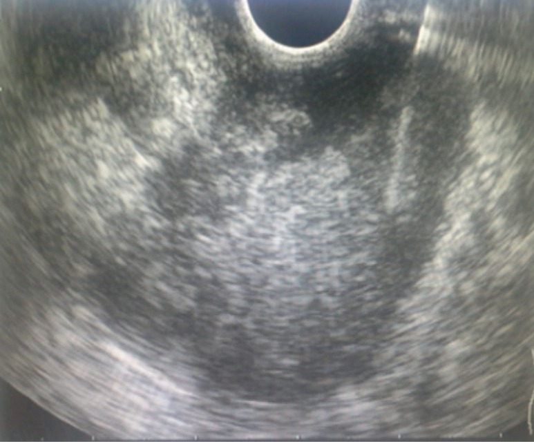
Hodgkin's lymphoma is a group of malignant lymphoid which involve various organs including gastrointestinal tract. Stomach and small intestine are commonly involved more; however, pancreas can be primarily involved as well. The secondary involvement of pancreas caused by Hodgkin's lymphoma is more prevalent than the primarily involvement (1 .25-2.2% vs. <1%). Primary pancreatic lymphomas (PPLs) consist of 1-2% of all lymphoma outside nods. The symptoms and findings of PPL imaging can be akin to that of pancreas adenocarcinoma and differentiating them is difficult without examining the tissue sample. The prognosis and treatment of PPL are different from those of adenocarcinoma and due to the superior prognosis of PPL compared to pancreas adenocarcinoma, the proper diagnosis of the disease is important.

Keywords: Pancreas; adenocarcinoma; lymphoma.


Conflict of interest statement

Conflicts of Interest: The authors have no conflicts of interest to declare.

Figures

Figure 1
Figure 1
Low grade B cell lymphoma (hematoxylin-eosin stain; original magnification ×200).
Figure 2
Figure 2
CD20-BCL2-CD10-CD43-CD3 positive atypical lymphocytic cells (immunohistochemistry; magnification, ×25).
Figure 3
Figure 3
Evaluating pancreatic mass. EUS-FNA, endoscopic ultrasound guided fine needle aspiration; FNB, fine-needle core biopsy.
Figure 4
Figure 4
Diffusely enlarged of pancreas and echoic mass lesion 37–50 mm at the head of pancreas with adhesion to portal vein and SMV. SMV, superior mesenteric vein.
Figure 5
Figure 5
EUS-FNA indicated echoic lesion adhered to head of pancreas with invasion to portal vein, SMA and SMV. EUS-FNA, endoscopic ultrasound guided fine needle aspiration; SMA, superior mesenteric artery; SMV, superior mesenteric vein.

References

    1. Zucca E, Roggero E, Bertoni F, et al. Primary extranodal non-Hodgkin's lymphomas. Part 1: Gastrointestinal, cutaneous and genitourinary lymphomas. Ann Oncol 1997;8:727-37. 10.1023/A:1008282818705 - DOI - PubMed
    1. Fukita Y, Asaki T, Adachi S, et al. Non-Hodgkin Lymphoma Mimicking Pancreatic Adenocarcinoma and Peritoneal Carcinomatosis. J Clin Oncol 2013;31:e373-6. 10.1200/JCO.2012.45.2904 - DOI - PubMed
    1. Alexander RE, Nakeeb A, Sandrasegaran K, et al. Primary pancreatic follicle center-derived lymphoma masquerading as carcinoma. Gastroenterol Hepatol (N Y) 2011;7:834-8. - PMC - PubMed
    1. Rad N, Heidarnezhad A, Soheili S, et al. A Man with Pancreatic Head Mass Lesion on Endoscopic Ultrasound and Granuloma on Cytopathology. Case Rep Gastroenterol 2016;10:760-8. 10.1159/000448875 - DOI - PMC - PubMed
    1. Merkle EM, Bender GN, Brambs HJ. Imaging findings in pancreatic lymphoma: differential aspects. AJR Am J Roentgenol 2000;174:671-5. 10.2214/ajr.174.3.1740671 - DOI - PubMed

LinkOut - more resources