Review
. 2017 Aug 25:7:373.
doi: 10.3389/fcimb.2017.00373. eCollection 2017.

Neutrophils to the ROScue: Mechanisms of NADPH Oxidase Activation and Bacterial Resistance

Affiliations
Review

Neutrophils to the ROScue: Mechanisms of NADPH Oxidase Activation and Bacterial Resistance

Giang T Nguyen et al. Front Cell Infect Microbiol. .

Abstract

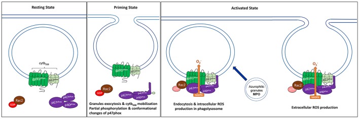
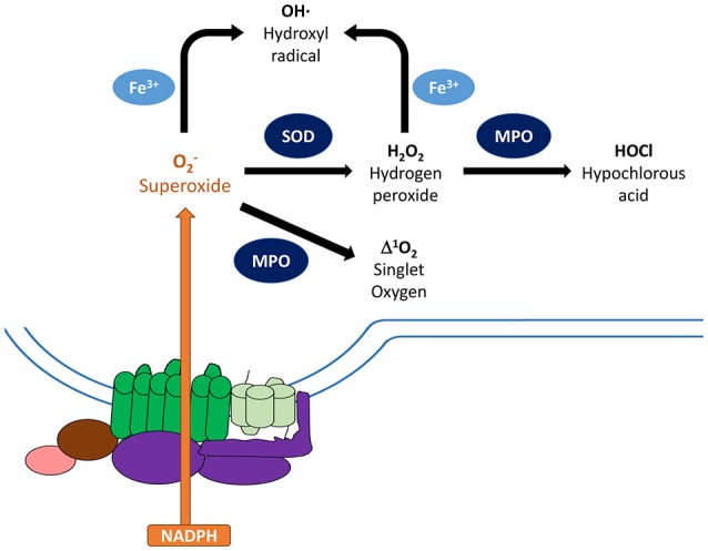
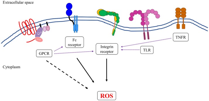
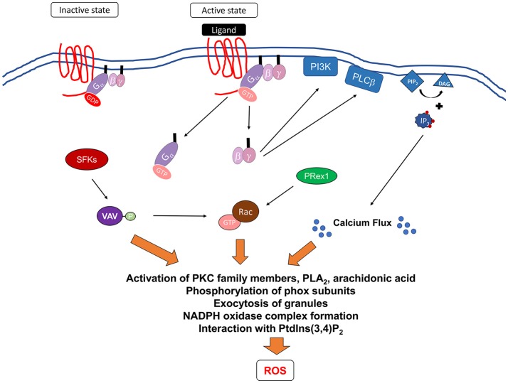
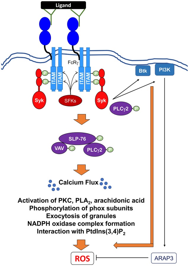
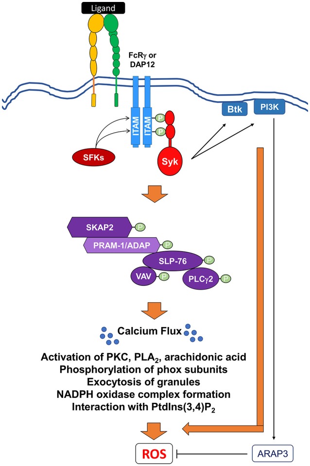
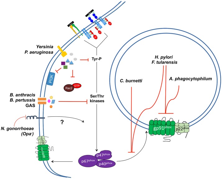
Reactive oxygen species (ROS) generated by NADPH oxidase play an important role in antimicrobial host defense and inflammation. Their deficiency in humans results in recurrent and severe bacterial infections, while their unregulated release leads to pathology from excessive inflammation. The release of high concentrations of ROS aids in clearance of invading bacteria. Localization of ROS release to phagosomes containing pathogens limits tissue damage. Host immune cells, like neutrophils, also known as PMNs, will release large amounts of ROS at the site of infection following the activation of surface receptors. The binding of ligands to G-protein-coupled receptors (GPCRs), toll-like receptors, and cytokine receptors can prime PMNs for a more robust response if additional signals are encountered. Meanwhile, activation of Fc and integrin directly induces high levels of ROS production. Additionally, GPCRs that bind to the bacterial-peptide analog fMLP, a neutrophil chemoattractant, can both prime cells and trigger low levels of ROS production. Engagement of these receptors initiates intracellular signaling pathways, resulting in activation of downstream effector proteins, assembly of the NADPH oxidase complex, and ultimately, the production of ROS by this complex. Within PMNs, ROS released by the NADPH oxidase complex can activate granular proteases and induce the formation of neutrophil extracellular traps (NETs). Additionally, ROS can cross the membranes of bacterial pathogens and damage their nucleic acids, proteins, and cell membranes. Consequently, in order to establish infections, bacterial pathogens employ various strategies to prevent restriction by PMN-derived ROS or downstream consequences of ROS production. Some pathogens are able to directly prevent the oxidative burst of phagocytes using secreted effector proteins or toxins that interfere with translocation of the NADPH oxidase complex or signaling pathways needed for its activation. Nonetheless, these pathogens often rely on repair and detoxifying proteins in addition to these secreted effectors and toxins in order to resist mammalian sources of ROS. This suggests that pathogens have both intrinsic and extrinsic mechanisms to avoid restriction by PMN-derived ROS. Here, we review mechanisms of oxidative burst in PMNs in response to bacterial infections, as well as the mechanisms by which bacterial pathogens thwart restriction by ROS to survive under conditions of oxidative stress.

Keywords: CGD; Fc receptors; G protein coupled receptors; NADPH oxidase; integrin receptors; neutrophils; reactive oxygen species; type 3 secreted effectors.


Figures

Figure 1
Figure 1
Components of the NADPH oxidase at resting and activated state. NADPH oxidase, also commonly referred to as the phagocyte oxidase (phox) complex, is a multi-protein electron transfer system that is made up of five components and Rac2. The catalytic core, also known as flavocytochrome b558 (cytb558), is a heterotrimeric dimer made up of two transmembrane proteins, gp91phox and gp22phox. (Left) At resting state, cytb558 resides at the membranes of phagosomes, secretory vesicles, specific granules, and the plasma membrane and catalyzes the transfer of electrons from NADPH to molecular oxygen generating superoxide anions (O2) as by-products. Regulatory subunits, p40phox, p47phox, and p67phox, reside in the cytosol of resting cells. (Center) Priming induces several changes such as translocation of cytb558 to plasma membrane via granule exocytosis, partial phosphorylation of p47phox leading to conformational changes. (Right) When PMNs are activated, the regulatory cytosolic complex translocates to the membrane and interacts with cytb558; this is required for NADPH activation. Another factor that regulates the recruitment of regulatory complex to the membranes and the overall activation of NADPH oxidase is small GTPase protein, Rac2. Activated GTP-bound Rac2 binds directly to gp91phox and p67phox, and is also required for ROS production. For intracellular ROS production in the phagolysosome, this occurs after endocytosis of the complex. Meanwhile, extracellular ROS occurs at the plasma membrane.
Figure 2
Figure 2
Metabolism of reactive oxygen species. Activated NADPH oxidase catalyzes the transfer of electrons from NADPH to molecular oxygen generating superoxide anions (O2) as the primary product. To minimize damage, cells are equipped with antioxidant scavenging enzymes, such as superoxide dismutase (SOD), which dismutates O2 to non-radical species hydrogen peroxide (H2O2), and catalase. SOD and glutathione peroxidase can further convert these species into water, which limit damages to the host. On the other hand, O2 can be converted to other reactive oxygen species that can damage nucleic acids, proteins, and cell membranes. Granule-localized myeloperoxidase (MPO) can convert H2O2 to hypochlorous acid (HOCl), which can enhance clearance of invading pathogens. MPO can also directly convert O2 into singlet oxygen (1O2*). In addition, ferric iron can convert O2 and H2O2 into hydroxyl radical (OH). Components of the NADPH oxidase: gp91phox (green), gp22phox (light green), regulatory factors (purple).
Figure 3
Figure 3
Neutrophils express several groups of receptors that can induce the formation and generation of reactive oxygen species. Activation of integrin and Fc receptors leads to complex intracellular signal transduction pathways that can robustly activate the NADPH oxidase complex (solid black arrows). Some members of G-protein-coupled receptors (GPCRs) family, specifically formyl receptors, can directly activate NADPH oxidase, although to a lesser extent than to what has been observed in integrin and Fc receptors (dotted black arrow). Ligand binding to TLRs, TNFRs, and some members of GPCRs can transform the neutrophils into an “primed” state, whereby the NADPH oxidase is more susceptible to activation by a secondary stimulus (purple dotted arrows). This is presumably another level of regulation to ensure that reactive oxygen species are produced at the right time and place that is only during an active infection.
Figure 4
Figure 4
Signaling pathways mediating formyl receptor (GPCR)-induced NADPH oxidase activation. Ligation of G-protein-coupled receptors leads to changes in the receptor conformation resulting in the exchange of GDP for GTP bound to the G protein. This leads to the dissociation of the G proteins subunits, Gα and Gβγ from the membrane to activate downstream effectors. It is currently unclear how Gα contributes to the activation of NADPH oxidase. Gβγ can activate PI3K, which can act to mediate PRex1-dependent Rac2 activation, and PLCβ, which leads to the breakdown of membrane phospholipid, PIP2, into DAG and IP3. DAG induces calcium flux, while IP3 can act on further downstream proteins. In addition, Src family kinases (SFKs) have been shown to be important and may activate Vav proteins leading to the activation of p38 MAPK and potentially Rac2. Activation of these proximal signaling molecules lead to exocytosis of granules, activation of various PKC family members, phospholipase A2 (PLA2), and release of arachidonic acid, a lipid messenger. All of these secondary messengers are required for phosphorylation of phox subunits, formation of NADPH oxidase, and interaction with phosphatidylinositol 3,4-biphosphate (PtdIns(3,4)P2).
Figure 5
Figure 5
Signaling pathways mediating Fc receptor-induced NADPH oxidase activation via IgG immune complexes. Ligation and crosslinking of Fc receptors leads to the phosphorylation of the ITAMs by Src family kinases (SFKs) resulting in the recruitment and the tyrosine phosphorylation of the Src homology domain of Syk. Activated Syk can then recruit and activate Btk (Bruton's tyrosine kinase), class I PI3K (phosphoinositide 3-kinase). A class I PI3K effector, ARAP3, has been shown to negatively regulate ROS production (Gambardella et al., 2013). Syk also induces the formation and activation of the SLP76 signaling complex, which includes SLP76, Vav, and PLCγ2. Activation of this complex leads to further downstream effectors resulting in the release of intracellular calcium stores (Ca2+ flux), which is critical for ROS production. In addition, PLCγ2 can potentially interact directly with Syk to perpetuate the signal for ROS production. Activation of these proximal signaling molecules lead to exocytosis of granules, activation of various PKC family members, phospholipase A2 (PLA2), and release of arachidonic acid, a lipid messenger. All of these secondary messengers are required for phosphorylation of phox subunits, formation of NADPH oxidase, and interaction with phosphatidylinositol 3,4-biphosphate (PtdIns(3,4)P2).
Figure 6
Figure 6
Signaling pathways mediating integrin-induced NADPH oxidase activation. Ligation and crosslinking of integrin receptors leads to the phosphorylation of the ITAM-containing proteins, DAP12 and FcRγ, by (SFKs), resulting in the recruitment and the tyrosine phosphorylation of the Src homology domain of Syk. Activated Syk can then act to recruit and activate Bruton's tyrosine kinase (Btk) and class I phosphoinositide 3-kinase (PI3K). A class I PI3K effector, ARAP3, has been shown to negatively regulate ROS production. Syk also induces the activation of SH2-domain-containing leukocyte protein of 76 kDa (SLP76) to form a multi-protein signaling complex. This SLP76 complex can then recruit and activate downstream effectors proteins like SKAP2, SLP76, the Vav GEF family, and PLCγ2. Activation of this complex leads to further downstream effectors resulting in the release of intracellular calcium stores (Ca2+ flux) and ultimate ROS production. Activation of these proximal signaling molecules lead to exocytosis of granules, activation of various PKC family members, phospholipase A2 (PLA2), and release of arachidonic acid, a lipid messenger. All of these secondary messengers are required for phosphorylation of phox subunits, formation of NADPH oxidase, and interaction with phosphatidylinositol 3,4-biphosphate (PtdIns(3,4)P2).
Figure 7
Figure 7
Mechanisms of NADPH oxidase inhibition by bacterial pathogens in PMNs. Several bacterial pathogens employ mechanisms to interfere with the activation and/or localization of the NADPH complex of PMNs. These include strategies to prevent oxidative burst in the phagosomal compartment. Three pathogens, F. tularensis, A. phagocytophilum, and H. pylori, exclude one or both components of the cytb558 complex from the phagosomal membrane. Three pathogens, F. tularensis H. pylori, and C. burnetti, exclude or prevent p67phox/p40phox/p47phox from binding to the phagosomal membrane. A number of extracellular pathogens also employ mechanisms to inhibit oxidative burst. P. aeruginosa inhibits the oxidative burst of PMNs through the activities of two T3SS effectors, ExoS and ExoT. Both effectors inhibit activation of PI3K signaling pathways upstream of p67phox/p40phox/p47phox activation. The pathogenic Yersinia sp. inhibit respiratory burst in PMNs, though their activities have been largely modeled in other phagocytic cell types. Y. pseudotuberuclosis translocates the effector protein YopE through a T3SS to block activation of Rac in HL-60 cells. Y. pseudotuberculosis also translocates another T3SS effector protein, YopH, which interferes with oxidative burst in macrophages. The effects of YopH on oxidative burst have not been examined in PMNs; it dismantles the SLP-76/SKAP2 signal transduction pathway in these cells, suggesting that interference of this pathway in PMNs could prevent oxidative burst. Three pathogens, B. anthracis, B. pertussis, and Group A Streptococcus (GAS), also secrete toxins into PMNs that interfere with signaling pathways required for oxidative burst. Finally, strains of N. gonorrhoeae lacking opacity-associated proteins do not activate oxidative burst in PMNs, though the mechanism by which this occurs remains unclear. It is hypothesized that the failure of opacity-negative strains to engage CEACAM receptors could result in a failure to stimulate kinase signaling upstream of p47phox activation. Alternatively, it is possible that opacity-negative strains may actively block trafficking of NADPH oxidase components to membrane sites. Additionally, three other pathogens, L. monocytogenes, S. typhimurium, and V. parahaemolyticus, are capable of inhibiting the oxidative burst in cultured cells; however, their effects on neutrophils have not been examined in detail.

References

    1. Abram C. L., Lowell C. A. (2009). The ins and outs of leukocyte integrin signaling. Annu. Rev. Immunol. 27, 339–362. 10.1146/annurev.immunol.021908.132554 - DOI - PMC - PubMed
    1. Ago T., Nunoi H., Ito T., Sumimoto H. (1999). Mechanism for phosphorylation-induced activation of the phagocyte NADPH oxidase protein p47(phox). Triple replacement of serines 303, 304, and 328 with aspartates disrupts the SH3 domain-mediated intramolecular interaction in p47(phox), thereby activating the oxidase. J. Biol. Chem. 274, 33644–33653. - PubMed
    1. Alenghat F. J., Baca Q. J., Rubin N. T., Pao L. I., Matozaki T., Lowell C. A., et al. (2012). Macrophages require Skap2 and Sirpalpha for integrin-stimulated cytoskeletal rearrangement. J. Cell. Sci. 125(Pt. 22), 5535–5545. 10.1242/jcs.111260 - DOI - PMC - PubMed
    1. Ali H., Sozzani S., Fisher I., Barr A. J., Richardson R. M., Haribabu B., et al. (1998). Differential regulation of formyl peptide and platelet-activating factor receptors. Role of phospholipase Cbeta3 phosphorylation by protein kinase A. J. Biol. Chem. 273, 11012–11016. - PubMed
    1. Allen L. A., Beecher B. R., Lynch J. T., Rohner O. V., Wittine L. M. (2005). Helicobacter pylori disrupts NADPH oxidase targeting in human neutrophils to induce extracellular superoxide release. J. Immunol. 174, 3658–3667. 10.4049/jimmunol.174.6.3658 - DOI - PubMed

Publication types