. 2018 Jan 1;526(1):133-145.
doi: 10.1002/cne.24327. Epub 2017 Sep 20.

A critical period for the trophic actions of leptin on AgRP neurons in the arcuate nucleus of the hypothalamus

Affiliations

A critical period for the trophic actions of leptin on AgRP neurons in the arcuate nucleus of the hypothalamus

Anna Kamitakahara et al. J Comp Neurol. .

Abstract

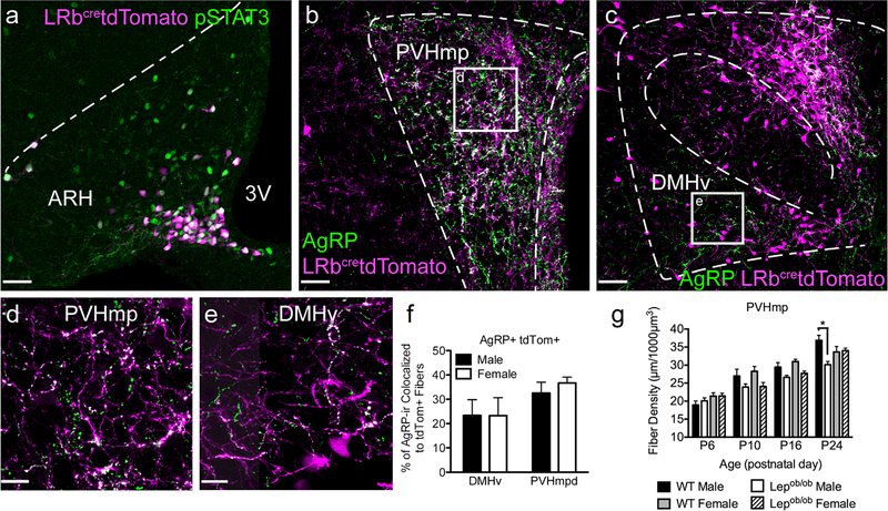
In the developing hypothalamus, the fat-derived hormone leptin stimulates the growth of axons from the arcuate nucleus of the hypothalamus (ARH) to other regions that control energy balance. These projections are significantly reduced in leptin deficient (Lepob/ob ) mice and this phenotype is largely rescued by neonatal leptin treatments. However, treatment of mature Lepob/ob mice is ineffective, suggesting that the trophic action of leptin is limited to a developmental critical period. To temporally delineate closure of this critical period for leptin-stimulated growth, we treated Lepob/ob mice with exogenous leptin during a variety of discrete time periods, and measured the density of Agouti-Related Peptide (AgRP) containing projections from the ARH to the ventral part of the dorsomedial nucleus of the hypothalamus (DMHv), and to the medial parvocellular part of the paraventricular nucleus (PVHmp). The results indicate that leptin loses its neurotrophic potential at or near postnatal day 28. The duration of leptin exposure appears to be important, with 9- or 11-day treatments found to be more effective than shorter (5-day) treatments. Furthermore, leptin treatment for 9 days or more was sufficient to restore AgRP innervation to both the PVHmp and DMHv in Lepob/ob females, but only to the DMHv in Lepob/ob males. Together, these findings reveal that the trophic actions of leptin are contingent upon timing and duration of leptin exposure, display both target and sex specificity, and that modulation of leptin-dependent circuit formation by each of these factors may carry enduring consequences for feeding behavior, metabolism, and obesity risk.

Keywords: Critical period; RRID:AB_2313908; RRID:IMSR_JAX:000632; RRID:IMSR_JAX:006417; RRID:IMSR_JAX:007914; RRID:SCR_002668; agouti-related peptide (AgRP); arcuate nucleus of the hypothalamus (ARH); leptin; sexual dimorphism.


Figures

FIGURE 1
FIGURE 1
The trophic effect of leptin on AgRP axonal growth is restricted to the first 4 postnatal weeks. On P60, the density of AgRP projections to the DMHv was measured in male WT and Lepob/ob mice treated with either vehicle or leptin during one of several discrete time periods in early postnatal life. Representative images of AgRP innervation of the DMH in (a) WT vehicle injected, (b) Lepob/ob vehicle injected, and (c) Lepob/ob leptin treated mice in the treatment group spanning from P4–14. Dashed lines indicate the anatomical borders of the DMH. d, dorsal; c, central; v, ventral subregion of the DMH. 3V denotes the third ventricle. Scale bar, 70 μm. Square indicates the ROI in the DMHv used for quantification further illustrated in d-f using a high-magnification 63X oil corrected lens. Scale bar, 13 μm. (g) Quantification of AgRP axonal fiber density in the DMHv for all treatment groups revealed that after postnatal day 28, leptin no longer stimulates axonal growth from AgRP neurons. OB, Lepob/ob; Veh, Vehicle; Lep, Leptin. (WT Veh, n = 32, OB Veh, n = 22, OB Lep P4–8, n = 5, OB Lep P6–10, n = 4, OB Lep P8–12, n = 3, OB Lep P12–16, n = 4, OB Lep P4–14, n = 4, OB Lep P16–26, n = 5, OB Lep P28–38, n = 4) Data are expressed as mean ± SEM of the density of AgRP-containing axonal fibers within the DMHv in a set volume. Significance between groups was determined by one-way ANOVA and Holm-Sidak’s multiple comparisons test; * indicates p < .0001 compared to WT vehicle, ˆ indicates p < .05 compared to Lepob/ob vehicle
FIGURE 2
FIGURE 2
Leptin treatment is not sufficient to restore AgRP innervation to the PVHmp in leptin-deficient males. Representative images of AgRP innervation of the PVH in (a) WT mice injected with vehicle, (b) Lepob/ob mice injected with vehicle, and (c) Lepob/ob mice treated with leptin from P4–14. Dashed lines indicate the anatomical borders of the PVH. PVHmp, medial parvicellular part of the PVH. 3V, third ventricle. Scale bar, 40 μm. Square indicates the ROI in the PVHmp used for quantification, further illustrated in (d-f). Scale bar, 13 μm. (g) Quantification of AgRP axonal fiber density in the PVHmp for all treatment groups. OB, Lepob/ob; Veh, Vehicle; Lep, Leptin. (WT Veh, n = 32, OB Veh, n = 31, Lep P4–8, n = 5, OB Lep P6–10, n = 4, OB Lep P8–12, n = 3, OB Lep P12–16, n = 4, OB Lep P4–14, n = 4, OB Lep P16–26, n = 5, OB Lep P28–38, n = 4) Data are expressed as mean ± SEM of the density of AgRP-containing axonal fibers within each target region in a set volume. Significance between groups was determined by one-way ANOVA and Holm-Sidek multiple comparisons test; * indicates p < .0001 compared to WT vehicle
FIGURE 3
FIGURE 3
AgRP neurons in the ARH that project to the PVHmp and DMHv are leptin sensitive during early postnatal life. (a) Representative image of leptin-induced pSTAT3 (green) colocalized to tdTomato expressing neurons (magenta) in the ARH of a LRbcretdTomato animal on P10, 1 hr following leptin injection. 3V, third ventricle. Dotted lines illustrate the boundaries of the nucleus. Scale bar, 40 μm. (b, c) Immunohistochemical labeling of AgRP projections (green) targeting the PVH (b), and DMH (c) in LRbcretdTomato mice. Squares indicate the ROI in the PVHmp and DMHv quantified and illustrated in (d) and (e). Representative high-magnification images of AgRP immunoreactivity (green) in the PVHmpd (d) and DMHv (e) colocalized to axonal projections from leptin receptor expressing neurons (magenta). Scale bar, 13 μm. (f) Quantification of the percentage of AgRP immunoreactivity (ir) colocalized to tdTomato labeled fibers in male and female LRbcretdTomato mice on P10 (male, n = 3; female, n = 3). (g) The density of AgRP projections to the PVHmp measured in male and female, WT and Lepob/ob mice on P6, P10, P16, and P24 are graphed. Data are expressed as mean ± SEM of the density of AgRP-containing axonal fibers within the PVHmp in a defined volume. Significance between groups was determined by one-way ANOVA and Holm-Sidek multiple comparisons test performed at each age; * indicates p < .01 compared to WT Male. Data are expressed as mean ± SEM
FIGURE 4
FIGURE 4
Postnatal leptin treatment is sufficient to restore AgRP innervation to the PVHmp in leptin-deficient females. Representative images of the PVH in (a) WT vehicle treated, (b) Lepob/ob vehicle treated, and (c) Lepob/ob leptin treated female mice treated from P4–14. Dashed lines indicate the anatomical borders of the PVH. 3V denotes the third ventricle. Scale bar, 40 μm. Square indicates the ROI in the PVHmpd used for quantification, further illustrated in (d-f). Scale bar, 13 μm. (g) Quanification of AgRP axonal fiber density in the PVHmpd and DMHv in WT and Lepob/ob females treated with vehicle or leptin. OB, Lepob/ob; Veh, Vehicle; Lep, Leptin. (WT Veh, n = 18, OB Veh, n = 19, OB Lep P4–14, n = 4, OB Lep Leptin P16–24, n = 4, OB Lep P25–33, n = 5) Data are expressed as mean SEM of the density of AgRP-containing axonal fibers within each target region in a set volume. Significance between groups was determined by one-way ANOVA and Holm-Sidak multiple comparisons test; * indicates p < .0001 compared to WT vehicle, ˆindicates p <.05 compared to Lepob/ob vehicle
FIGURE 5
FIGURE 5
AgRP neurons do not express androgen receptor or estrogen receptor alpha during early postnatal development. Representative images of estrogen receptor alpha (a and c; ERα; magenta) or androgen receptor (b and d; AR; magenta) immunoreactivity in the ARH of male (a and b) and female (c and d) NPYGFP animals on P10. Square in (a) denotes the location of insets. 3V, third ventricle
FIGURE 6
FIGURE 6
Schematic model of the early postnatal trophic effects of leptin on development of AgRP neuronal projections. (a) The timing of the naturally occurring early postnatal surge in serum leptin, known to stimulate the growth of ARH projections, is graphed. Treatment of Lepob/ob mice with exogenous leptin during several discrete time periods enabled identification of the closure of a critical period for growth of ARH AgRP projections (Bars below graph indicate each leptin treatment period. Green bars indicate treatment periods that rescued innervation and red bars indicate treatment periods that were not effective). Short duration, 5-day, leptin treatment periods from P4–8, P6–10, P8–12, or P12–16 were not sufficient to rescue AgRP innervation density to the DMHv in Lepob/ob mice. By contrast, longer duration, 11-day, leptin treatment periods from P4–14 and P16–26 were able to rescue innervation deficits in the DMHv of Lepob/ob mice. However, leptin treatment of Lepob/ob mice from P28–38 was no longer effective, suggesting that the critical period for the trophic actions of leptin is closed at this time. (b) Early postnatal leptin treatment also revealed striking sexually dimorphic, target-dependent effects on innervation of the PVHmp. While treatment of Lepob/ob mice with leptin (for 9 or 11 days during the critical period) was sufficient to rescue deficits in projections to the DMHv in both males and females, deficits in innervation to the PVHmp were only rescued in female Lepob/ob mice treated with leptin

References

    1. Ahima RS, Prabakaran D, & Flier JS (1998). Postnatal leptin surge and regulation of circadian rhythm of leptin by feeding. Implications for energy homeostasis and neuroendocrine function. Journal of Clinical Investigation, 101(5), 1020-1027. 10.1172/JCI1176 - DOI - PMC - PubMed
    1. Arnold AP, & Gorski RA (1984). Gonadal steroid induction of structural sex differences in the central nervous system. Annual Review of Neuroscience, 7, 413-442. 10.1146/annurev.ne.07.030184.002213 - DOI - PubMed
    1. Atasoy D, Betley JN, Su HH, & Sternson SM (2012). Deconstruction of a neural circuit for hunger. Nature, 488, 172-177. 10.1038/nature11270 - DOI - PMC - PubMed
    1. Baquero AF, de Solis AJ, Lindsley SR, Kirigiti MA, Smith MS, Cowley MA, & Grove KL (2014). Developmental switch of leptin signaling in arcuate nucleus neurons. Journal of Neuroscience, 34 (30), 9982-9994. 10.1523/JNEUROSCI.0933-14.2014 - DOI - PMC - PubMed
    1. Bates SH, Stearns WH, Dundon TA, Schubert M, Tso AWK, Wang Y & Myers MG (2003). STAT3 signalling is required for leptin regulation of energy balance but not reproduction. Nature, 421 (6925), 856-859. 10.1038/nature01388 - DOI - PubMed

MeSH terms

LinkOut - more resources