. 2017 Oct 2;127(10):3770-3783.
doi: 10.1172/JCI94753. Epub 2017 Sep 11.

Fibroblast-specific TGF-β-Smad2/3 signaling underlies cardiac fibrosis

Affiliations

Fibroblast-specific TGF-β-Smad2/3 signaling underlies cardiac fibrosis

Hadi Khalil et al. J Clin Invest. .

Abstract

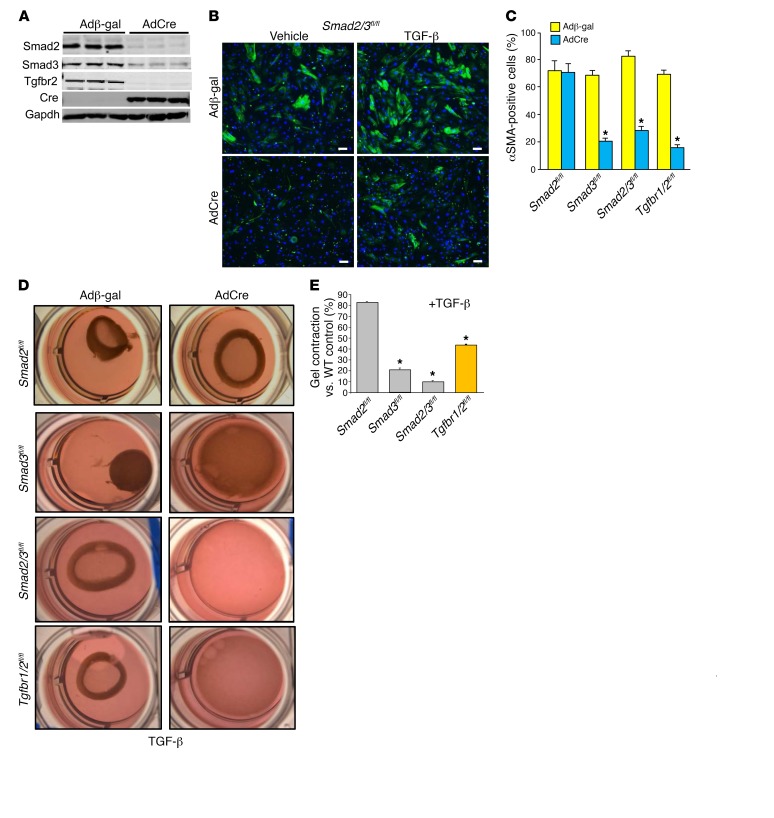
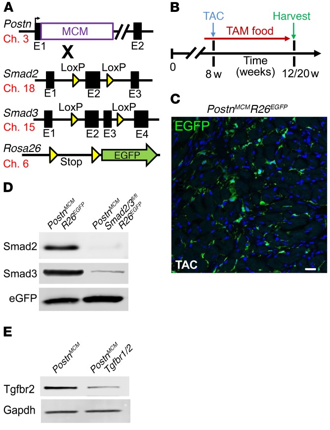
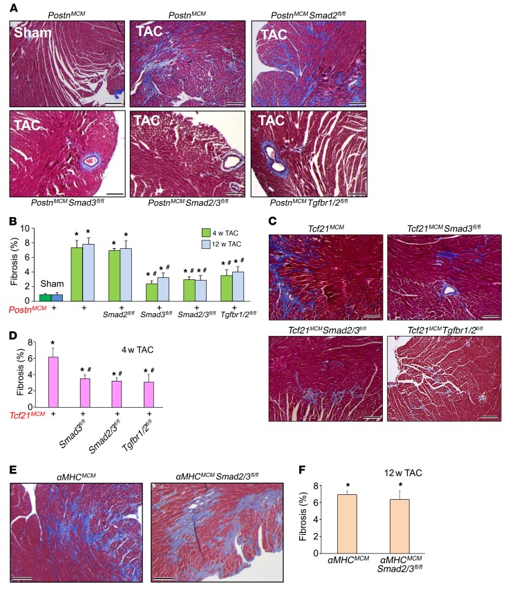
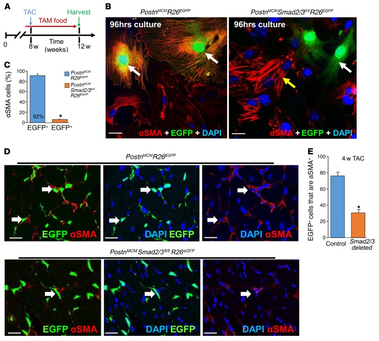
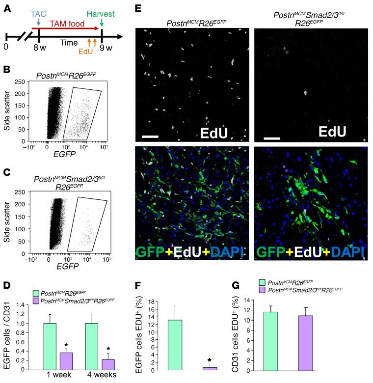
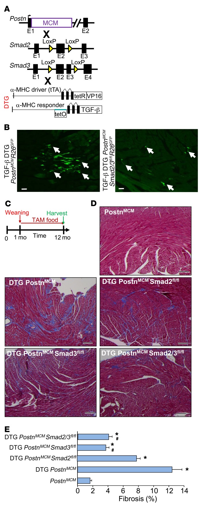
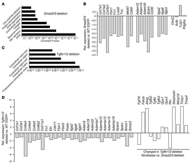
The master cytokine TGF-β mediates tissue fibrosis associated with inflammation and tissue injury. TGF-β induces fibroblast activation and differentiation into myofibroblasts that secrete extracellular matrix proteins. Canonical TGF-β signaling mobilizes Smad2 and Smad3 transcription factors that control fibrosis by promoting gene expression. However, the importance of TGF-β-Smad2/3 signaling in fibroblast-mediated cardiac fibrosis has not been directly evaluated in vivo. Here, we examined pressure overload-induced cardiac fibrosis in fibroblast- and myofibroblast-specific inducible Cre-expressing mouse lines with selective deletion of the TGF-β receptors Tgfbr1/2, Smad2, or Smad3. Fibroblast-specific deletion of Tgfbr1/2 or Smad3, but not Smad2, markedly reduced the pressure overload-induced fibrotic response as well as fibrosis mediated by a heart-specific, latency-resistant TGF-β mutant transgene. Interestingly, cardiac fibroblast-specific deletion of Tgfbr1/2, but not Smad2/3, attenuated the cardiac hypertrophic response to pressure overload stimulation. Mechanistically, loss of Smad2/3 from tissue-resident fibroblasts attenuated injury-induced cellular expansion within the heart and the expression of fibrosis-mediating genes. Deletion of Smad2/3 or Tgfbr1/2 from cardiac fibroblasts similarly inhibited the gene program for fibrosis and extracellular matrix remodeling, although deletion of Tgfbr1/2 uniquely altered expression of an array of regulatory genes involved in cardiomyocyte homeostasis and disease compensation. These findings implicate TGF-β-Smad2/3 signaling in activated tissue-resident cardiac fibroblasts as principal mediators of the fibrotic response.


Conflict of interest statement

Conflict of interest: The authors have declared that no conflict of interest exists.

Figures

Figure 1
Figure 1. Canonical TGF-β signaling mediates myofibroblast transformation in vitro.
(A) Western blot showing the expression levels of Smad2, Smad3, Tgfbr2, and Cre in skin fibroblasts 72 hours after infection with Adβ-gal and AdCre. (B) Representative immunofluorescent images of cells infected with Adβ-gal control or AdCre to delete Smad2/3 in skin fibroblasts from Smad2/3fl/fl mice. αSMA+ stress fibers are shown in green. Cells were treated with vehicle or 10 ng/ml of TGF-β for 24 hours. Scale bars: 10 μm. (C) Quantitation of the experiment shown in B except that additional genotypes of primary skin fibroblasts were used as indicated. Data are shown as ± SEM and are from 3 separate experiments. *P < 0.01 versus Adβ-gal. P values were calculated by 1-way ANOVA with post hoc Tukey’s HSD. (D and E) Pictures and quantification of collagen gel matrix contraction in culture dishes in Smad2-, Smad3-, Smad2/3-, and Tgfbr1/2-loxP–targeted cardiac fibroblasts infected with Adβ-gal or AdCre for 72 hours, then treated with 10 ng/ml of TGF-β for 24 hours. Adβ-gal–infected cells without TGF-β treatment showed no contraction in any of the genotypes at baseline (not shown). Data are shown as ± SEM and are from 3 separate experiments. *P < 0.01 versus WT Adβ-gal +TGF-β. P values were calculated with 1-way ANOVA with post hoc Tukey’s HSD. The wells in D are 28 mm across.
Figure 2
Figure 2. Generation of fibroblast-specific canonical TGF-β–deleted mice.
(A) Schematic representation of different mouse lines used, including the Postn genetic locus containing a tamoxifen-regulated MCM cDNA cassette inserted into exon 1 (E1), which was crossed with Smad2- and/or Smad3-loxP–containing gene-targeted lines, along with the Rosa26 reporter allele(R26EGFP). The mouse chromosome associated with each allele is shown. (B) Experimental scheme whereby mice were subjected to TAC injury or sham procedure for 4 and 12 weeks. Mice were fed tamoxifen diet 48 hours before surgery and then maintained on tamoxifen until harvesting. (C) Representative histological section showing EGFP-labeled interstitial cells in hearts of PostnMCM/+ R26EGFP/+ mice after 4 weeks of TAC injury (n = 4). Scale bar: 10 μm. (D) Western blot analysis for Smad2, Smad3, and EGFP from 500,000 cells isolated from hearts of the 2 genotypes of mice shown. (E) Western blot for Tgfbr2 in total heart mesenchymal cells lacking myocytes, CD31, and CD45 cells and conditionally targeted for Tgfbr1/2-loxP with PostnMCM in cardiac-activated fibroblasts after TAC stimulation.
Figure 3
Figure 3. Fibroblast-specific deletion of canonical TGF-β signaling reduces myocardial fibrosis in mice.
(A and B) Masson’s trichrome–stained histological pictures and quantitation of the area of fibrosis (blue) in hearts from the indicated genotypes of mice with PostnMCM/+ after 12 weeks of TAC injury and tamoxifen treatment. Average fibrotic area ± SEM. n = 8–10 mice in each group. *P < 0.05 versus sham operated; #P < 0.05 versus PostnMCM/+ TAC at 4 or 12 weeks. P values were calculated by 1-way ANOVA with post hoc Tukey’s HSD. (C and D) Masson’s trichrome–stained histological photographs and quantification of the area of fibrosis in hearts from the indicated mice crossed with Tcf21MCM/+ after 4 weeks of TAC injury and tamoxifen treatment. Average fibrotic area ± SEM. n = 6–9 mice in each group, *P < 0.05 versus sham operated; #P < 0.05 versus Tcf21MCM/+ TAC at 4 weeks. P values were calculated by 1-way ANOVA with post hoc Tukey’s HSD. (E and F) Masson’s trichrome–stained histological photographs and quantification of the area of fibrosis in αMHCMCM/+ and cardiomyocyte-specific deletion of Smad2/3 from hearts after 12 weeks of TAC injury. Mice were injected 5 times with 100 μl of 0.5 mg/ml of tamoxifen 15 days before injury. Average fibrotic area ± SEM. n = 7–9 mice in each group. *P < 0.05 versus sham operated. P values were calculated using 1-way ANOVA with post hoc Tukey’s HSD. Scale bars: 150 μm.
Figure 4
Figure 4. Fibroblast-specific Smad2/3 deletion alters myofibroblast activity in vivo.
(A) Experimental scheme whereby mice were subjected to TAC for 4 weeks. Mice were fed tamoxifen-laden chow 48 hours before surgery and then until harvesting. (B and C) Representative immunostaining images and quantitation for EGFP+ cellular expression (green) and costaining for αSMA (red) and DAPI (blue) in isolated cardiac interstitial cells from the indicated genotypes of mice that were cultured for 96 hours in low serum medium. The white arrows indicate EGFP+ cells and the yellow arrow shows EGFP cells that are αSMA+ (n = 4). *P < 0.05 versus PostnMCM/+ R26EGFP/+ cells. P values were calculated with Student’s t test. Scale bars: 10 μm. (D and E) Representative immunohistochemistry cryosections and quantitation of EGFP-labeled (green) interstitial cells along with αSMA (red) staining from hearts of the indicated genotypes of mice after 4 weeks of TAC. The white arrows show EGFP+ cells that are also αSMA+ in each of the 2 genotypes of mice, although in the Smad2/3-deleted hearts, most of the EGFP+ fibroblasts are deficient in αSMA expression (n = 4 mice in each group). *P < 0.05 versus PostnMCM R26EGFP hearts. P values were calculated with Student’s t test. Scale bars: 10 μm. Additional images of immunohistochemistry as shown in D are shown in Supplemental Figure 5.
Figure 5
Figure 5. Fibroblast-specific Smad2/3 deletion reduces the number of activated myofibroblasts in vivo.
(A) Experimental schematic whereby mice were subjected to TAC injury for 7 days. Mice were fed tamoxifen-laden chow 48 hours before surgery and then until harvesting. EdU was injected into mice 24 hours and 4 hours before harvest. (B and C) Representative flow cytometry plots of isolated EGFP+ interstitial cells (rightward scatter) from hearts of the indicated genotypes of mice. (D) The ratio of total GFP+ activated fibroblasts normalized to CD31+ cells from the heart taken from the indicated genotypes of mice after 1 or 4 weeks of TAC. Error bars represent SEM. n = 3 mice in each group. *P < 0.05 versus PostnMCM/+ R26EGFP/+. P values were calculated with Student’s t test. (E and F) Representative immunohistological images and quantitation of the percentage of EGFP+ interstitial cells costained for EdU (white) according to the schematic in A. DAPI was used to show nuclei (blue). n = 3 mice in each group. *P < 0.05 versus PostnMCM/+ R26EGFP/+. P values were calculated with Student’s t test. Scale bars: 50 μm. (G) Quantitation of CD31-positive cells that were also EdU positive in heart histological sections from mice subjected to TAC of the indicated genotypes. Representative images of the CD31-positive cells with EdU staining along with GFP-positive fibroblasts from histological heart sections are shown in Supplemental Figure 6.
Figure 6
Figure 6. Fibroblast-specific deletion of Smad2/3 reduces TGF-β–induced myocardial fibrosis.
(A) Schematic representation of PostnMCM mice crossed to Smad2- and/or Smad3-loxP mice in conjunction with an inducible heart-specific latency-escaping TGF-β mutant cDNA driven by a cardiomyocyte-specific DTG system. (B) Representative histological heart sections with GFP+ interstitial cells (white arrows) showing that the TGF-β DTG effectively mediated Cre-dependent induction of PostnMCM/+activity, facilitating R26EGFP recombination and expression, while deletion of Smad2/3 reduced total EGFP+ cells. Hearts were harvested 4 weeks after tamoxifen administration. n = 4 mice harvested. Scale bars: 10 μm. (C) Experimental schematic whereby data shown in D and E were from mice fed tamoxifen through 12 months of age. (D and E) Pictures of Masson’s trichrome–stained cardiac histological sections and quantitation of the area of fibrosis (blue staining) in the indicated genotypes of mice in the presence of the TGF-β mutant transgenic system (DTG) for 12 months. Data are shown as average fibrotic area ± SEM. n = 6–8 in each group. *P < 0.05 versus PostnMCM/+; #P < 0.05 versus DTG PostnMCM/+. P values were calculated by 1-way ANOVA with post hoc Tukey’s HSD. Scale bars: 150 μm.
Figure 7
Figure 7. Smad2/3 deletion in activated fibroblasts reduces fibroblast and ECM-related gene expression.
(A) Gene expression cluster analysis showed a profile of altered ECM and fibrosis-related gene expression in EGFP+ fibroblasts obtained from hearts of Smad2/3-activated fibroblast-deleted mice versus control WT hearts, isolated 4 weeks after TAC injury. (B) Quantification of selected mRNAs in Smad2/3-deleted EGFP+-activated fibroblasts from hearts of Smad2/3 PostnMCM/+ R26EGFP/+ allele–containing mice. (C) Gene expression cluster analysis shows a profile of altered ECM- and fibrosis-related gene expression in EGFP+ fibroblasts obtained from hearts of Tgfbr1/2 fibroblast–deleted mice versus control WT hearts, isolated 4 weeks after TAC injury. (D) Quantification of selected mRNAs in Tgfbr1/2-deleted EGFP+ fibroblasts from hearts of Tgfbr1/2 Tcf21MCM/+ R26EGFP/+ allele–containing mice. The white bars in D represent genes altered in expression compared with those with deletion of Smad2/3.

References

    1. Benjamin EJ, et al. Heart disease and stroke statistics-2017 update: a report from the American Heart Association. Circulation. 2017;135(10):e146–e603. doi: 10.1161/CIR.0000000000000485. - DOI - PMC - PubMed
    1. Bowers SL, Banerjee I, Baudino TA. The extracellular matrix: at the center of it all. J Mol Cell Cardiol. 2010;48(3):474–482. doi: 10.1016/j.yjmcc.2009.08.024. - DOI - PMC - PubMed
    1. Fan D, Takawale A, Lee J, Kassiri Z. Cardiac fibroblasts, fibrosis and extracellular matrix remodeling in heart disease. Fibrogenesis Tissue Repair. 2012;5(1):15. doi: 10.1186/1755-1536-5-15. - DOI - PMC - PubMed
    1. Xie M, Burchfield JS, Hill JA. Pathological ventricular remodeling: therapies: part 2 of 2. Circulation. 2013;128(9):1021–1030. doi: 10.1161/CIRCULATIONAHA.113.001879. - DOI - PMC - PubMed
    1. Souders CA, Bowers SL, Baudino TA. Cardiac fibroblast: the renaissance cell. Circ Res. 2009;105(12):1164–1176. doi: 10.1161/CIRCRESAHA.109.209809. - DOI - PMC - PubMed

MeSH terms

Substances