. 2017 Sep 10;9(9):121.
doi: 10.3390/cancers9090121.

The PI3Kδ Inhibitor Idelalisib Inhibits Homing in an in Vitro and in Vivo Model of B ALL

Affiliations

The PI3Kδ Inhibitor Idelalisib Inhibits Homing in an in Vitro and in Vivo Model of B ALL

Etai Adam et al. Cancers (Basel). .

Abstract

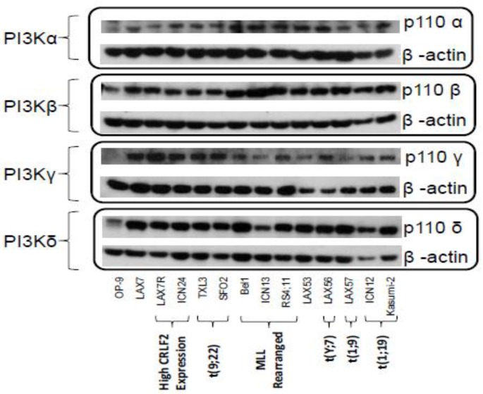
The quest continues for targeted therapies to reduce the morbidity of chemotherapy and to improve the response of resistant leukemia. Adhesion of acute lymphoblastic leukemia (ALL) cells to bone marrow stromal cells triggers intracellular signals that promote cell-adhesion-mediated drug resistance (CAM-DR). Idelalisib, an U.S. Food and Drug Administration (FDA)-approved PI3Kδ-specific inhibitor has been shown to be effective in CLL in down-regulating p-Akt and prolonging survival in combination with Rituximab; herein we explore the possibility of its use in B ALL and probe the mechanism of action. Primary B ALL in contact with OP9 stromal cells showed increased p-Aktser473. Idelalisib decreased p-Akt in patient samples of ALL with diverse genetic lesions. Addition of idelalisib to vincristine inhibited proliferation when compared to vincristine monotherapy in a subset of samples tested. Idelalisib inhibited ALL migration to SDF-1α in vitro and blocked homing of ALL cells to the bone marrow in vivo. This report tests PI3Kδ inhibitors in a more diverse group of ALL than has been previously reported and is the first published report of idelalisib inhibiting homing of ALL cells to bone marrow. Our data support further pre-clinical evaluation of idelalisib for the therapy of B ALL.

Keywords: ALL; CAM-DR; PI3K; drug resistance; idelalisib; leukemia; migration; mouse model.


Conflict of interest statement

Gilead Sciences provided the drugs and some funding for this research; aside from that the authors declare no conflict of interest.

Figures

Figure 1
Figure 1
PI3K isoforms in various B ALL cells demonstrated by Western blot (WB). WB performed for p110 subunit of each of the four class I isotypes of PI3K. OP9 cells serve as a co-culture control. Characteristics of each ALL sample are included below the name.
Figure 2
Figure 2
p-Akt activation of B ALL cells by integrin ligands or stromal cells. (A) Timeline of starvation and stroma/ligand stimulation assay. Primary B ALL cells LAX7R, ICN24, TXL3, and SFO2 were starved in medium for 4 h, then cultured with plates coated with integrin ligands, stromal cells, or a control. (B) Western blot of Akt phosphorylation in these cells.
Figure 3
Figure 3
Treatment with the PI3Kδ inhibitors idelalisib and GS-649443 decreases p-Akt levels in B ALL. (A) Timeline of starvation/stimulation assay. (B) Western blot showing relative phosphorylation of Akt after treatment with either Idelalisib or GS-649443.
Figure 4
Figure 4
Treatment with PI3Kδ inhibitors idelalisib and GS-649443 decreases migration toward SDF-1α. These are the results of a transwell migration assay wherein ALL cells move across a porous membrane towards SDF-1α (500 ng/mL). We compared the percentage of migrated ALL cells treated with Idelalisib or GS-649443 to DMSO control. Each condition was run in triplicate and the mean of triplicates ±95% CI is graphed above. All treated samples show significant decrease (p < 0.001) in migration compared to untreated samples, except TXL3 with GS-649443 which was significant to p < 0.01.
Figure 5
Figure 5
Inhibition of ALL homing to bone marrow after treatment of cells with idelalisib. (A) Timeline of experiment. (B) Scatterplot of CFUs of LAX56 cells recovered from tissues of mice after ex vivo treatment with idelalisib or DMSO control. Each dot represents the number of CFUs counted on a single plate that was seeded with 5 × 10⁴ mononuclear cells from the tissue specified.
Figure 6
Figure 6
Effect of different concentrations of idelalisib (Idela) on proliferation of ALL cells alone and in combination with Vincristine (5 nM). The mean of three counts under trypan blue exclusion is graphed ± SD for days 3 and 5, day 0 is plotted based on amount of cells first plated. Additionally, the inset is a table of p values from the t-test comparisons of results of different conditions at day 5. p values > 0.05 are listed as not significant (N/S). p values ≤ 0.005 are highlighted to show they meet our cutoff for statistical significance.

References

    1. Hunger S.P., Lu X., Devidas M., Camitta B.M., Gaynon P.S., Winick N.J., Reaman G.H., Carroll W.L. Improved survival for children and adolescents with acute lymphoblastic leukemia between 1990 and 2005: A report from the children’s oncology group. J. Clin. Oncol. 2012;30:1663–1669. doi: 10.1200/JCO.2011.37.8018. - DOI - PMC - PubMed
    1. Bhojwani D., Pui C.H. Relapsed childhood acute lymphoblastic leukaemia. Lancet Oncol. 2013;14:e205–e217. doi: 10.1016/S1470-2045(12)70580-6. - DOI - PubMed
    1. Shishido S., Bonig H., Kim Y.M. Role of integrin alpha4 in drug resistance of leukemia. Front. Oncol. 2014;4:99. doi: 10.3389/fonc.2014.00099. - DOI - PMC - PubMed
    1. Vivanco I., Sawyers C.L. The phosphatidylinositol 3-kinase akt pathway in human cancer. Nat. Rev. Cancer. 2002;2:489–501. doi: 10.1038/nrc839. - DOI - PubMed
    1. Chantry D., Vojtek A., Kashishian A., Holtzman D.A., Wood C., Gray P.W., Cooper J.A., Hoekstra M.F. P110delta, a novel phosphatidylinositol 3-kinase catalytic subunit that associates with p85 and is expressed predominantly in leukocytes. J. Biol. Chem. 1997;272:19236–19241. doi: 10.1074/jbc.272.31.19236. - DOI - PubMed

LinkOut - more resources