Review
. 2017 Sep 11;10(9):1066.
doi: 10.3390/ma10091066.

Review on the Antimicrobial Properties of Carbon Nanostructures

Affiliations
Review

Review on the Antimicrobial Properties of Carbon Nanostructures

Ahmed Al-Jumaili et al. Materials (Basel). .

Abstract

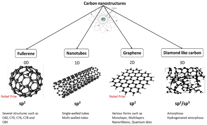
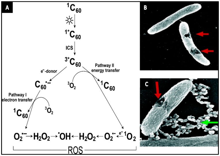
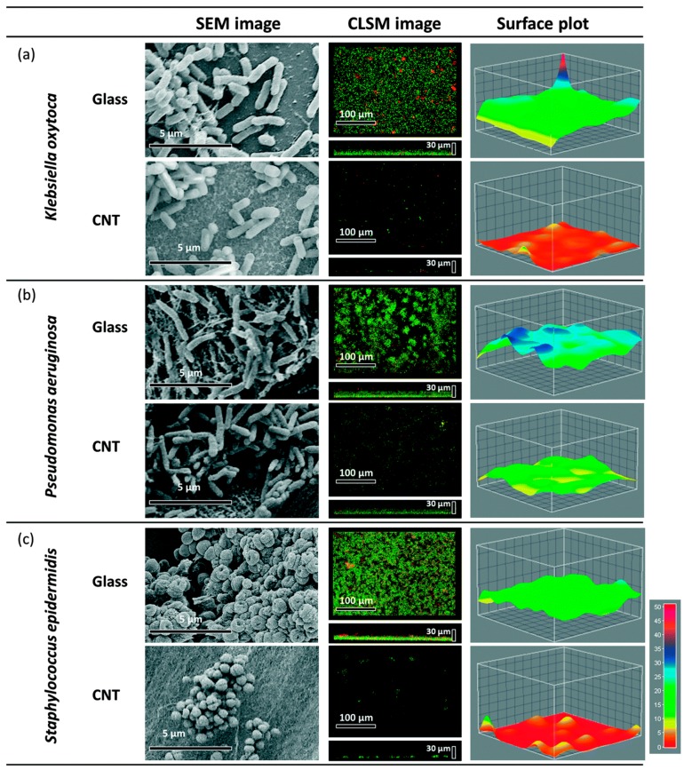
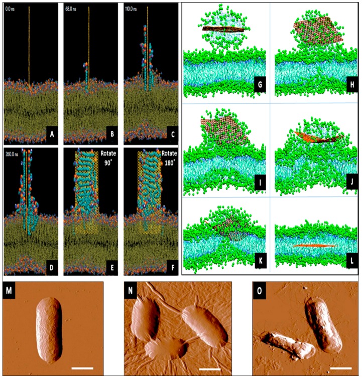
Swift developments in nanotechnology have prominently encouraged innovative discoveries across many fields. Carbon-based nanomaterials have emerged as promising platforms for a broad range of applications due to their unique mechanical, electronic, and biological properties. Carbon nanostructures (CNSs) such as fullerene, carbon nanotubes (CNTs), graphene and diamond-like carbon (DLC) have been demonstrated to have potent broad-spectrum antibacterial activities toward pathogens. In order to ensure the safe and effective integration of these structures as antibacterial agents into biomaterials, the specific mechanisms that govern the antibacterial activity of CNSs need to be understood, yet it is challenging to decouple individual and synergistic contributions of physical, chemical and electrical effects of CNSs on cells. In this article, recent progress in this area is reviewed, with a focus on the interaction between different families of carbon nanostructures and microorganisms to evaluate their bactericidal performance.

Keywords: antimicrobial properties; carbon nanostructures; carbon nanotubes; diamond-like carbon; fullerene; graphene.


Conflict of interest statement

The authors declare no conflict of interest.

Figures

Figure 1
Figure 1
Several forms of carbon nanostructures.
Figure 2
Figure 2
(A) Schematic representation of fullerene C60 photochemical pathways leading to reactive oxygen species (ROS) generation. Reprinted with permission from Reference [46]; (B,C) SEM images of S. oneidensis MR-1 cells treated with C60−NH2 show cellular damage. Cell samples were fixed for SEM images approximately 1 h after exposure to 20 mg/L C60−NH2. Green arrow points to nanoparticle aggregations and red arrow points to the damaged part of the cell. Reprinted with permission from Reference [56].
Figure 3
Figure 3
Scanning Electron Microscope (SEM), Confocal Scanning Laser Microscopy (CLSM) and surface plots of biofilm formation of K. oxytoca (a); P. aeruginosa (b) and S. epidermidis (c) on multi-wall carbon nanotubes (MWCNT) (tube length 540 μm) and glass control. Reprinted with permission from Reference [100].
Figure 4
Figure 4
Simulation 1 (AF) shows lipid extraction by a graphene nanosheet. An illustrative route of a fully restrained graphene docked at the surface of the outer membrane. The simulation time is shown in each snapshot; e and f are rotated counterclockwise by angle (90° and 180°) from its previous view. Reprinted with permission from Reference [37]; simulation 2 (GL) describes the process of self-insertion of graphene sheet into the phospholipid membrane. A graphene sheet merges with the membrane and releases the monolayer that enters the membrane. The snaps are taken at t GL = 2.9, 52.4, 120.0, 299.2, 356.4, and 516.4 ns, respectively. Reprinted with permission from Reference [120]; representative AFM images showing E. coli cells after incubation with: (M) deionized water without GO for 2 h; (N) 40 μg/mL GO-0 suspension for 2 h, and (O) the 40 μg/mL GO-240 suspension for 2 h. These images reveal the lateral dimension-dependent antibacterial performance of GO nanosheets. Larger GO sheets are covering most of the bacterial cell surface during the interaction compared to smaller nanosheets. The scale bars are 1 μm for all images. Reprinted with permission from Reference [131].

References

    1. Peng J., Gao W., Gupta B.K., Liu Z., Romero-Aburto R., Ge L., Song L., Alemany L.B., Zhan X., Gao G. Graphene quantum dots derived from carbon fibers. Nano Lett. 2012;12:844–849. doi: 10.1021/nl2038979. - DOI - PubMed
    1. Hahm M.G., Leela Mohana Reddy A., Cole D.P., Rivera M., Vento J.A., Nam J., Jung H.Y., Kim Y.L., Narayanan N.T., Hashim D.P. Carbon nanotube—Nanocup hybrid structures for high power supercapacitor applications. Nano Lett. 2012;12:5616–5621. doi: 10.1021/nl3027372. - DOI - PubMed
    1. Novoselov K.S., Fal V., Colombo L., Gellert P., Schwab M., Kim K. A roadmap for graphene. Nature. 2012;490:192–200. doi: 10.1038/nature11458. - DOI - PubMed
    1. Cao X., Shi Y., Shi W., Lu G., Huang X., Yan Q., Zhang Q., Zhang H. Preparation of novel 3D graphene networks for supercapacitor applications. Small. 2011;7:3163–3168. doi: 10.1002/smll.201100990. - DOI - PubMed
    1. Li H., He X., Liu Y., Huang H., Lian S., Lee S.-T., Kang Z. One-step ultrasonic synthesis of water-soluble carbon nanoparticles with excellent photoluminescent properties. Carbon. 2011;49:605–609. doi: 10.1016/j.carbon.2010.10.004. - DOI