

Simple Elimination of Background Fluorescence in Formalin-Fixed Human Brain Tissue for Immunofluorescence Microscopy
- PMID: 28892031
- PMCID: PMC5614400
- DOI: 10.3791/56188
Simple Elimination of Background Fluorescence in Formalin-Fixed Human Brain Tissue for Immunofluorescence Microscopy
Abstract
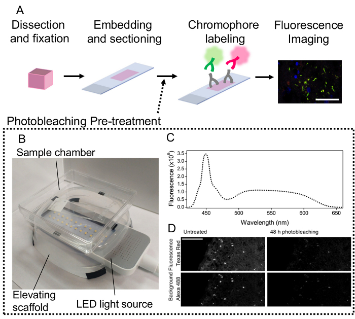
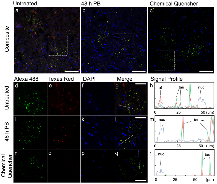
Immunofluorescence is a common method used to visualize subcellular compartments and to determine the localization of specific proteins within a tissue sample. A great hindrance to the acquisition of high quality immunofluorescence images is endogenous autofluorescence of the tissue caused by aging pigments such as lipofuscin or by common sample preparation processes such as aldehyde fixation. This protocol describes how background fluorescence can be greatly reduced through photobleaching using white phosphor light emitting diode (LED) arrays prior to treatment with fluorescent probes. The broad-spectrum emission of white phosphor LEDs allow for bleaching of fluorophores across a range of emission peaks. The photobleaching apparatus can be constructed from off-the-shelf components at very low cost and offers an accessible alternative to commercially available chemical quenchers. A photobleaching pre-treatment of the tissue followed by conventional immunofluorescence staining generates images free of background autofluorescence. Compared to established chemical quenchers which reduced probe as well as background signals, photobleaching treatment had no effect on probe fluorescence intensity while it effectively reduced background and lipofuscin fluorescence. Although photobleaching requires more time for pre-treatment, higher intensity LED arrays may be used to reduce photobleaching time. This simple method can potentially be applied to a variety of tissues, particularly postmitotic tissues that accumulate lipofuscin such as the brain and cardiac or skeletal muscles.

References
-
- Banerjee B, Miedema BE, Chandrasekhar HR. Role of basement membrane collagen and elastin in the autofluorescence spectra of the colon. J Investig Med. 1999;47(6):326–332. - PubMed
-
- Terman A, Brunk UT. Lipofuscin. Int J Biochem Cell Biol. 2004;36(8):1400–1404. - PubMed
-
- Del Castillo P, Llorente AR, Stockert JC. Influence of fixation, exciting light and section thickness on the primary fluorescence of samples for microfluorometric analysis. Basic Appl Histochem. 1989;33(3):251–257. - PubMed
-
- Ottis P, Koppe K, et al. Human and rat brain lipofuscin proteome. Proteomics. 2012;12(15-16):2445–2454. - PubMed
-
- Sun Y, Chakrabartty A. Cost-effective elimination of lipofuscin fluorescence from formalin-fixed brain tissue by white phosphor light emitting diode array. Biochem Cell Biol. 2016;94(6):545–550. - PubMed
Publication types
MeSH terms
Substances
Grants and funding
LinkOut - more resources
Full Text Sources
Other Literature Sources
