. 2017 Oct;24(10):825-833.
doi: 10.1038/nsmb.3466. Epub 2017 Sep 11.

Guide-bound structures of an RNA-targeting A-cleaving CRISPR-Cas13a enzyme

Affiliations

Guide-bound structures of an RNA-targeting A-cleaving CRISPR-Cas13a enzyme

Gavin J Knott et al. Nat Struct Mol Biol. 2017 Oct.

Abstract

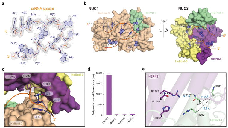
CRISPR adaptive immune systems protect bacteria from infections by deploying CRISPR RNA (crRNA)-guided enzymes to recognize and cut foreign nucleic acids. Type VI-A CRISPR-Cas systems include the Cas13a enzyme, an RNA-activated RNase capable of crRNA processing and single-stranded RNA degradation upon target-transcript binding. Here we present the 2.0-Å resolution crystal structure of a crRNA-bound Lachnospiraceae bacterium Cas13a (LbaCas13a), representing a recently discovered Cas13a enzyme subtype. This structure and accompanying biochemical experiments define the Cas13a catalytic residues that are directly responsible for crRNA maturation. In addition, the orientation of the foreign-derived target-RNA-specifying sequence in the protein interior explains the conformational gating of Cas13a nuclease activation. These results describe how Cas13a enzymes generate functional crRNAs and how catalytic activity is blocked before target-RNA recognition, with implications for both bacterial immunity and diagnostic applications.


Figures

Figure 1
Figure 1. Overall structure of the LbaCas13a:crRNA complex
a, Phylogenetic tree of all Cas13a family members with colored circles highlighting the A- and U-cleaving subfamilies (adapted from ). The homolog described in this study, LbaCas13a, is highlighted in yellow. b, Schematic representation of the crRNA repeat scaffold crystallized in this study. Nucleotides at the 5′ and 3′ end that were disordered in the structure are shown in semitransparent text. The ribose and nucleobase of G(14) were disordered. Only the phosphate of G(14) was modeled in the structure and is represented here as a dashed circle. c, Domain organization schematic of LbaCas13a shown with the REC, NUC, NUC1, and NUC2 lobes annotated. The color code used is consistent throughout the manuscript. d, Two views of the LbaCas13a:crRNA complex are shown related by a 180° rotation as cartoon (top) or surface (bottom) representations. The crRNA backbone is shown in orange.
Figure 2
Figure 2. Structure and recognition of the crRNA 5′ handle
a, Multiple sequence alignment of select type VI-A Cas13a crRNA 5′ handles representing both subfamilies adapted from . Nucleotides conserved across all Cas13a homologs, amongst just the U-cleaving, or A-cleaving subfamily are shown in grey, teal or purple respectively. The sites of pre-crRNA processing for each Cas13a homolog are noted by red arrows. LbaCas13a, is highlighted in yellow with the repeat nucleotides numbered negatively from the start of the spacer. b, Side by side comparison of the crRNA crystallized in this study (LbaCas13a, left) and previously (LshCas13a, right). c–f, Highlights of specific interactions between the crRNA 5′ handle and LbaCas13a. Purine stacks within the loop region of the 5′ handle interacting with Helical-1 and the NTD (c–d). Residues involved in conserved contacts between LbaCa13a and the crRNA loop are shown as sticks, with hydrogen-bonding interactions denoted by dashed lines. Binding of the 5′ flank (A(-28)-U(-24)) of LbaCas13a within the shallow Helical-1/HEPN2 groove via F422 and F1300 stacking with G(-26) and A(-25) respectively (e), and sequence-specific recognition of the dinucleotide 3′ AA-bulge (f).
Figure 3
Figure 3. Structure and recognition of the crRNA spacer
a, Simulated annealing mFo-DFc omit electron density map of the crRNA spacer contoured at 2σ. b, Opposing perspectives of the two halves of the NUC lobe that distort the crRNA spacer into a stabilized U-turn conformation. The half of the NUC lobe projecting out of the page is omitted in each image for clarity. c, The 3′ spacer emerging from the NUC lobe where it interacts with HEPN2. The spacer is shown in cartoon representation (orange) with the A-form nucleobases C(11)-G(13) shown as sticks. The amino acids F1338 and F1293 are shown as sticks stacking with C(11) gating access to the upstream spacer. d, Endpoint total fluorescence values for ssRNA-targeting assay by LbaCas13a HEPN mutants (background-corrected mean ± st. dev, n=3). Fitted apparent rates for these reactions are presented in Supplementary Fig. S4e. e, View of the composite HEPN nuclease active-site with the catalytic residues (RX4H) shown in stick representation. Notably, H605 of HEPN1-I is removed from the surface exposed composite site with distances shown.
Figure 4
Figure 4. Structural basis for crRNA maturation by LbaCas13a
a, The pre-crRNA processing groove formed between Helical-1 and HEPN2 domains of wild-type LbaCas13a (left). Close-up view of the pre-crRNA processing site in LbaCas13a (right). The 5′-hydroxyl and amino acids implicated in processing are shown as sticks with hydrogen-bonding interactions denoted by dashed lines. b, LbaCas13a-mediated pre-crRNA processing measured under single-turnover conditions for alanine substitutions within the pre-crRNA processing groove. For clarity, 60 min endpoints (mean ± st. dev, n=3) are graphed as the percentage of substrate remaining (uncleaved pre-crRNA). Time course data are plotted and rates calculated in Supplementary Fig. S4. c, Bar graph of LbaCas13a-mediated pre-crRNA processing under single-turnover conditions in the presence of EDTA or a modified pre-crRNA dG(-29). Percentage of uncleaved substrate was measured after 60 min (background correct mean ± st. dev, n=3). d, Close-up view of the pre-catalytic state of LbaCas13a (H328A) showing the pre-crRNA processing groove formed between Helical-1 and HEPN2 domains. The amino acids surrounding the scissile phosphate between A(-28) and G(-29) are shown as sticks with hydrogen-bonding interactions denoted by dashed lines. H328, marked with an asterisk, is shown in a ball-and-stick representation modeled in the conformation observed in the structure of wild-type LbaCas13a. e, Proposed model for acid-base-catalyzed maturation of pre-crRNA by LbaCas13a.

References

    1. Dy RL, Richter C, Salmond GP, Fineran PC. Remarkable Mechanisms in Microbes to Resist Phage Infections. Annu Rev Virol. 2014;1:307–31. - PubMed
    1. Barrangou R, Marraffini LA. CRISPR-Cas systems: Prokaryotes upgrade to adaptive immunity. Mol Cell. 2014;54:234–44. - PMC - PubMed
    1. Marraffini LA. CRISPR-Cas immunity in prokaryotes. Nature. 2015;526:55–61. - PubMed
    1. Hsu PD, Lander ES, Zhang F. Development and applications of CRISPR-Cas9 for genome engineering. Cell. 2014;157:1262–78. - PMC - PubMed
    1. Wright AV, Nunez JK, Doudna JA. Biology and Applications of CRISPR Systems: Harnessing Nature’s Toolbox for Genome Engineering. Cell. 2016;164:29–44. - PubMed

MeSH terms

Substances