. 2017 Oct;24(10):866-869.
doi: 10.1038/nsmb.3464. Epub 2017 Sep 11.

Structures of the human mitochondrial ribosome in native states of assembly

Affiliations

Structures of the human mitochondrial ribosome in native states of assembly

Alan Brown et al. Nat Struct Mol Biol. 2017 Oct.

Abstract

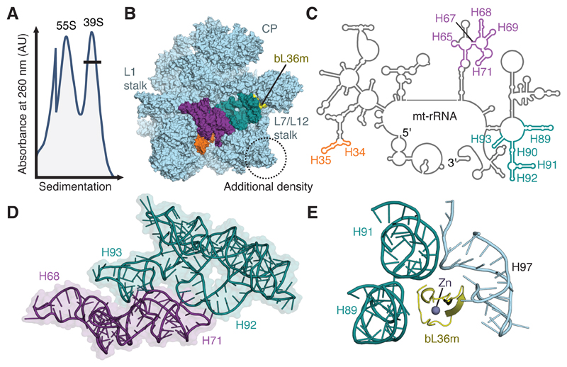
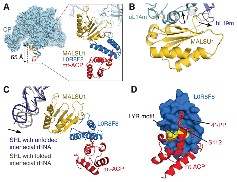
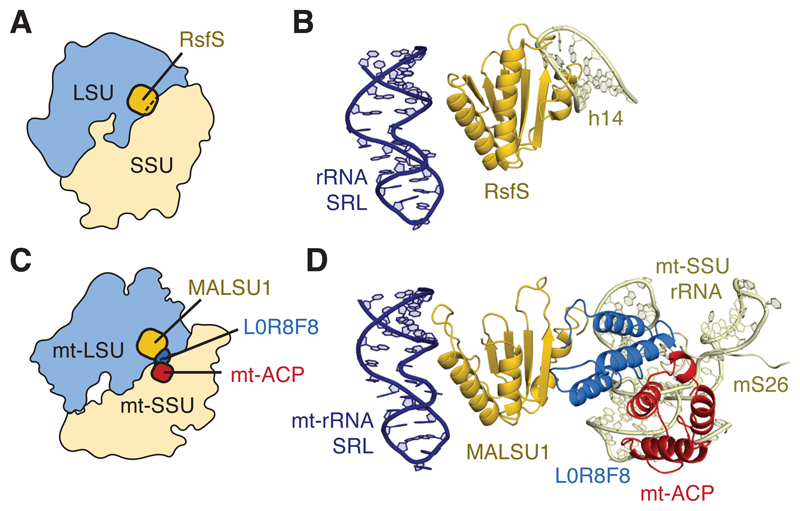
Mammalian mitochondrial ribosomes (mitoribosomes) have less rRNA content and 36 additional proteins compared with the evolutionarily related bacterial ribosome. These differences make the assembly of mitoribosomes more complex than the assembly of bacterial ribosomes, but the molecular details of mitoribosomal biogenesis remain elusive. Here, we report the structures of two late-stage assembly intermediates of the human mitoribosomal large subunit (mt-LSU) isolated from a native pool within a human cell line and solved by cryo-EM to ∼3-Å resolution. Comparison of the structures reveals insights into the timing of rRNA folding and protein incorporation during the final steps of ribosomal maturation and the evolutionary adaptations that are required to preserve biogenesis after the structural diversification of mitoribosomes. Furthermore, the structures redefine the ribosome silencing factor (RsfS) family as multifunctional biogenesis factors and identify two new assembly factors (L0R8F8 and mt-ACP) not previously implicated in mitoribosomal biogenesis.


Conflict of interest statement

Competing financial interests

The authors declare no competing financial interests.

Figures

Figure 1
Figure 1. Purification and structural characterization of native mitoribosomal assembly intermediates
(A) Differential centrifugation separates intact mitoribosomes (55S) from a pool of mt-LSU-like complexes (39S). This pool contains two well-defined assembly intermediates that differ in the presence of folded interfacial rRNA. (B) View of the assembly intermediate with folded interfacial rRNA viewed from the intersubunit interface. Both intermediates feature additional density (circled) relative to the mt-LSU of intact 55S mitoribosomes. However, one class displays unfolded interfacial rRNA with density for H34-35 (orange), H65 and H67-71 (purple), and H89-93 (teal) absent, along with protein bL36m (yellow). Landmark features of the mitoribosome are labeled, including the two stalks and central protuberance (CP). (C) Secondary structure diagram for mt-rRNA with sections of unfolded rRNA colored according to B. (D) Interconnectivity of mt-rRNA helices H67-71 (purple) and H89-93 (teal). (E) In the mature mitoribosome, bL36m coordinates H89 and H91 that are absent in the reconstruction with H97.
Figure 2
Figure 2. A module of MALSU1–L0R8F8–mt-ACP binds both mt-LSU assembly intermediates.
(A) Location of the MALSU1–L0R8F8–mt-ACP module, shown here bound to mt-LSU with unfolded interfacial rRNA (viewed from the side). The module extends from the surface of the mt-LSU by ~65 Å. (B) Binding of MALSU1 induces conformational changes in uL14m and bL19m from their positions in the 55S mitoribosome (shown in grey, with the direction of movement indicated with arrows). Residues of uL14 (T97, R98, and K114) that, when mutated to alanine, disrupt binding of RsfS are mapped to the uL14m structure (T117, R118, and K136) and are shown in stick representation. (C) MALSU1 interacts electrostatically with the SRL (H95) of the mt-rRNA. The SRL makes a closer association with MALSU1 when the interfacial rRNA is folded. (D) The interaction between L0R8F8 and mt-ACP involves the LYR motif of L0R8F8 (colored yellow) and the 4´-phosphopantetheine modification of mt-ACP serine 112.
Figure 3
Figure 3. Models of anti-association activity.
(A) Schematic showing the position of RsfS relative to the bacterial ribosome. RsfS overlaps with the position of the ribosomal small subunit (SSU). (B) RsfS would clash with rRNA helix 14 (h14) of the SSU. (C) Schematic showing that MALSU1 alone cannot inhibit mitoribosomal subunit joining by steric hindrance: only together with L0R8F8–mt-ACP does the module bridge the distance between the mt-LSU and mt-SSU in the mature human mitoribosome. (D) L0R8F8–mt-ACP would clash with regions of mt-rRNA around helices h5 and h15 and the N-terminus of mS26 in the mt-SSU, thereby preventing subunit association.

References

    1. Amunts A, Brown A, Toots J, Scheres SHW, Ramakrishnan V Ribosome. The structure of the human mitochondrial ribosome. Science. 2015;348:95–98. - PMC - PubMed
    1. Greber BJ, et al. Ribosome. The complete structure of the 55S mammalian mitochondrial ribosome. Science. 2015;348:303–308. - PubMed
    1. Adilakshmi T, Bellur DL, Woodson SA. Concurrent nucleation of 16S folding and induced fit in 30S ribosome assembly. Nature. 2008;455:1268–1272. - PMC - PubMed
    1. Davis JH, et al. Modular Assembly of the Bacterial Large Ribosomal Subunit. Cell. 2016;167:1610–1622.e15. - PMC - PubMed
    1. Bogenhagen DF, Martin DW, Koller A. Initial steps in RNA processing and ribosome assembly occur at mitochondrial DNA nucleoids. Cell Metab. 2014;19:618–629. - PubMed

MeSH terms