. 2018 Mar;20(3):571-581.
doi: 10.1111/dom.13113. Epub 2017 Oct 10.

Sitagliptin and Roux-en-Y gastric bypass modulate insulin secretion via regulation of intra-islet PYY

Affiliations

Sitagliptin and Roux-en-Y gastric bypass modulate insulin secretion via regulation of intra-islet PYY

Claudia Guida et al. Diabetes Obes Metab. 2018 Mar.

Abstract

Aims: The gut hormone peptide tyrosine tyrosine (PYY) is critical for maintaining islet integrity and restoring islet function following Roux-en-Y gastric bypass (RYGB). The expression of PYY and its receptors (NPYRs) in islets has been documented but not fully characterized. Modulation of islet PYY by the proteolytic enzyme dipeptidyl peptidase IV (DPP-IV) has not been investigated and the impact of DPP-IV inhibition on islet PYY function remains unexplored. Here we have addressed these gaps and their effects on glucose-stimulated insulin secretion (GSIS). We have also investigated changes in pancreatic PYY in diabetes and following RYGB.

Methods: Immunohistochemistry and gene expression analysis were used to assess PYY, NPYRs and DPP-IV expression in rodent and human islets. DPP-IV activity inhibition was achieved by sitagliptin. Secretion studies were used to test PYY and the effects of sitagliptin on insulin release, and the involvement of GLP-1. Radioimmunoassays were used to measure hormone content in islets.

Results: PYY and DPP-IV localized in different cell types in islets while NPYR expression was confined to the beta-cells. Chronic PYY application enhanced GSIS in rodent and diabetic human islets. DPP-IV inhibition by sitagliptin potentiated GSIS; this was mediated by locally-produced PYY, and not GLP-1. Pancreatic PYY was markedly reduced in diabetes. RYGB strongly increased islet PYY content, but did not lead to full restoration of pancreatic GLP-1 levels.

Conclusion: Local regulation of pancreatic PYY, rather than GLP-1, by DPP-IV inhibition or RYGB can directly modulate the insulin secretory response to glucose, indicating a novel role of pancreatic PYY in diabetes and weight-loss surgery.

Keywords: Roux-En-Y gastric bypass; dipeptidyl peptidase IV; glucagon-like peptide-1; insulin; islets; peptide tyrosine tyrosine.


Conflict of interest statement

No potential conflicts of interest relevant to this article were reported.

Figures

Figure 1
Figure 1
PYY protein localization in rat, mouse and human islets. A, Representative confocal microscopy images from rat (upper panel) and human sections (lower panel) stained with PYY (green) and insulin, glucagon, somatostatin or PP. B, Immungold labelling for PYY on mouse islets (upper panels) and human islets (lower panels). The density of gold labelling was much higher in mouse islets compared to human islets. In the mouse, labelling was present over glucagon granules (GGN, electron dense with no halo and an additional very dense core), somatostatin granules (SST, electron pale and slightly angular) and pancreatic polypeptide granules (PP, similar to glucagon but smaller and more variable in size) but not on insulin granules (Ins, dense core and clear halo). In insulin‐containing β‐cells primary and secondary lysosomes (Ly, inset in middle panel) were densely labelled with immunogold for PYY. In human islets no convincing labelling of any granule type was found. Lysosomes were identified by their pale electron density and different sizes, usually bounded by a clear membrane. M, mitochondrion; n, nucleus. Scale bars, 0.5 μm
Figure 2
Figure 2
NPY receptor expression in rodent islets. Representative confocal microscopy images of NPY1R staining in rat islets co‐stained with insulin, glucagon or somatostatin
Figure 3
Figure 3
NPY receptor expression in human islets. Representative confocal microscopy images of A, NPY1R and B, NPY4R staining in human islets co‐stained with insulin, glucagon or somatostatin
Figure 4
Figure 4
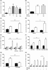
Time and dose‐dependent modulation of insulin secretion by PYY. A, Insulin secretion from mouse islets cultured without (black bars) or with (white bars) 100 nM PYY (1‐36) for the indicated time points. Secretion was measured from islets stimulated with 1 mM or 20 mM glucose. Data are presented as percentage of basal secretion (mean ± SEM as percentage of content; 1 hour, control: 0.0196 ± 0.0045, with PYY: 0.0190 ± 0.0018; 6 hours, control: 0.073 ± 0.016, with PYY: 0.121 ± 0.02; 24 hours, control: 0.0189 ± 0.0063, with PYY: 0.0198 ± 0.0028; 72 hours, control: 0.043 ± 0.006, with PYY: 0.033 ± 0.0003). B, Insulin secretion from mouse islets chronically cultured (72 hours) with PYY (1‐36) or (3‐36) at the indicated concentration. Secretion was measured from islets stimulated with 1 mM (black bars) or 20 mM glucose (white bars). Data are presented as percentage of basal secretion (mean ± SEM as percentage of content; control: 0.478 ± 0.073, with PYY (1‐36; 100pM): 0.244 ± 0.033, PYY (1‐36; 100 nM): 0.278 ± 0.023, PYY (3‐36; 100pM): 0.471 ± 0.072, PYY (3‐36; 100 nM): 0.249 ± 0.036). C, Insulin secretion from human islets stimulated with 1 mM (black bars), 6 mM (grey bars) or 20 mM (white bars) glucose in absence or presence of 100 nM PYY (1–36). D, Insulin secretion from human islets isolated from 2 diabetic donors cultured for 72 hours with or without 100 nM PYY (1‐36). Insulin release was measured from islets stimulated with 1 mM (black bars) or 20 mM glucose (white bars); for each diabetic donor, experiments were done in groups of 3 to 4, totalling 7 to 8 replicates and statistical analysis was performed on these. *P < .05, **P < .01 for indicated comparisons
Figure 5
Figure 5
Effects of sitaglipin on insulin secretion are mediated by islet PYY. A and B, Insulin secretion from mouse (A) and human (B) islets stimulated acutely with 1 mM or 20 mM glucose, with varying doses of sitagliptin (STG). C and D, Insulin secretion from mouse (C) and human (D) islets chronically cultured with or without 100 nM sitagliptin. Secretion was measured from islets stimulated with 1 mM (black bars) or 20 mM glucose (white bars). Data are presented as percentage of basal secretion (mean ± SEM as percentage of content; control: 0.123 ± 0.059, sitagliptin: 0.112 ± 0.031 (for mouse islets), control: 0.378 ± 0.055, sitagliptin: 0.558 ± 0.123 (for human islets). E, Insulin secretion from rodent islets chronically cultured in the presence of sitagliptin, sitagliptin + exendin (9‐39) or sitagliptin + anti‐PYY antibody. Secretion was measured in islets stimulated with 1 mM (black bars), 6 mM (grey bars) or 20 mM glucose (white bars). Data are presented as percentage of basal secretion (mean ± SEM as percentage of content; control: 0.105 ± 0.004, sitagliptin: 0.08 ± 0.014, sitagliptin +exendin (9‐39): 0.09 ± 0.012, sitagliptin + anti‐PYY: 0.09 ± 0.008). F, Insulin secretion at 1 mM (black bars) and 20 mM (white bars) glucose from islets isolated from wildtype (WT) and GLP‐1 receptor knockout (GLP‐1R KO) mice and cultured chronically with 100 nM sitaglipin. Data are presented as percentage of basal secretion (mean ± SEM as percentage of content; WT with no sitagliptin: 0.006 ± 0.002, WT + sitagliptin: 0.049 ± 0.005, GLP‐1R KO: 0.046 ± 0.008). G, Insulin secretion from mouse islets chronically treated with sitagliptin in the absence or presence of 1 μM BIBP. Data are presented as percentage of basal secretion (mean ± SEM as percentage of content; sitagliptin: 0.049 ± 0.005, sitagliptin + BIBP: 0.049 ± 0.005). H, Insulin secretion from mouse islets chronically treated with 100 nM PYY (1‐36) or PYY (3‐36) or 100 nM sitagliptin. Secretion was measured in islets stimulated with 1 mM (black bars) or 20 mM glucose (white bars). Data are presented as percentage of basal secretion (mean ± SEM as percentage of content; control: 0.478 ± 0.073, PYY (1‐36): 0.278 ± 0.023, PYY (3‐36): 0.249 ± 0.036, sitagliptin: 0.333 ± 0.019, sitagliptin + PYY (1‐36): 0.344 ± 0.026, sitagliptin + PYY (3‐36): 0.325 ± 0.048). *P < .05, **P < .01 for indicated comparisons
Figure 6
Figure 6
Islet PYY levels increase after RYGB. A, Plasma PYY levels in control rats (n = 4) and diabetic GK rats (n = 7). B, PYY content from islets isolated from control rats (n = 9), diabetic sham‐operated GK rats (n = 12) and RYGB‐operated GK rats (n = 11). PYY content is presented in pg/islet. Data are presented as mean ± SEM. *P < .05 vs control rats, ***P < .001 vs control rat islets, ###P < .001 for indicated comparison

Similar articles

Cited by

References

    1. Ramracheya RD, McCulloch LJ, Clark A, et al. PYY‐dependent restoration of impaired insulin and glucagon secretion in type 2 diabetes following Roux‐En‐Y gastric bypass surgery. Cell Rep. 2016;15:944–950. - PMC - PubMed
    1. Sam AH, Gunner DJ, King A, et al. Selective ablation of peptide YY cells in adult mice reveals their role in beta cell survival. Gastroenterology. 2012;143:459–468. - PubMed
    1. Khan D, Vasu S, Moffett RC, Irwin N, Flatt PR. Islet distribution of Peptide YY and its regulatory role in primary mouse islets and immortalised rodent and human beta‐cell function and survival. Mol Cell Endocrinol. 2016;436:102–113. - PubMed
    1. Shi YC, Loh K, Bensellam M, et al. Pancreatic PYY is critical in the control of insulin secretion and glucose homeostasis in female mice. Endocrinology. 2015;156:3122–3136. - PubMed
    1. Persaud SJ, Bewick GA. Peptide YY: more than just an appetite regulator. Diabetologia. 2014;57:1762–1769. - PubMed

Publication types