. 2017 Nov 17;8(8):1820-1832.
doi: 10.1080/21505594.2017.1377881. Epub 2017 Nov 24.

Susceptibility of Mycobacterium tuberculosis-infected host cells to phospho-MLKL driven necroptosis is dependent on cell type and presence of TNFα

Affiliations

Susceptibility of Mycobacterium tuberculosis-infected host cells to phospho-MLKL driven necroptosis is dependent on cell type and presence of TNFα

Rachel E Butler et al. Virulence. .

Abstract

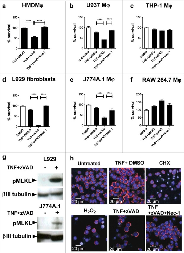
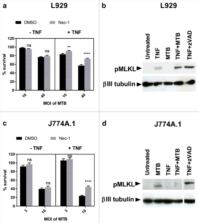
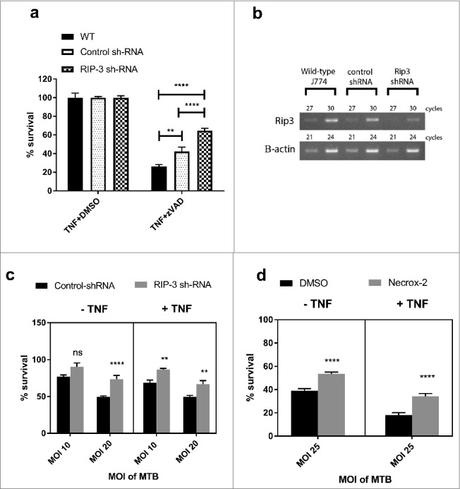
An important feature of Mycobacterium tuberculosis pathogenesis is the ability to control cell death in infected host cells, including inhibition of apoptosis and stimulation of necrosis. Recently an alternative form of programmed cell death, necroptosis, has been described where necrotic cell death is induced by apoptotic stimuli under conditions where apoptotic execution is inhibited. We show for the first time that M. tuberculosis and TNFα synergise to induce necroptosis in murine fibroblasts via RIPK1-dependent mechanisms and characterized by phosphorylation of Ser345 of the MLKL necroptosis death effector. However, in murine macrophages M. tuberculosis and TNFα induce non-necroptotic cell death that is RIPK1-dependent but independent of MLKL phosphorylation. Instead, M. tuberculosis-infected macrophages undergo RIPK3-dependent cell death which occurs both in the presence and absence of TNFα and involves the production of mitochondrial ROS. Immunocytochemical staining for MLKL phosphorylation further demonstrated the occurrence of necroptosis in vivo in murine M. tuberculosis granulomas. Phosphorylated-MLKL immunoreactivity was observed associated with the cytoplasm and nucleus of fusiform cells in M. tuberculosis lesions but not in proximal macrophages. Thus whereas pMLKL-driven necroptosis does not appear to be a feature of M. tuberculosis-infected macrophage cell death, it may contribute to TNFα-induced cytotoxicity of the lung stroma and therefore contribute to necrotic cavitation and bacterial dissemination.

Keywords: MLKL; Mycobacterium tuberculosis; RIPK1; RIPK3; fibroblast; macrophage; necroptosis.


Figures

Figure 1.
Figure 1.
Necroptosis of macrophage cell lines, human MDMs and murine fibroblasts treated with TNFα+zVAD. (a) Human monocyte-derived macrophages (HMDMφ) were treated for 20 hours with 50ng/mL TNF+DMSO, TNF+30 µM zVAD and TNF+30 µM zVAD + 30 µM Nec-1, before measuring cell survival using a crystal violet assay (normalised to TNFα+DMSO treated control cells). Results are mean +/- SEM n = 4, and are representative of 3 independent experiments. (b) U937 macrophages were treated for 20 hours with 50ng/mL TNF, TNF+30 µM zVAD and TNF+30 µM zVAD + 30 µM Nec-1, before measuring cell survival using a crystal violet assay (normalised to untreated control cells). Results are mean +/- SEM n = 10, and are representative of at least 2 independent experiments. (c) THP-1 were treated for 20 hours with 50ng/mL TNF+DMSO, TNF+30 µM zVAD, and TNF + 30 µM zVAD + 30 µM Nec-1, before measuring cell survival using a crystal violet assay (normalised to DMSO treated control cells). Results are mean +/- SEM n = 10. (d) L929 fibroblasts, (e) J774A.1 macrophages and (f) RAW macrophages were treated for 20 hours with 10ng/ml TNF+ DMSO, TNF + 30 µM zVAD, and TNF + 30 µM zVAD + 30 µM Nec-1. Results are mean +/- SEM n = 10, are expressed as a percentage of DMSO treated controls, and representative of 2–3 independent experiments. Statistics are one way ANOVA with Tukey's post-test. ****p<0.0001. (g) Western blot of lysates of L929 fibroblasts and J774A.1 macrophages that had been treated with 10ng/ml TNFα and 25ng/ml TNFα respectively in the presence of 30 µM zVAD.fmk for 18 hours, developed with antibodies against MLKL phosphorylated at Ser345, or βIII tubulin as a loading control. Results are representative of 2–3 independent experiments. (h) J774A.1 macrophages were seeded on glass slide flasks and untreated or treated with cyclohexamide 2.5 µg/ml, 0.5mM H2O2, TNFα 25ng/ml, TNF + 30 µM zVAD, and TNF + 30 µM zVAD + 30 µM Nec-1. After 20 hours, cells were stained with Alexa-568-phalloidin (displayed as red) and counterstained with DRAQ5 (displayed as blue) before viewing by confocal microscopy. Results are representative of 2 independent experiments.
Figure 2.
Figure 2.
M. tuberculosis induces RIPK1-dependent cell death in the presence of excess TNFα in fibroblasts and macrophages, but only induces MLKL phosphorylation in fibroblasts. (a) L929 murine fibroblasts were infected with M. tuberculosis for 24 hours, then treated with DMSO or 30 µM Nec-1 in the presence or absence of 10ng/ml TNFα for 18 hours, before measuring cell survival using a crystal violet assay. Results are mean +/- SEM of n = 6 samples, and are expressed as a percentage of the uninfected control of each treatment. Statistics are two way ANOVA with Sidak post-test. Ns not significant; **p<0.01; ****p<0.0001. (b) Western blot of L929 fibroblasts infected with MOI 20 M. tuberculosis for 24 hours, then treated with TNFα for 24 hours, or with TNFα + 3 µM zVAD for 18 hours, developed with anti-pMLKL antibody and anti beta-III tubulin antibody. (c) J774A.1 murine macrophages were infected with M. tuberculosis for 3 hours, then treated with DMSO or 30 µM Nec-1 in the presence or absence of 25ng/ml TNFα for 48 hours. Results are mean +/- SEM of n = 10 samples, and are expressed as a percentage of the uninfected control of each treatment. Statistics are two way ANOVA with Sidak post-test. Ns not significant; ****p<0.0001. (d) Western blot of J774A.1 cells infected with MOI 10 M. tuberculosis for 3 hours and subsequently treated with 25ng/ml TNFα for 24 hours, or with TNF + 30 µM zVAD for 18 hours, developed with anti-pMLKL antibody and anti beta-III tubulin antibody. (a-d) All results are representative of at least 2 similar experiments.
Figure 3.
Figure 3.
RIPK3 and mitochondrial ROS mediate M. tuberculosis-induced cell death both in the presence and absence of TNFα. (a) WT J774A.1 macrophages, RIP3K shRNA knockdown J774A.1 macrophages, and control shRNA J774A.1 macrophages were treated for 20 hours with DMSO control or 30 µM zVAD.fmk in the presence of 25ng/mL TNF, before measuring cell survival using a crystal violet assay. Results are mean +/- SEM of n = 10 samples and are expressed as a percentage of the TNFα+DMSO-treated control for each condition. Statistics are two way ANOVA with Sidak post-test. **p<0.01; ****p<0.0001. Results are representative of 3 independent experiments. (b) Knockdown of RIP3K mRNA was confirmed by RT-PCR, using murine RIP-3 and beta-actin primers (sc-61483-PR and sc-29192-PR, Santa Cruz). (c) RIP3K hRNA knockdown J774A.1 macrophages, and control shRNA J774A.1 macrophages were infected with M. tuberculosis for 3 hours, and subsequently incubated in the absence or presence of 25ng/ml TNF for 48 hours, before measuring cell survival using a crystal violet assay. Results are mean +/- SEM of n = 10 samples, and are expressed as a percentage of the uninfected control of each treatment. Statistics are two way ANOVA with Sidak post-test. *p>0.05; ***p<0.001; ****p<0.0001. Results are representative of 2 independent experiments (d) J774A.1 macrophages were infected with M. tuberculosis for 3 hours and incubated with Necrox-2 in the presence and absence of TNFα for 24 hours, before measuring cell survival using a crystal violet assay. Results are mean +/- SEM of n = 10 samples, and are expressed as a percentage of the uninfected control of each treatment. Statistics are one way ANOVA with Tukey's post-test. *p>0.05; ***p<0.001; ****p<0.0001.
Figure 4.
Figure 4.
Murine tuberculosis granulomas contain non-macrophage cells undergoing pMLKL driven necroptosis. Mice were infected with M. tuberculosis by intranasal challenge and lung pathology analysed at 21d.p.i. (A) H&E stain. Granulomatous inflammation within the lung with abundant foamy macrophages and small areas of necrosis (asterisk). Original magnification: 100X. Inset shows Ziehl-Neelsen staining revealing numerous acid fast bodies (AFBs) present within the lesion. Original magnification: 400X. (B) Non-immune 1 ͦ antibody isotype control staining for immunohistochemistry. Original magnification: 400X. (C) pMLKL immunohistochemistry. Positive staining within the cytoplasm of elongated cells within the granulomatous inflammation and necrosis. Original magnification: 400X. (D) pMLKL immunohistochemistry. Positive staining is observed predominantly in cells with fusiform-nuclei (black arrow) adjacent to non-immunoreactive foamy macrophages (white arrow) and necrotic cell debris. Original magnification: 400X.

Similar articles

Cited by

References

    1. Bermudez LE, Goodman J. Mycobacterium tuberculosis invades and replicates within type II alveolar cells. Infect Immunity. 1996;64:1400–6 - PMC - PubMed
    1. Hernandez-Pando R, Jeyanathan M, Mengistu G, Aguilar D, Orozco H, Harboe M, Rook GA, Bjune G. Persistence of DNA from Mycobacterium tuberculosis in superficially normal lung tissue during latent infection. Lancet. 2000;356:2133–8. doi: 10.1016/S0140-6736(00)03493-0. PMID:11191539 - DOI - PubMed
    1. Mariotti S, Sargentini V, Pardini M, Giannoni F, De Spirito M, Gagliardi MC, Greco E, Teloni R, Fraziano M, Nisini R. Mycobacterium tuberculosis may escape helper T cell recognition by infecting human fibroblasts. Hum Immunol. 2013;74:722–9. doi: 10.1016/j.humimm.2013.02.005. PMID:23459076 - DOI - PubMed
    1. Harding CV, Boom WH. Regulation of antigen presentation by Mycobacterium tuberculosis: a role for Toll-like receptors. Nat Rev Microbiol. 2010;8:296–307. doi: 10.1038/nrmicro2321. PMID:20234378 - DOI - PMC - PubMed
    1. Russell DG. Mycobacterium tuberculosis and the intimate discourse of a chronic infection. Immunol Rev. 2011;240:252–68. doi: 10.1111/j.1600-065X.2010.00984.x. PMID:21349098 - DOI - PMC - PubMed

Publication types

MeSH terms