. 2017 Sep 11;13(9):e1006981.
doi: 10.1371/journal.pgen.1006981. eCollection 2017 Sep.

Self-fertility in Chromocrea spinulosa is a consequence of direct repeat-mediated loss of MAT1-2, subsequent imbalance of nuclei differing in mating type, and recognition between unlike nuclei in a common cytoplasm

Affiliations

Self-fertility in Chromocrea spinulosa is a consequence of direct repeat-mediated loss of MAT1-2, subsequent imbalance of nuclei differing in mating type, and recognition between unlike nuclei in a common cytoplasm

Sung-Hwan Yun et al. PLoS Genet. .

Abstract

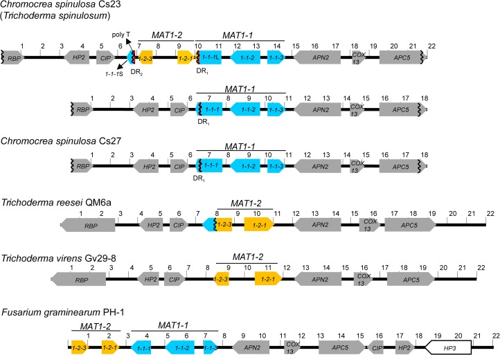
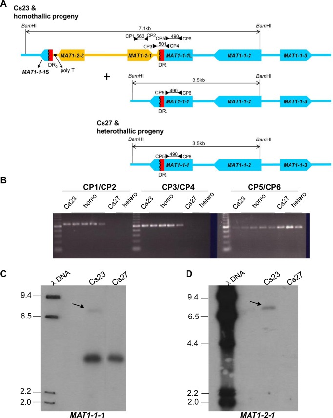
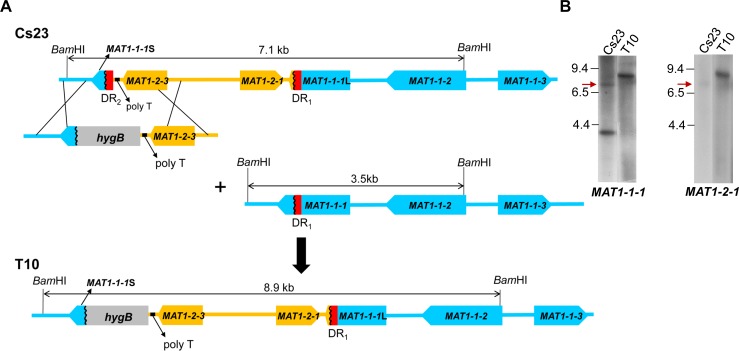
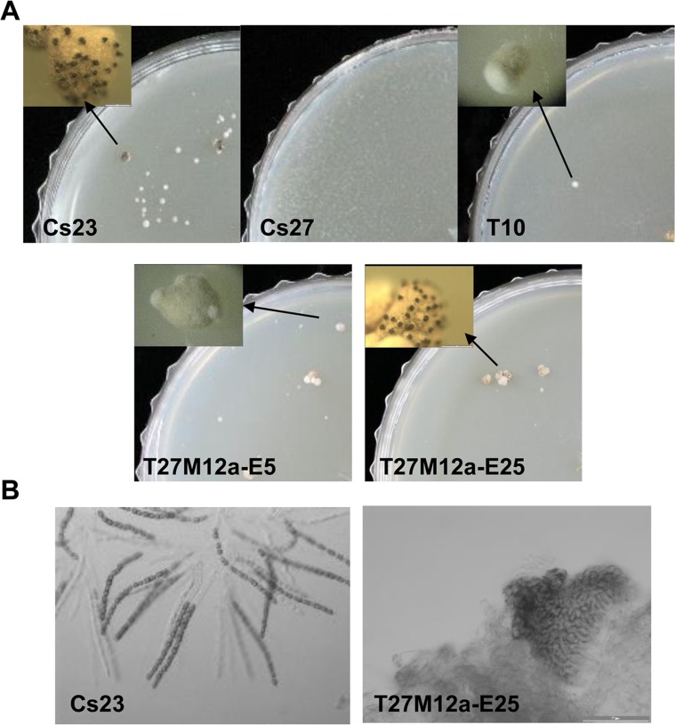
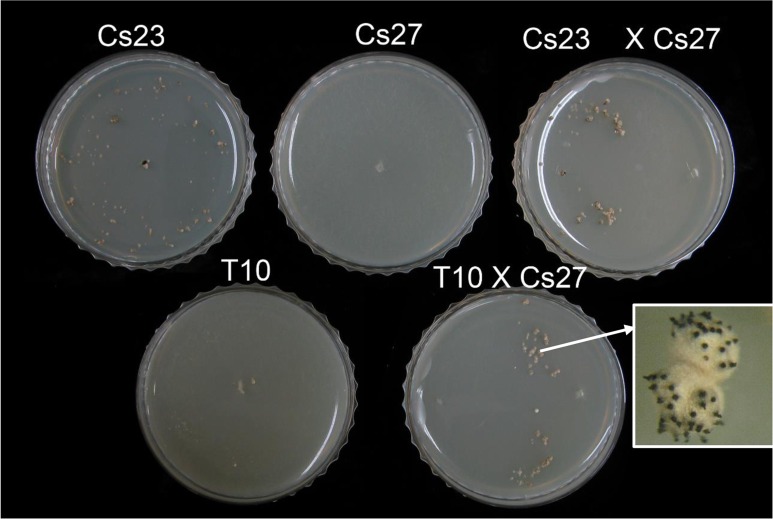
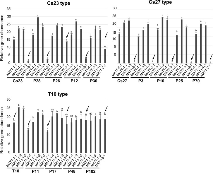
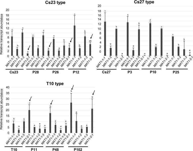
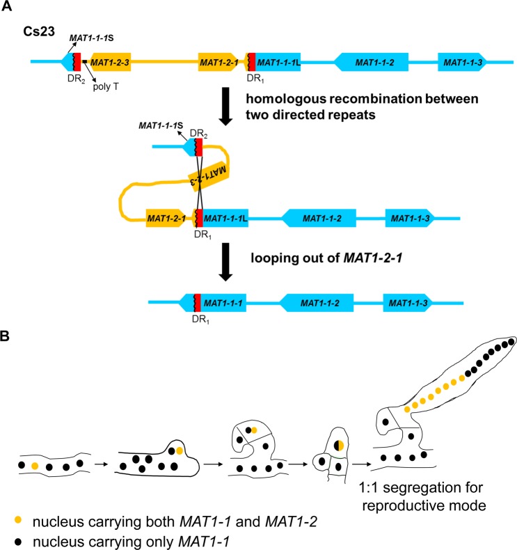
The filamentous fungus Chromocrea spinulosa (Trichoderma spinulosum) exhibits both self-fertile (homothallic) and self-sterile (heterothallic) sexual reproductive behavior. Self-fertile strains produce progeny cohorts that are 50% homothallic, 50% heterothallic. Heterothallic progeny can mate only with homothallic strains, and progeny also segregate 50% homothallic, 50% heterothallic. Sequencing of the mating type (MAT) region of homothallic and heterothallic strains revealed that both carry an intact MAT1-1 locus with three MAT1-1 genes (MAT1-1-1, MAT1-1-2, MAT1-1-3), as previously described for the Sordariomycete group of filamentous fungi. Homothallic strains, however, have a second version of MAT with the MAT1-2 locus genetically linked to MAT1-1. In this version, the MAT1-1-1 open reading frame is split into a large and small fragment and the truncated ends are bordered by 115bp direct repeats (DR). The MAT1-2-1 gene and additional sequences are inserted between the repeats. To understand the mechanism whereby C. spinulosa can exhibit both homothallic and heterothallic behavior, we utilized molecular manipulation to delete one of the DRs from a homothallic strain and insert MAT1-2 into a heterothallic strain. Mating assays indicated that: i) the DRs are key to homothallic behavior, ii) looping out of MAT1-2-1 via intra-molecular homologous recombination between the DRs in self-fertile strains results in two nuclear types in an individual (one carrying both MAT1-1 and MAT1-2 and one carrying MAT1-1 only), iii) self-fertility is achieved by inter-nuclear recognition between these two nuclear types before meiosis, iv) the two types of nuclei are in unequal proportion, v) having both an intact MAT1-1-1 and MAT1-2-1 gene in a single nucleus is not sufficient for self-fertility, and vi) the large truncated MAT1-1-1 fragment is expressed. Comparisons with MAT regions of Trichoderma reesei and Trichoderma virens suggest that several crossovers between misaligned parental MAT chromosomes may have led to the MAT architecture of homothallic C. spinulosa.


Conflict of interest statement

The authors have declared that no competing interests exist.

Figures

Fig 1
Fig 1. Structural organization of the MAT regions in homothallic and heterothallic C. spinulosa strains and comparisons with MAT in closely related heterothallic Trichoderma spp., and homothallic F. graminearum.
Homothallic Cs23 has two MAT structures, while heterothallic Cs27 has one. Open reading frames (ORF) are depicted as large arrows indicating the direction of transcription. Homologs are indicated by the same gene name and color. Direct repeats (DRs) are shown as vertical red bars. 1-1-1, 1-1-2, 1-1-3, 1-2-1, 1-2-3: MAT1-1-1, MAT1-1-2, MAT1-1-3, MAT1-2-1, MAT1-2-3, respectively. MAT1-1-1 partial ORFs are indicated by a vertical wavy line. The MAT1-1 genes (MAT1-1-1, MAT1-1-2, MAT1-1-3) and MAT1-2 genes (MAT1-2-1, MAT1-2-3) are indicated by a horizontal bar. HP: encodes a hypothetical protein; APN2: similar to DNA lyase 2; COX13: similar to cytochrome c oxidase polypeptide VIa; APC5: similar to anaphase-promoting complex subunit 5; CIP: similar to complex I intermediate-associated protein 30; RBP: similar to a protein carrying RNA-binding motif. Note MAT1-2-3 is fused to a fragment of MAT1-1-1 in T. reesii QM6a. Numbers represent kilobases.
Fig 2
Fig 2. Structural organization of MAT in C. spinulosa homothallic and heterothallic strains.
(A). Homothallic strains carry two versions of the MAT region, as shown. In the version with four MAT genes, two identical 115-bp direct repeats (designated DR1 and DR2) are present. DR1 is at the 3’ end of the larger truncated fragment of the MAT1-1-1 ORF (MAT1-1-1L). DR2 is associated with the smaller truncated MAT1-1-1 ORF (MAT1-1-1S) that is adjacent to MAT1-2-3. A homopolymeric tract (poly T, indicated by a black bar) is found in the region between MAT1-1-1 and the MAT1-2-3 ORF. In contrast, heterothallic strain Cs27 has only one (DR1) of the two direct repeats and no poly T tract. A second version of MAT with a structure similar to Cs27 MAT is also found in Cs23. MAT1-1-1 is intact in this version. Primer positions, expected sizes of amplified fragments, and direction (arrowheads) are indicated. BamHI sites and expected fragment sizes are shown.(B). PCR amplification of MAT genes from Cs23, Cs27, and progeny of a self of Cs23 using MAT-specific primers (indicated in A). Primer pairs CP1/2 and CP3/4 amplified products from homothallic strains only. Agarose gels: leftmost lanes in each panel: 100 bp ladder, homo: homothallic progeny, hetero: heterothallic progeny. (C-D). DNA gel blot of genomic DNAs from Cs23 and Cs27 hybridized with either MAT1-1-1 (C) or MAT1-2-1 (D) probes. λ DNA: lambda DNA digested with HindIII, Cs23 and Cs27: BamHI-digested genomic DNA from C. spinulosa Cs23 and Cs27 respectively. The faint 7.1 kb band (C, arrows) was visible in DNA from Cs23 probed with either MAT1-1-1 or MAT1-2-1. Cs27 hybridized (3.5 kb) to the MAT1-1-1 probe but not the MAT1-2-1 probe. The 3.5 kb fragment is also evident in Cs23, supporting the notion that there are two MAT structures in homothallic strains. The lane Cs23 (D) was from the same gel as the other lanes, but with intervening lanes removed. Sizes (in kb) are indicated to the left of the gel.
Fig 3
Fig 3. Targeted deletion of DR2 from Cs23.
(A). A construct carrying the selectable marker hygB plus regions of homology flanking DR2 was introduced into WT self-fertile strain Cs23. Double crossover by homology would replace DR2 with hygB, otherwise the genomic architecture of the region is unchanged. T10, a strain with this type of replacement, also lacks the second version of MAT carrying only MAT1-1.(B) Gel blot of BamHI-digested genomic DNA of Cs23 and candidate DR2-deletion transformant T10, hybridized with MAT1-1-1 or MAT1-2-1. A band at 8.9 kb is evident in T10 with either probe and is of equivalent intensity. The original BamHI-digested Cs23 MAT1-2-1 fragment (7.1 kb) shown in (A) is indicated by red arrows. The 3.5 kb band is present only in Cs23 and only when probed with MAT1-1-1. Sizes (in kb) are indicated to the left of the gel. All lanes are from the same gel; irrelevant lanes between the lanes Cs23 and T10 were cut out of the image.
Fig 4
Fig 4. Self-fertility of WT and transgenic strains.
(A). Self-fertile Cs23 forms stroma bearing perithecia (left panel inset, black dots at arrow) when grown on CMA, while self-sterile Cs27 does not. Strain T10 was able to form some stroma-like hyphal aggregates but no perithecia (top row inset, at arrow). Strains T27M12a-E5 and T27M12a-E25 formed stroma-like structures similar to those in Cs23, and, in the latter case, produced black perithecia immersed in the stroma, (but no asci/ascospores, B). (B). Self-fertile Cs23 forms stroma bearing perithecia with asci with 16 ascospores. Transgenic T27M12a-E25 forms stroma with perithecia but no ascospores.
Fig 5
Fig 5. Introduction of the MAT1-2 region from Cs23 into the genome of C. spinulosa Cs27.
(A and C). Schemes to introduce MAT1-2-1. Plasmids pMAT2 and pM2M1 are described in the text. T27M12a-H1 (B, lane H1, asterisk) and T27M12b-H3 (D, lane H3, asterisk), represent homologous integrants from transformation with each type of plasmid. (B). DNA gel blot of BamHI-digested DNA from control strains Cs23 and Cs27, six transgenic strains (lanes E1, E25, 1–4) carrying MAT1-2-1 at an ectopic position (ectopic), and five transgenic strains (lanes H1, 5–8) generated by homologous recombination as drawn in (A) (homologous), hybridized with a MAT1-2-1 probe. Lane E1: T27M12a-E1; lane E25: T27M12a-E25; lane 1: T27M12a-E7; lane 2: T27M12a-E5; lane 3: T27M12a-E9; lane 4: T27M12a-E18; lane H1: T27M12a-H1; lane 5: T27M12a-H3; lane 6: T27M12a-H4; lane 7: T27M12a-H12; lane 8: T27M12a-H15. All homologous recombinants showed a single hybridizing band of expected size (5.4 kb). Asterisks highlight strains specifically described in the text. (D). Gel blots of the SphI-digested genomic DNAs from the control strain Cs27 and four transgenic strains carrying MAT1-2-1, hybridized with a hygB probe. Lane 1: T27M12b-H1; lane 2: T27M12b-H4; lane H3: T27M12b-H3; lane 3: T27M12b-H7. Integration by homologous recombination as drawn in (C) should generate a single hybridizing band of 13.0 kb (e.g., lane H3). Note some lanes had more complicated patterns likely reflecting both homologous and ectopic integration events. Sizes (in kb) are indicated on the left.
Fig 6
Fig 6. Outcrosses of self-sterile strain T10 to self-sterile Cs27 are fertile.
Top row: Cross plates showing selfs of control Cs23 and Cs27 strains and a cross between them. Note stroma with perithecia formed on the Cs23 and the cross plate, as in Fig 4. Bottom row: Cross plates of a self of self-sterile T10 and a cross between T10 and self-sterile Cs27. Note formation of stroma bearing fertile perithecia on the cross plate.
Fig 7
Fig 7. Relative amounts of individual MAT genes determined by qPCR using genomic DNA.
Amplification level (amount) of MAT1-2-1 is indicated by arrows, with MAT1-2-1 level in Cs23 as a reference. The same letter above bars indicates no significant difference. The three panels represent Cs23 and Cs23-type progeny, C27 and Cs27-type progeny and T10 and T10-type progeny from the T10 by Cs27 crosses. Primer pairs used: for MAT1-1-1: qCsmat1-1for7 & qCsmat1-1rev7, for MAT1-1-2: qCsmat1-2for6 & qCsmat1-2rev6, for MAT1-1-3: qCsmat1-3for4 & qCsmat1-3rev4, for MAT1-2-1: qCsmat2for4 & qCsmat2rev4.
Fig 8
Fig 8. Relative transcript levels for individual MAT genes determined by qPCR using total RNA.
Amplification level (amount) of MAT1-2-1 is indicated by arrows, with MAT1-2-1 level in Cs23 as a reference (set to 1). The same letter above bars indicates no significant difference. The three panels represent Cs23 and Cs23-type progeny, C27 and Cs27-type progeny and T10 and T10-type progeny from the T10 by Cs27 crosses, as in Fig 7. Primer pairs used as in Fig 7.
Fig 9
Fig 9. Proposed model for elimination of MAT1-2 and acquisition of concomitant homothallic capability involving recognition between two types of nuclei in the same individual.
(A). Looping out of the MAT1-2 region from self-fertile C. spinulosa strain 23 by homologous recombination between the 115-bp direct repeats. (B). Model for C. spinulosa ascus development. The two types of nuclei in the ascogenous hyphae are present in unequal proportions, with nuclei carrying MAT1-1;MAT1-2 in the minority. A nucleus carrying MAT1-1;MAT1-2 (yellow) recognizes and pairs with a nucleus carrying MAT1-1 only (black) and migrates into the developing crozier. The nuclear pair are partitioned in the penultimate cell of the crozier where they undergo karyogamy, meiosis, and ascus development. The diploid nucleus undergoes meiosis followed by two rounds of mitosis, yielding 16 ascospores segregating 1:1 ratio for self-fertility and large size versus self-sterility and small size in a single ascus.

Similar articles

Cited by

References

    1. Coppin E, Debuchy R, Arnaise S, Picard M (1997) Mating types and sexual development in filamentous ascomycetes. Microbiol Mol Biol Rev 61: 411–428. - PMC - PubMed
    1. Metzenberg RL, Glass NL (1990) Mating type and mating strategies in Neurospora. Bioessays 12: 53–59. doi: 10.1002/bies.950120202 - DOI - PubMed
    1. Martin T, Lu SW, van Tilbeurgh H, Ripoll DR, Dixelius C, et al. (2010) Tracing the origin of the fungal alpha1 domain places its ancestor in the HMG-box superfamily: implication for fungal mating-type evolution. PLoS One 5: e15199 doi: 10.1371/journal.pone.0015199 - DOI - PMC - PubMed
    1. Nelson MA (1996) Mating systems in ascomycetes: a romp in the sac. Trends Genet 12: 69–74. - PubMed
    1. Turgeon BG (1998) Application of mating type gene technology to problems in fungal biology. Annu Rev Phytopathol 36: 115–137. doi: 10.1146/annurev.phyto.36.1.115 - DOI - PubMed

Substances